
Abstract
Momordica charantia L., known as bitter melon (BM), is a plant that belongs to the family Cucurbitaceae. Aims of this study are to investigate the anti-inflammatory effect of crude BM extract on 2,4,6-trinitrobenzene sulfonic acid (TNBS)-induced experimental colitis model in rat. It was also aimed to determine the content and bioaccessibility of carotenoids of BM. BM was purchased from local markets in Izmir, Turkey. Fruits of BM were lyophilized, powdered, and used in the experiment. Carotenoids were determined by high-performance liquid chromatography. To determine the bioaccessibility of β-carotene, in vitro digestion was performed. Wistar albino rats were divided into four groups: group A (BM+TNBS), group B (BM), group C (TNBS), and group D (control). BM solution was given 300 mg/(kg·day) for 6 weeks orally. Colitis was induced by 0.25 mL of a solution containing 100 mg/kg 5% (w/v) TNBS in 50% ethanol (w/v) intrarectally after 6 weeks. After sacrification, macroscopic and microscopic evaluations were performed. Myeloperoxidase, cytokines levels (interleukin-17 [IL-17], TNF-alpha, and interleukin-10 [IL-10]) were measured in serum and colonic samples by ELISA test. Institutional Animal Ethics Committee approval was obtained. Total carotenoid content of BM was determined 11.7 mg/g dry weight as β-carotene equivalents. Bioaccessibility of total carotenoids was determined as 2.1% with in vitro digestion. Pretreatment with crude BM extract significantly reduced weight loss, macroscopic, and microscopic colitis damages in colonic samples (P = .000), (P = .015), and (P = .026), respectively. Serum anti-inflammatory cytokine IL-10 increased significantly in both treatment groups (P = .000). BM is a rich source of carotenoids, but the bioaccessibility of its carotenoids is low. This study displays that BM has protective anti-inflammatory effects on TNBS-induced colitis.
Introduction
Inflammatory bowel diseases (IBDs) are chronic inflammatory conditions of the intestinal tract. Current medical therapies in IBDs are still far from being curative. Therefore, there is a great effort to find new and effective agents for IBD treatment. 1 Momordica charantia L., also known as bitter melon (BM), bitter apple or bitter gourd, is a yellow-colored member of the family Cucurbitaceae. It has been used in traditional medicine worldwide as a remedy for the management of hyperglycemia, oxidative stress, cancer, and inflammatory conditions such as colitis. 2 –8 The health benefits and biological activity of BM have been usually attributed to the compounds such as triterpenoids, saponins, proteins, and carotenoids it contains. 9 –11
Carotenoids are a large group of yellow to red natural pigments. So far, >700 carotenoids have been discovered. The major dietary carotenoids are β-carotene, α-carotene, lycopene, β-cryptoxanthin, lutein, and zeaxanthin. 12 Studies have shown that carotenoid consumption and tissue levels are associated to the prevention of some kind of cancer, 13 diabetes, 2,14 and IBDs. 12,15 Therefore, the carotenoid contents of BM may be the responsible bioactive compounds due to their antioxidant and anti-inflammatory properties. 16 –18 However, there are a few studies related to the carotenoid contents of BM and there are no data about the bioaccessibility of its carotenoids in the literature. In those studies, nutritionally important phytoconstituents of BM, including carotenoids, were quantified. 10,11,18
Since not all the consumed amount of carotenoids enter the systemic circulation, absorbed amount of carotenoids is more important than the consumed amount within the food. A prerequisite for the carotenoid absorption is its bioaccessibility in the intestine. The carotenoid bioaccessibility is the fraction of ingested carotenoids released from the food matrix in the gastrointestinal tract during digestion and thus becomes available for intestinal absorption. Food matrix, localization of carotenoids in plant tissues, emulsification into lipid droplets in the stomach, and incorporation into mixed micelles affect the bioaccessibility of the carotenoids. 19 Determination of the bioaccessibility of carotenoids has a great importance because this is a sign that carotenoids enter into the colon and may release from the food matrix and may interact with the intestinal medium and colonocytes.
Consequently, the aims of this study are to investigate the anti-inflammatory effect of crude BM extract on 2,4,6-trinitrobenzene sulfonic acid (TNBS)-induced experimental colitis model in rats. It is also aimed to demonstrate the in vitro bioaccessibility of total carotenoids and β-carotene of BM.
Materials and Methods
Plant material
Matur fruits of BM were purchased from local market in Seferihisar, a town located 48 km away from Izmir in Turkey. Plants were washed, separated fruits from their seeds, and taken to pieces. Fruits of the plants were stored in −80°C until been lyophilized. Frozen fruits were lyophilized at −48°C ± 2°C, 13.33 kPa absolute pressure (FT33; Armfield, UK). Lyophilized fruits were powdered by a grinder and were stored at +4°C in refrigerator until the experiment.
Static in vitro digestion and bioaccessibility of BM
Chemicals
4-Nitrophenyl α-
Methods
Determination of β-carotene and total carotenoids
β-Carotene and total carotenoids were determined in lyophilized BM. Total carotenoids were calculated as β-carotene equivalents. Results were given as milligram carotenoid per gram dry weight BM.
Solutions of β-carotene standard (1 mg/mL) were freshly prepared in n-hexane. Diluted solutions (1–500 μg/mL) were prepared from this stock solution. Carotenoid extraction of undigested and digested samples was performed by the addition of extraction solvent containing hexane, acetone, and ethanol (50:25:25, v/v/v) to the sample. Then sample solution homogenized in Ultra-Turrax (T25 Digital Ultra-Turrax; IKA, Germany) at 2590 g for 2 min to obtain a homogenous sample. After this step, the certain amount of sample and extraction solvent were combined and allowed incubation at 5 g for 20 min. Then distilled water was added and the mixture was centrifuged at 9500 g and +4°C (IEC Cl312 Multispeed Centrifuge; Thermo Scientific, Germany). The hexane layer was taken for carotenoid analysis.
β-carotene and total carotenoids of the samples were determined by high-performance liquid chromatography (HPLC) proposed by Bhatnagar-Panwar et al. 20 For HPLC analysis aliquots of 20 μL were injected into ODS-2 reverse-phase C18 column (5 μm, 4.6 mm × 250 mm) at 30°C. The mobile phase consisted of methanol containing 0.5% triethylamine, acetonitrile, and chloroform. Elution was done using solution A containing methanol, acetonitrile, chloroform (50:40:10 v/v/v) and solution B containing methanol, acetonitrile, chloroform (35:35:30 v/v/v). The column was developed with solution A for the first 2 min, then a linear gradient with solution B was applied for a period of 8 min, after which solution A was pumped through the column for another 10 min. Flow rate was 1.2 mL/min.
In vitro gastrointestinal digestion
To determine the bioaccessibility of β-carotene, samples were in vitro digested according to the procedure described by Minekus et al. 21 In this consensus protocol, within the COST FA1005 INFOGEST network, the practical static digestion method is based on human gastrointestinal physiologically relevant conditions. Before digestion, the enzymatic activities of enzymes, as well as the bile salt concentration in the porcine bile extract were determined. The main steps of digestion are given as follows.
Five grams of lyophilized sample was mixed with 5 mL of simulated gastric fluid electrolyte stock solution containing pepsin solution (20,000 U/mL) for 2 h and finalized with intestinal digestion by addition of simulated intestinal fluid electrolyte stock solution containing bile extract solution and pancreatin solution (800 U/mL, based on trypsin activity) under stirring for 2 h at 37°C. The soluble fraction at the end of intestinal digestion was obtained by centrifugation (10,000 g at 5°C for 20 min) and frozen at −20°C until further analysis.
Bioaccessibility of β-carotene and total carotenoids
The proportion of β-carotene and total carotenoids released from the food matrix into the digestive fluids after digestion was calculated as follows:
Bioaccessibility (%) = ([β-carotene digesta]/[β-carotene matrix]) × 100 where β-carotene digesta is the concentration of β-carotene collected in the supernatant phase at the end of the intestinal digestion, and β-carotene matrix is the concentration of β-carotene in matrices after chemical extraction.
Bioaccessibility (%) = ([total carotenoids digesta]/[total carotenoids matrix]) × 100 where total carotenoids digesta is the concentration of total carotenoids collected in the supernatant phase at the end of the intestinal digestion, and total carotenoids matrix is the concentration of β total carotenoids in matrices after chemical extraction.
Animals and experiment groups
The study was approved by Ege University Animal Ethic Committee (2016-087).
Wistar albino rats were provided from Ege University Laboratory Animal Research Center (Izmir, Turkey). The rats were housed at the Experimental Animal Center Laboratory of Ege University in single cages individually in air-conditioned room, which was maintained on 12 h light/12 h dark cycle at constant temperature 22°C ± 2°C with 48–51% humidity.
After 1-week acclimation period, the rats were divided randomly into four groups. Each group contains five males and five females with mean body weight 290 ± 10 g.
Group A (BM+TNBS group) and group B (BM group) were treatment groups and animals received BM solution with water, whereas group C (TNBS group) and group D (Control group) received normal water without BM for 6 weeks orally. After 6 weeks, animals in group A and group C were exposed with TNBS intrarectally (IR) for induction of colitis, whereas group B and group D were exposed with saline IR for sham. BM solution was administered 300 mg/(kg·day). The calculated amount of crude BM extract was dissolved with 1:1 lecithin in 50 mL water to prepare BM solution. Animals in all groups were fed a standard pellet diet with similar calorie intake and allowed free access to water ad libitum throughout the entire study. The animals fed with standard pellets diet. Body weights of rats were measured at the beginning, then before induction of colitis at the sixth week and before sacrification at the seventh week. The design and animal groups of the experiment are shown in Figure 1.

The design and animal groups of the experiment. BM, bitter melon; IR, intrarectally; TNBS, 2,4,6-trinitrobenzenesulfonic acid.
Induction of TNBS-induced colitis
TNBS was purchased from Sigma Chemical Co. On the sixth week of the experiment all animals were fasted for 12 h but given free access to water. The rats were lightly anesthetized with 10% chloral hydrate by an intraperitoneal route. A flexible catheter with a 2 mm diameter was inserted 6–8 cm rectally into the distal colon of the rats. Experimental colitis was induced by 0.25 mL solution containing 100 mg/kg of 5% (w/v) TNBS dissolved in 50% ethanol (w/v) as described in the literature by Morris et al. 22 An equal volume of saline was administered IR for sham groups (BM and control groups).
Collection of blood and colonic tissue samples
After 7 days of induction of colitis, rats were anesthetized with 0.4 mL 10% alfamine and 2% alfazyne (1:1) by intraperitoneal route after overnight fasting. Blood samples were collected intracardiac route. After blood collection, the distal colon was harvested, opened longitudinally, rinsed with saline and stored at −80°C for cytokines and myeloperoxidase (MPO) analysis, and after macroscopic scoring, colonic tissue was stored in 10% formaldehyde for histopathological examination.
Assessment of colitis in colonic samples
Macroscopic scores
The macroscopically visible lesions were assessed by two blinded observers of the experimental group. Lesions were scored on a scale ranging from “0” to “10” based on the criteria of colonic adhesions to surrounding tissues, strictures, mucosal ulcerations, and wall thickening, which were described by Vilaseca et al. 23
Microscopic scores
Colonic tissue samples were assessed by a blinded pathologist. Routine techniques were used for the samples before embedding in paraffin. For the histological assessment, sections were stained with hematoxylin and eosin and coded for blindness. Lesions were scored on a scale ranging from “0” to “10” based on the presence of ulcerations, the degree of inflammation, depth of the lesions and presence of fibrosis. 23
Measurement of cytokines and MPO in serum and colon by ELISA
Rat colon tissue homogenization
Rat colon samples were incised and weighed 1 mL of phosphate buffer saline with pH = 7.4 were added to 200 mg of rat colon tissue sample and immediately were frozen with liquid nitrogen. On the day of measurements, samples were thawed and were conserved in 2–8°C and homogenized thoroughly by a homogenizer (Ultra-Turrax® T25; Janke & Kunkel IKA Labortechnik). Homogenized samples were centrifuged at 640 g for 20 min. The supernatant was collected carefully and examinations of tests were performed.
ELISA test procedures
Interleukin-17 (IL-17), interleukin-10 (IL-10), tumor necrosis factor alpha (TNF-α), and MPO were measured both in serum and in the supernatant of homogenized colonic tissue of rats. They were measured in duplicate by sandwich ELISA on the basis of the biotin double antibody sandwich technology (Cat. No. E0108Ra, Cat. No. E0115Ra, and Cat. No. E0764Ra, respectively) (Bioassay Technology Laboratory, Shanghai Korean Biotech Co. Ltd, Shanghai, China). ELISA was carried out as described in the manufacturer's instructions. A multiplate photometer is used for ELISA plate reading (Multiskan® EX Microplate Photometer; Thermo Scientific, Waltham, MA, USA). All samples were checked in duplicate.
Statistical analysis
Statistical analysis was performed with SPSS 20.0 statistical software. Data were analyzed by using one-way analysis of variance test and Mann–Whitney U test. P-values <.05 were accepted statistically significant.
Results
Carotenoids content of BM and in vitro bioaccessibility of carotenoids
Total carotenoid content of BM was 11.73 ± 0.35 mg/g dry weight as β-carotene equivalents. This value shows that BM is a rich source of carotenoids (541.4 ± 16.8 μg/g wet weight) compared with the other carotenoid-rich foods such as carrot (57.2 μg/g), and spinach (121.3 μg/g). 24 β-Carotene content of BM was 1.57 mg/g dry weight. The in vitro bioaccessibility of total carotenoids and β-carotene of BM was found as 2.12% ± 0.07% and 0.82% ± 0.04%, respectively.
Induction of colitis and body weight changes
MPO is a good indicator for the induction and severity of colitis. The mean colonic MPO levels were measured as follows: 7.5 ± 1.8 ng/mL in BM+TNBS group, 5.9 ± 0.8 ng/mL in BM group, 7.9 ± 2.8 ng/mL in TNBS group, and 6.0 ± 0.9 ng/mL in control. There were not found any statistically significant differences between the two colitis groups and between the two noncolitis groups compared with mean colonic MPO levels (P < .05). The mortality rates in colitis groups, namely BM+TNBS and TNBS groups were 40% and 30%, respectively. There was not any statistically significant difference between two groups according to mortality rates (P > .05). The body weight changes of groups between precolitis and presacrification period are shown in Table 1. Statistically significant differences were found between group A and C, group B and C, as well as between group C and D according to mean body weights (P = .000) (Table 1).
Body Weights Changes of Animals
P < .05 versus TNBS group, # P < .05 versus BM group, ** P < .05 versus control.
BM, bitter melon; SEM, standard error of the mean; TNBS, 2,4,6-trinitrobenzene sulfonic acid.
Macroscopic and microscopic colitis scores
Distal colon of rats was macroscopically evaluated (Fig. 2). Median macroscopic scores were as follows: 5 points (3–7) in group A, 1 point (1–1) in group B, 8 points (6–10) in group C, and group D had “0” point. There was a statistically significant difference between groups A and C according to median macroscopic scores (Mann–Whitney U test, U = 3.5, P = .015) (Fig. 3).
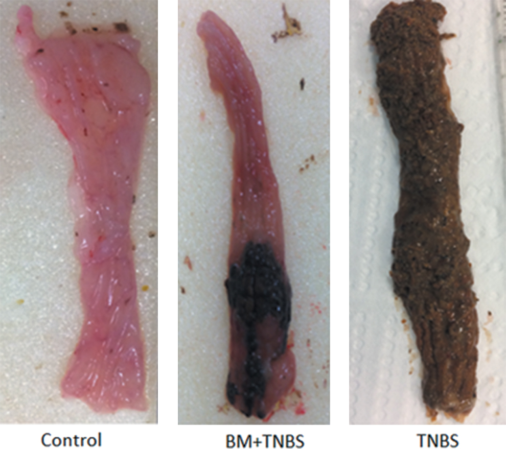
Macroscopic appearances of colonic samples. Color images are available online.

Macroscopic activity scores of colitis in colon. *There is statistically significant difference between BM+TNBS and TNBS groups compared with macroscopic colitis scores according to Vileseca (Mann–Whitney U test, P < 0.05). Color images are available online.
Sections of paraffin-embedded distal colon of rats were histologically evaluated (Fig. 4). Median microscopic colitis scores (min–max) were found as follows: 7.5 points (1–8) in BM+TNBS group and 9 points (4–9) in TNBS group. Median microscopic colitis scores were “0” point in BM and control groups. A statistically significant difference was found between BM+TNBS and TNBS groups according to microscopic scores (Mann–Whitney U test, U = 4.0, P = .026) (Fig. 5).
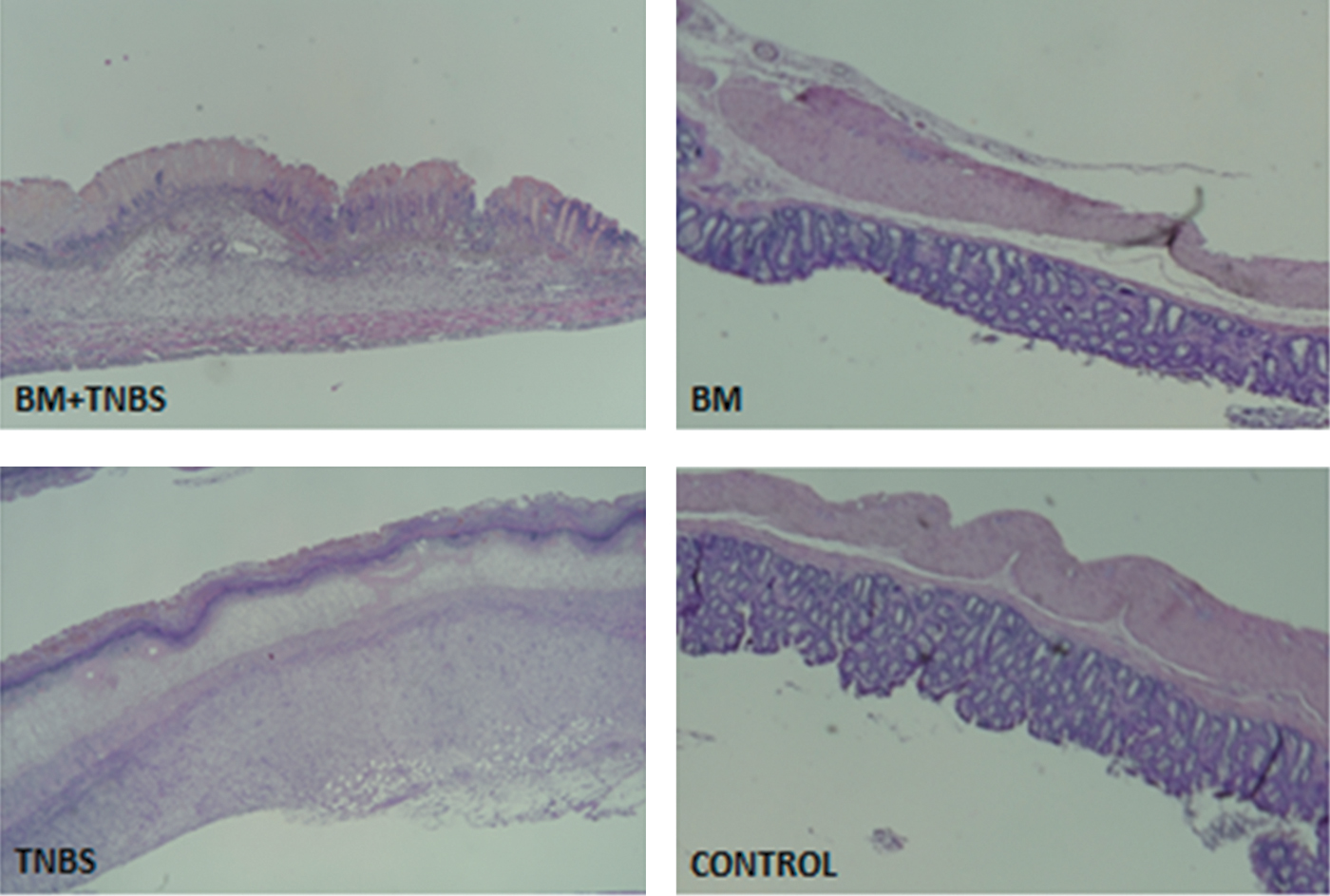
Histological appearances of colonic samples with hematoxylin and eosin stain. Color images are available online.
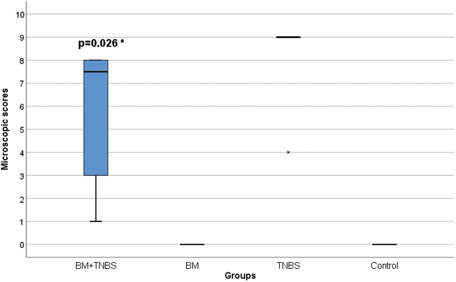
Microscopic activity scores of colitis in colon. *There is statistically significant difference between BM+TNBS and TNBS groups compared with microscopic colitis scores according to Vileseca (Mann–Whitney U test, P < .05). Color images are available online.
Cytokines levels in serum and colonic tissue of rats
Proinflammatory cytokines; IL-17, TNF-α, and anti-inflammatory cytokine IL-10 were measured both in serum and colonic samples. The mean cytokines levels are shown in Table 2. There were not found any statistically significant differences among groups according to mean serum and colonic proinflammatory cytokines levels. Likewise, there was no statistically significant difference according to mean colonic anti-inflammatory cytokine IL-10 levels (P < .05). But statistically significant difference was found among groups according to mean serum IL-10 levels (P = .03) (Table 2).
The Mean Serum and Colon Cytokines Levels of Groups
P < .05 versus TNBS group and # P < .05 versus control group (one-way analysis of variance).
IL-17, interleukin 17; IL-10, interleukin 10; NS, not significant; SEM, standard error of the mean; TNF-α, tumor necrosis factor alpha.
Discussion
This is the first study that evaluates the anti-inflammatory effect of crude BM extract on TNBS-induced colitis in rats and determines the in vitro bioaccessibility of carotenoids in BM. Our study revealed that crude BM extract significantly reduced colitis-induced weight loss and alleviated colonic damage scores of colitis in a TNBS-induced colitis model. Also crude BM extract exhibited a significant increase in the serum anti-inflammatory cytokine IL-10 levels but did not provide any significant reduction in proinflammatory cytokines levels; IL-17 and TNF-α both in serum and colonic tissue. There are few studies in the literature that address anti-inflammatory effects of BM. 4,5,25 –27 Lu and Linet al. 5 investigated the anti-inflammatory effects of extracts of BM on DSS-induced colitis model in mice. In that study oral administration of ethyl acetate and ethanol extracts of BM caused a decrease in the proinflammatory cytokines levels, especially IL-6 and IL-1β and an increase in IL-10 both in serum and colonic tissue.
Our results with crude BM extract showed an increase in IL-10 levels as the systemic response but no decrease in the proinflammatory cytokines levels in serum or colon. It is very well known that IL-10 plays potent anti-inflammatory and immunomodulatory roles in the inflammatory balance by reducing the production of proinflammatory molecules and the activity of Th1 cells. 28 Therefore, the significant improvement in colonic damage scores and colitis-induced weight losses may be associated with the increased systemic IL-10 levels. Even though the mean colonic IL-10 level in BM+TNBS group was higher than those in TNBS and control groups, but the differences did not reach statistical significance.
Contradictory results between the two studies in terms of proinflammatory cytokines may have been caused by different colitis models and animal species. Although DSS and TNBS colitis belong to the group of chemically induced colitis animal models, DSS-induced colitis reflects acute, whereas TNBS-induced colitis is a hapten-induced colitis model that leads to Th1-mediated immune response such as Crohn's disease mimicking IBD. 29 –31
We preferred to use in this study crude BM extract instead of purified bioactive molecules of BM because it is still in debate which bioactive component of BM, including alkaloids, terpenoids, saponins, tannins, flavonoids, and carotenoids, has the most anti-inflammatory effects. 1,32 In addition, there were not any studies on bioaccessibility of carotenoids, which are the potent candidate of anti-inflammatory effect of BM. We found total carotenoid 11.7 mg/g dry weight as β-carotene equivalents and β-carotene 1.57 mg/g dry weight in BM fruits. This value shows that BM is a rich source of carotenoids compared with the other carotenoid-rich foods such as carrot and spinach. 24 This value is higher than that of reported in the literature. 33,34 These variations among the carotenoids content of BM can explained by the differences in species, genotypes, and different maturation stage of the fruit. 35
Bioaccessibilities of total carotenoids and β-carotene of BM were found as 2.12% ± 0.07% and 0.82% ± 0.04%, respectively. The bioaccessibility is also a sign of the percentage of carotenoids that can enter into the colon after in vitro digestion. Once they reach the colon, they can exert their specific health benefits, including antioxidant activities, and may interact with the intestinal medium and colonocytes. Generally, bioaccessibility of carotenoids is low due to their lipophilic nature and their localization in the plant food matrix. They can be released from the food matrix if they can incorporate into the mixed micelles during the digestion step. The low bioaccessibility of carotenoids indicates that carotenoids in the food matrix can enter the colon and can be available for the fermentation by gut microflora. In addition, carotenoids that reach to the colon can exert their antioxidant activity. Goñi et al. reported that >70% of lycopene was released during colonic fermentation of indigestible portion of fruits. 36 They found a similar result for β-carotene, for which at least 50% was released by colonic bacteria metabolism. They suggested that the high release of β-carotene from the food matrix during colonic fermentation might help to counteract the oxidizing compounds generated during colonic fermentation, especially in the distal colon.
This study has the following limitations: only standard dosage and just crude BM extract without any purified bioactive compounds extracted from BM. Longer usage with higher doses of BM may improve its protective effects on colitis. Further studies are needed to determine the anti-inflammatory effect potency and toxicity in different dosages. Using BM as a pretreatment, which actually does not fit in the actual life, was another limitation of this study.
In conclusion, finally, it should also be stated that the studies on the anti-inflammatory effect of purified bioactive molecules of BM are needed to find out the most important anti-inflammatory components. According to this study results we suggest that BM plays a protective role on TNBS-induced experimental colitis. This effect might be related to both carotenoids and the other bioactive compounds of BM. There has been an increasing interest in experimenting the medicinal plants to find new horizons in IBD treatment and BM can be one of the potent candidates of this target.
Footnotes
Author Disclosure Statement
No competing financial interests exist.
Funding Information
This study was funded by the Ege University Scientific Research Projects (Project ID/number: 80/2017-TIP-007).