
Abstract
Rosa canina is a well-known medicinal plant used in folk remedy that alleviates various disorders, including inflammation, gastritis, and diarrhea. The objective of this investigation was to identify and quantify the phenolic components of R. canina methanolic extract (RCME) and to determine its protective action with dextran sulfate sodium (DSS)-generated mice colitis model. RCME chemical analysis was done using Liquid Chromatography–Electrospray Ionization–Tandem Mass Spectrometry, and experimental animals received RCME at different doses before colitis induction by oral DSS administration during 7 days. Another group received sulfasalazine as a positive control. Colitis damages and RCME benefits were assessed using histopathological and biochemical changes and improvements. Many phenolic compounds have been identified. In addition, the DSS intoxication induced an alteration of colonic epithelium associated with an oxidative stress state. DSS administration led to an increase or decrease of intracellular mediators such as free iron and ionizable calcium. RCME consumption effectively protected against colonic histological/biochemical alterations induced by DSS intoxication providing support for the traditional use of this plant.
Introduction
Inflammatory bowel disease (IBD) is designed by the chronic inflammatory disorders that involve the gastrointestinal tract and it is distinguished by alternating stages of clinical relapse and remission. 1 IBD includes two main subtypes, ulcerative colitis (UC) and Crohn's disease (CD), which affect more than five million people in the world. 2 In UC, the pathogenesis starts in most of the cases with a rectal-mucosal inflammation; then, it may extend and affect the whole colon. 3 The related manifestations were reported to be abdominal pain symptoms, rectal bleeding, and frequent watery stools. 4 Currently, the exact cause of UC is not entirely clarified, but it seems to be related with an immunoregulation dysfunction in the gut. Various immunoregulatory factors are involved in the disease constancy; in this regard, previous researches have demonstrated that during this immune dysregulation process there is an excessive production of reactive oxygen species (ROS). 5 Consequently, oxidative stress plays a potential role in the pathogenesis of UC. Oxidative stress is defined as an imbalance between the biochemical processes of ROS generation and those responsible for their control and elimination. 6 This imbalance can occur when the antioxidant defense system is overworked by the increase of oxidants or when the defenses are weakened by a lack of intake and/or production of antioxidants. The equilibrium or redox homeostasis is disrupted, and cells become vulnerable to ROS attacks. 7
Despite the extensive research, until now there is no available medical therapy for UC pathogenesis. 8 As a result of the increase in the number of patients suffering from ulcerative colitis in the world, it's an urgent call or a challenge to find a cure to diverse inflammatory disorders using a suitable potential antioxidant. Alternative medicine is the recent clue to prevent patient from UC. Natural products and traditional medicines are of great importance for drug discovery to treat/prevent the IBDs and their associated disruptions. 9
Rosa canina L., belonging to the Rosaceae family, is widespread in the temperate and subtropical zones of the Northern hemisphere. 10
According to the British Herbal Pharmacopoeia (1983), R. canina is frequently used in traditional medicine and widely used as an ornamental plant. 11 It is reported that due to its therapeutic properties, R. canina has many uses such as medicinal, pharmaceutical, and cosmetic usages. 12 Nadpal et al. 13 demonstrated that R. canina extract exerts antioxidant and anti-inflammatory activities. In addition, rose hips are shown to have a wide range of pharmacological characteristics. Őiseth and Nordal 14 reported for the first time the isolation of Tiliroside from hips of R. canina, and the current state of research shows that it has been identified in 172 plant species to date, coming from 35 different families, mostly Rosaceae. 15 As reported by Nowak, 16 the essential oil of R. canina, as analyzed using gas chromatography (GC)/mass spectrometry (MS) method, contained 97 chemical components—mainly vitispiran (isomer), α-E-acaridial, dodecanoic acid, hexadecanoic acid, docosane (C22), β-ionone, 6-methyl-5- hepten-2-one, myristic acid, and linoleic acid. 17
Indeed, it is used in the prevention/treatment of several diseases such as cold, flu, vitamin C deficiency, diabetes, arthritis, sciatica, poor peripheral circulation, gastrointestinal, kidney, and lower urinary tract disturbances. 18 All these biological properties are due to its richness in bioactive components. R. canina is a valuable source of unsaturated fatty acids, 19 carotenoids, 20 vitamin C, polyphenols, 21,22 proanthocyanidins, tannins, 23 minerals, 18 flavonoids, and sugars. 20
The aim of this study was to qualitatively/quantitatively characterize native phenolic profile of R. canina flowers methanolic extract and to examine the possible mechanisms of its effect on DSS-induced inflammatory bowel disease (IBD) model of mice, as well as the confirmation of ethnopharmacological use of this plant as an anti-IBD remedy.
Materials and Methods
Chemicals and reagent
Dextran sulfate sodium (DSS), methanol, formic acid, trichloroacetic acid (TCA), s-butyrylcholine, butylhydroxytoluene (BHT), ethanol, 5.5′-dithiobis-(2-nitrobenzoic acid) (DTNB), and eosin stain were purchased from Sigma-Aldrich Co. (St. Louis, United States). All other chemicals and reagents were obtained from Biomaghreb (Ariana, Tunisia) and are ISO 9001 certified.
Plant material, methanol extract preparation, and choice
R. canina flowers were collected during the month of May (2016) from Zaghouan governorate (North of Tunisia). The collection site was selected according to the Flora of Tunisia. 24 A voucher specimen was identified and authenticated by Mrs Mouhiba Ben-Naceur, professor of taxonomy in the Higher Institute of Biotechnology of Beja, Tunisia and deposited at the herbarium in the Faculty of Sciences, University of El Manar, Tunis, Tunisia. The plant material was oven dried at 40°C for 72 h then ground with an electric blunder (Moulinex Ovatio 2, Serris, France). The extraction of the plant powder was made in 80% methanol (0.1 g/mL) constantly shaken during 24 h at room temperature. After centrifugation, the methanol extract was rotor evaporated and finally freeze-dried. Finally, the Rosa canina methanol extract (RCME) was stored at −80°C until used. The yield of the extract was calculated in grams and converted into percentage. The extraction yield was 11.23% ± 0.12%. Before in vivo manipulation, we evaluate the chemical composition of different extracts formed using various solvents such as water, methanol, ethanol, and acetone. Our preliminary results showed that the methanolic extract contains more phenolic compounds than the other solvents.
Characterization of RCME phenolic compounds by liquid chromatography–electrospray ionization–tandem mass spectrometry analysis
Liquid chromatography–electrospray ionization–tandem mass spectrometry (LC-ESI-MS) profile of RCME (4 mg/mL), filtered through MF-Millipore™ membrane filter with pore size of 0.45 μm (Merck, Darmstadt, Germany), was analyzed using a LC-MS-2020 quadrupole mass spectrometer (Shimadzu, Kyoto, Japan) equipped with an electrospray ionization source and operated in negative ionization mode as described by Chahbani et al. 25 The spectra were monitored in selected ion monitoring mode and processed using Shimadzu LabSolutions software (version 5.32). The mass spectrometer was operated in negative ion mode using a capillary voltage of −3.5 V, a nebulizing gas flow rate of 1.5 L/min, a dry gas flow rate of 12 L/min, a dissolving line temperature of 250°C, a block source temperature of 400°C, a voltage detector of 1.2 V, and the full scan spectra from 50 to 2000 m/z.
Acute toxicity study
A total of 42 mice were divided into 7 groups of 6 each. The methanol extract of R. canina (RCME) was orally administrated to mice in graded doses (100, 200, 400, 800, 1200, 1600, and 3200 mg/kg). The mice were observed for toxic signs and mortality for 7 days.
Experimental procedure
This study was performed in strict accordance with the NIH guidelines for the care and use of laboratory animals and was approved by the Institutional Animal Care and Use Committee of National Institute of Health. 26 Healthy adult male mice (18–22 g) were purchased from the Pasteur Institute of Tunis. They were provided with standard food (standard pellet diet, Badr-Utique, Bizerte, Tunisia) and water ad libitum and maintained in animal house at controlled temperature (22°C ± 2°C) with a 12-h light/12-h dark cycle. All experimental studies were done between 08.00 and 11.00 am to avoid the potential influence of circadian rhythm. For the different experiments, the DSS and sulfasalazine doses were chosen based on previous references. 27,28 However, R. canina methanol extract various doses were obtained following a toxicity study.
A total of 70 mice were randomly divided into 7 groups, a control group and 6 treated groups, of 10 each.
The first received normal drinking water and served as a control group. The second group was given daily an oral administration of RCME (400 mg/kg, b.w., p.o.); group 3 received only 3% DSS (w/v) in distilled water. The groups 4, 5, and 6 were daily treated with DSS and RCME (100, 200, 400 mg/kg, b.w., p.o.) and group 7 which received DSS + Sulfasalazine (100 mg/kg, b.w., p.o.). The RCME was orally administrated to mice for 2 weeks, while the DSS was dissolved in drinking water and given to mice for 1 week (day 7–14). The DSS solution was prepared freshly every other day. Body weight should be measured daily. Twenty four hours before sacrifice, the mice should be deprived of food.
Tissue preparation
After sacrifice, the colons were quickly removed and washed in 0.9% NaCl and their weights were measured. The colons were homogenized with an Ultra-Turrax homogenizer in a phosphate buffered saline solution, centrifuged at 9000 g for 20 min, and stored at 80°C for biochemical analysis.
Histological examination
Colons were removed immediately after sacrifice and emptied of fecal material. Distal colonic fragments were fixed in 10% formalin, dehydrated in graded ethanol solutions, and embedded in paraffin. Sections of 5 mm were deparaffinized, hydrated, and mounted in glass slides. All sections were stained with hematoxylin–eosin and examined under light microscopy.
Protein measurement
The protein measurement was conducted according to Hartree, which is a slight modification of the Lowry method. 29
Lipid peroxidation determination
Lipid peroxidation in colon samples was estimated by the determination of malondialdehyde (MDA) production using the thiobarbituric acid reactive substances (TBARS) test. 30 In this assay, the reaction of MDA with thiobarbituric acid (TBA) forms a colorimetric product proportional to the MDA present in the tested samples. Therefore, 250 μL sample and 250 μL TCA-BHT were vortexed and centrifuged for 10 min at 3000 g. Then, 400 μL supernatant, 80 μL HCl, and 320 μL tris-TBA were incubated at 80°C for 10 min. After cooling, the absorbance of the mixture was measured at 532 nm using a spectrophotometer. The MDA concentration is expressed in nmol MDA/mg of protein.
Nonenzymatic antioxidant measurement
Reduced glutathione (GSH) concentration was performed according to Sedlak and Lindsay method. 31 First, the colon homogenate prepared in EDTA was mixed well with cold water and TCA (50%) in a total volume of 1 mL. After 15 min centrifugation at 1200 g, an aliquot of supernatant was mixed with tris buffer (400 mM) and DTNB (10 mM). Finally, after 5 min incubation, the absorbance was measured at 412. The GSH concentration was expressed as nmol of GSH consumed/min/mg protein.
Sulfhydryl group (-SH) concentrations were done according to Ellman's method 32 ; first, the mixture of colon homogenate, tris-base (250 mM), and EDTA with 100 μL of 20 mM ethylenediaminetetra-acetic acid (EDTA) pH 8.2. Then, the reaction mixture was vortexed, and its optical density was measured at 412 nm (A1). Then, 100 μL of 10 mM DTNB was added and incubated during 15 min, and the absorbance of the sample was measured at 412 nm (A2). The sulfhydryl group concentration was calculated using this expression: (A2 − A1 − B) × 1.57 Mm. The results were expressed as μmo of thiol groups per mg of protein.
Antioxidant enzyme activity assays
Superoxide dismutase (SOD) activity was conducted according to Misra and Fridovich method. 33 The reaction system contained colon homogenate, sodium carbonate/bicarbonate buffer (62.5 mM), bovine catalase (0.4 U/mL), and epinephrine (5.5 mg/ML) in a total volume of 2 mL. Changes in absorbance were measured at 480 nm, and the results have been expressed as units (U) of SOD activity per mg protein. Catalase (CAT) activity was determined according to the method described by Aebi. 34 The reaction mixture contained colon homogenate, phosphate buffer (100 mM), and H2O2 (30 mM) in a total volume of 1 mL. The decrease of absorbance was measured at 240 nm. CAT activity was calculated using the extinction coefficient of 40 mM−1cm−1 for H2O2, and the results are expressed as nmol min−1 mg−1 protein. The glutathione peroxidase (GPx) activity was quantified following the method described by Flohé and Günzler. 35 A 1 mL reaction mixture that contained colon sample, phosphate buffer (100 mM), GSH (4 mM), and H2O2 (5 mM) was incubated for 1min at 37°C. After stopping the reaction with TCA (5%), the mixture was centrifuged for 5 min at 1500 g. An aliquot from supernatant was mixed with phosphate buffer (100 mM) and DTNB (10 mM) and vortexed. The absorbance was measured spectrophotometrically at 412 nm. The GPx activity was expressed as nmol of GSH consumed/min/mg proteins.
ROS production measurement
The colon H2O2 level was carried out following Dingeon et al. 36 method. Briefly, the reaction between p-hydroxybenzoic acid and 4-aminoantipyrine in the presence of peroxidase led to the formation of a quantitative quinone imine detected at 505 nm.
The hydroxyl radical level was determined using Jainu and Shyamala method. 37 Briefly, after oxidation of deoxyribose by hydroxyl radical generated by the Fe3+-ascorbate-EDTA-H2O2 pathway and incubation with intestine homogenate at 37°C for 1 h, the reaction mixture was stopped by adding of TCA (2.8%) and TBA (1%) and boiled at 100°C for 20 min. Changes in absorbance were recorded at 532 nm against blank containing deoxyribose and buffer.
Calcium and free iron measurement
Colonic-mucosa calcium level was performed following Stern and Lewis method. 38 The reaction between calcium and cresolphthalein led to the formation of a colored complex measurable at 570 nm. Colonic-mucosa nonhaem iron was measured following Leardi method. 39 The dissociated iron (Fe3+) from transferrin-iron complex was reduced, by ascorbic-acid action, to form (Fe2+). The newly formed iron (Fe2+) reacts with ferrozine forming a measurable complex at 562 nm.
Statistical analysis
The results were expressed as the mean ± standard error of the mean (SEM). The statistical analysis was carried out using one-way ANOVA software. Values of P < .05 were considered significant.
Results
LC-ESI-MS phenolic profile of RCME
The LC-ESI-MS analyses of RCME were realized (Table 1). In fact, LC-ESI-MS analysis of RCME allows the identification of 15 phenolic compounds, which were classified into 7 phenolic acids and 8 flavonoids. The compound identification was made by comparing retention times and mass spectra with those of the authentic standards. Flavonoids constituted the important group of the identified phenolics of RCME, among which quercitrin was found to be the major compound followed by rutin. Table 1 also shows that quinic acid and gallic acid were the major phenolic acids for RCME. In contrast many researches were reported that R. canina extracts generally contain a high tiliroside level. 16,40,41
Liquid Chromatography–Electrospray Ionization–Tandem Mass Spectrometry Analysis of the Rosa canina Methanolic Extract
The numbering refers to elution order of compounds from an Aquasil C18 column.
Identification was confirmed using 32 authentic commercial standards.
Acute oral toxicity of RCME
In oral toxicity study, mice given graded doses appeared normal, and neither abnormal behavior nor mortality was detected during the experimentation period. Thus, the LD50 value was higher than 3.2 g/kg for the RCME.
Histological estimation
The colon histopathological features of DSS group included massive necrotic injuries in the mucosa and the submucosa, edema, areas of hemorrhages, and inflammatory cellular infiltration (Fig. 1). In addition, the inflammation extended through the mucosa, submucosa, and muscularis mucosa, while RCME alone at 400 mg/kg; b.w. has no effect on the histological structure of the colon. However, RCME treatment greatly reduced the histopathological modifications caused by DSS intoxication. Sulfasalazine also restored the integrity of colonic epithelium.
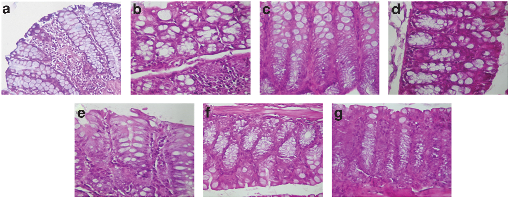
Effect of RCME and sulfasalazine on the microscopic appearances in mice colon with inflammation induced by DSS. Animals were treated with various doses of the RCME (100, 200, and 400 mg/kg, b.w., p.o.), the reference molecule, sulfasalazine (100 mg/kg, b.w., p.o.), or bidistilled water and challenged with oral intake of DSS (3%, w/v) or NaCl 0.9% during 7 days.
Effect of RCME and DSS body weight gain, colon weight, and length
The changes of body weight, as well as colon weight/length, were the main parameters for evaluating the degree of colonic damage in DSS-induced IBD model. As shown in Table 2, normal control mice treated with bidistilled water alone or those who have received only RCME gained body weight. In the DSS group, body weight gradually decreased and did not recover at the end of the experimental period. In contrast, the gained body weight in mice treated with RCME (0.38 ± 0.21 g) or sulfasalazine (−0.6 ± 0.23 g) significantly (P < .05) recovered compared with DSS group (−1.38 ± 0.38 g) and control group (2.18 ± 0.22 g). The DSS-induced colitis reduced also the colonic weight/length compared to the controls (Table 2). However, RCME (100, 200, and 400 mg/kg, b.w., p.o.) significantly (P < .05) and dose dependently protected against DSS-induced colonic weight and length changes.
Effect of Rosa canina Methanolic Extract and Sulfasalazine on Dextran Sulfate Sodium-Induced Changes in Body Weight Gain, as Well as Colon Weight and Length
Animals were treated with various doses of the RCME (100, 200, and 400 mg/kg, b.w., p.o.), the reference molecule, sulfasalazine (100 mg/kg, b.w., p.o.), or bidistilled water and challenged with oral intake of DSS (3%, w/v) or NaCl 0.9% during 7 days. The data are expressed as mean ± SEM (n = 10).
P < .05 compared to control group.
P < .05 compared to colitis group.
DSS, dextran sulfate sodium; RCME, Rosa canina methanolic extract.
Effect of RCME pretreatment on DSS-induced lipid peroxidation
The colonic lipoperoxidation rate was estimated by the MDA content. The DSS exposure induced a significant (P < .05) increase of MDA level (13.5 nmol/mg protein) in the colon compared to the control group (5.7 nmol/mg protein). However, the sulfasalazine administration corrected the effect of DSS. Added to that, the RCME consumption prevented the colonic lipid peroxidation in a dose dependent manner (Fig. 2).
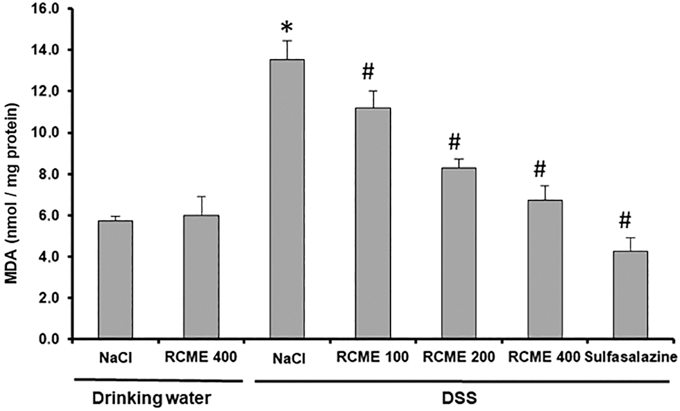
Effect of RCME and sulfasalazine on DSS-induced disturbance in colonic peroxidation of lipids and the generation of MDA. Animals were treated with various doses of the RCME (100, 200, and 400 mg/kg, b.w., p.o.), the reference molecule, sulfasalazine (100 mg/kg, b.w., p.o.), or bidistilled water and challenged with oral intake of DSS (3%, w/v) or NaCl 0.9% during 7 days. The data are expressed as mean ± SEM (n = 10). *P < .05 compared to control group and # P < .05 compared to colitis group. MDA, malondialdehyde.
Effect of RCME and DSS on sulfhydryl groups (-SH) and GSH levels
The results of analyses of nonenzymatic antioxidants are summarized in Figure 3. DSS in drinking water caused also an antioxidant defense system disturbance, as evidenced by the significant (P < .05) decrease of sulfhydryl group (1.8 nmol/mg protein vs. 3.3 nmol/mg protein for the control group) and reduced glutathione levels (64.2 vs. 85.6 nmol/mg protein for the control group). However, pretreatment with the RCME significantly (P < .05) prevented DSS-induced decreases in the -SH groups (3.6 nmol/mg protein) and GSH level (130.1 nmol/mg protein). Noticeably, administration of sulfasalazine at the dose of 100 mg/kg b.w., p.o has also effectively protected against nonenzymatic antioxidant depletion.

Effect of RCME and sulfasalazine on DSS-induced disturbance in colon sulfhydryl groups
Effect of RCME and DSS on antioxidant enzyme activities
The antioxidant enzyme activities such as SOD, CAT, and GPx have been changed following the DSS intoxication (7.7 U SOD/min/mg protein, 1.8 U CAT/min/mg protein, and 0.03 nmol GSH consumed/min/mg protein, respectively) compared to the control group (4.1 U SOD/min/mg protein, 0.9 U CAT/min/mg protein, and 0.02 nmol GSH consumed/min/mg protein, respectively). RCME or sulfasalazine pretreatment showed their efficiencies and have restored these activities at the basal levels (Fig. 4).
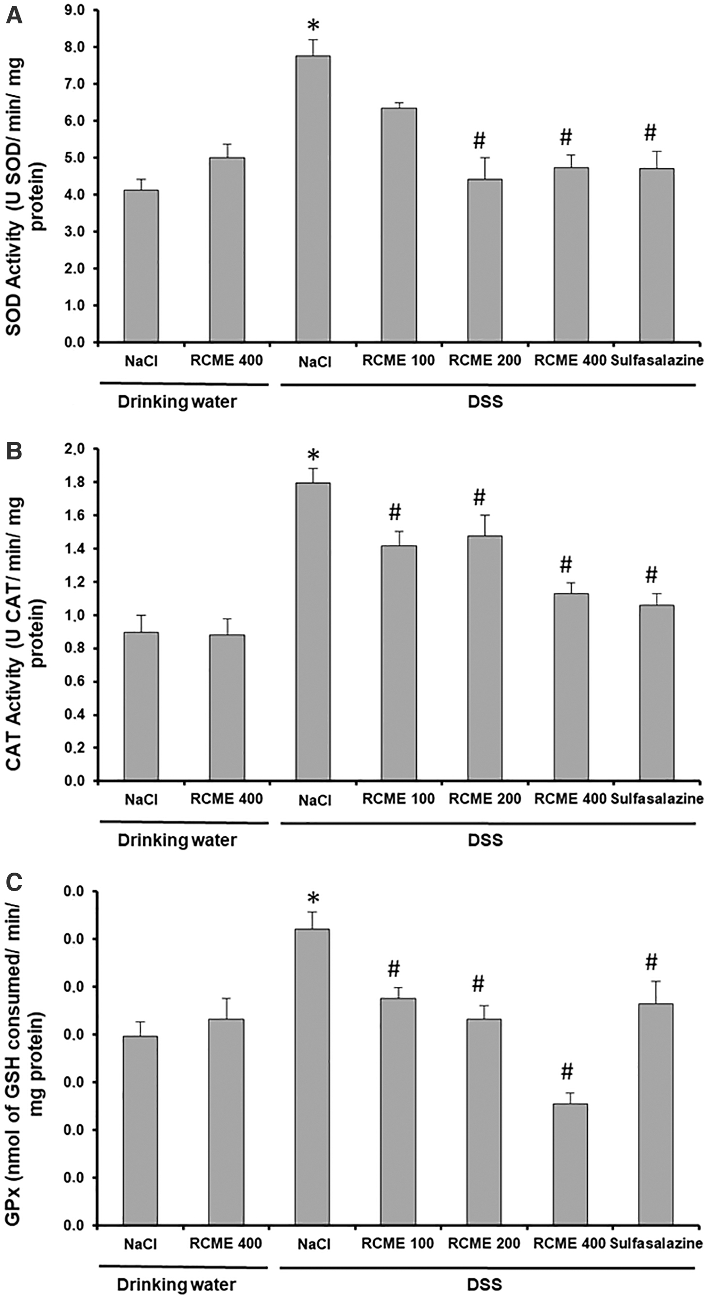
Effect of RCME and sulfasalazine on DSS-induced disturbance in colonic antioxidant enzyme activities like SOD
Effect of RCME and DSS on ROS, free iron, and calcium levels
To explore the oxidative consequences of DSS treatment in the colon and to determine the possible protective effects of RCME, hydroxyl radical (OH
Effect of Rosa canina Methanolic Extract and Sulfasalazine on Dextran Sulfate Sodium-Induced Disturbance in Colon Hydroxyl Radical, Hydrogen Peroxide, Free Iron, and Calcium Levels
Animals were treated with various doses of the RCME (100, 200, and 400 mg/kg, b.w., p.o.), the reference molecule, sulfasalazine (100 mg/kg, b.w., p.o.), or bidistilled water and challenged with oral intake of DSS (3%, w/v) or NaCl 0.9% during 7 days. The data are expressed as mean ± SEM (n = 10).
P < .05 compared to control group.
P < .05 compared to colitis group.
Discussion
The LC-ESI-MS analysis of RCME allows the identification of a wide range of phenolic compounds. It has already documented that R. canina is a valuable source of phenolic compounds, but most of the studies were carried out on rose hips or fruits. 42,43 It was previously reported that rutin, quinic acid, and gallic acid are identified in R. canina aerial parts. 40 The obtained results showed that quercitrin was the major compound in RCME as was previously reported for R. canina rose hips. 16 Besides, gallic acid content in RCME was found to be higher than that reported in R. canina fruits, 44 which could justify the powerful radical scavenging activity of R. canina flowers.
Interestingly, a survey of the literature shows that the major compounds, identified in the RCME, had potent antioxidant potential. In fact, quercitrin (compound 10), rutin (compound 9), and gallic acid (compound 2) presented IC50 values of 12.50, 5.53, and 0.71 μg/mL, respectively, in DPPH• radical scavenging activity. 45 –47 Therefore, R. canina flowers could serve as an important natural source of antioxidants that could exert beneficial pharmacological effects.
In folk medicine from different countries, the R. canina flowers are widely used as a cure for the gastrointestinal tract diseases. 48 In this context, this research was interested in studying the effect of RCME in a mouse model of IBD induced by DSS. Indeed, DSS caused UC is a well-established model. The administration of DSS to mice produced the development of a reproducible acute inflammation. The DSS-induced inflammation is characterized by ulcers accompanied by loss of crypts. Moreover, the DSS model is well used for its simplicity and its similarities with human UC. 28
In this respect, DSS intake in the drinking water of the mice resulted in highly reproducible colitis accompanied with diverse alterations, including the loss of body and colonic weights, rectorrhagia, and diarrhea. The wet weight of the inflamed colon tissue, macroscopic score, body weight, rectal bleeding, and morphological damages were well debated as a sensitive indicator for the severity of inflammation during colitis. In DSS groups, the severe weight loss was significantly and dose dependently prevented by RCME pretreatment in a similar way to the standard drug (sulfasalazine). In contrast, the mice that received only RCME showed the same body weight as the control group.
DSS-induced colitis showed many of the clinical and histopathological inflammatory features found in humans suffering from ulcerative colitis. 49,50 Indeed, DSS-induced colon inflammation begins in the distal colon and progresses to the entire colon and in severe cases the cecum. 49,51 In this context, histological examination of DSS-group colon tissues revealed an extensive UC of the epithelial layer, edema, and crypt variation structure of colon wall related with granulocytes and mononuclear cell infiltration in the mucosa and submucosa. In addition, the data showed a mucosal irregularity characterized by microvillar disturbance. In fact, the DSS toxicity is due to sulfate groups, since the administration of dextran alone does not cause any symptomatology in mice. 52 The observing irritation of the barrier turns out to be mostly of innate type. Indeed, the DSS caused the alteration of the intestinal barrier that brings into contact the flora and the underlying intestinal immunity. The activation of the intestinal immune system and the recruitment of inflammatory cells into the intestine contribute to the maintenance of inflammation and intestinal lesions. 53,54
Quinic acid found in water extracts of medicinal plants such as Uncaria tomentosa can exert an anti-inflammatory action through mechanisms involving reduction of the pro-inflammatory transcription factor nuclear factor kappa B. 55 Quercetin has also been reported to suppress in mice the generations of pro-inflammatory cytokines, such as IL-1b, IL-6, and TNF-α, from macrophages in lipopolysaccharide (LPS)-caused inflammation. 56 Similarly, gallic acid inhibited the levels of NO, PGE-2, and IL-6 formation in LPS-induced cell inflammation. 57
In contrast, we showed that the sulfasalazine treatment alleviated histological-colonic damage caused by DSS. RCME consumption also significantly and dose dependently protected against all histological damage induced by DSS intoxication by restoring the integrity of colonic epithelium. The anti-inflammatory action of RCME is already proved. 58 In this regard, various studies reported the beneficial effect of polyphenols on colonic histological structure. 59
In subjects with UC, it is reported that there is an excessive ROS production.
60
Although ROS are known to be beneficial species, the overproduction induces an alteration of redox homeostasis leading to oxidative stress state. However, it has been proved that chronic oxidative stress is strongly involved in the pathophysiology and perpetuation of UC.
60
In our study, the DSS exposition generated a colon overproduction of H2O2 and OH
However, this ferric imbalance was not observed in RCME pretreated mice and sulfasalazine treated mice. In addition, the calcium homeostasis was also affected in the same way as free iron. In this regard, several previous studies have shown that oxidative stress status provokes a marked increase in Ca2+ concentration in the cytoplasm of bowel cells. 59,62 This powerful ROS scavenging activity of RCME is may be due to its high level of quercetin and its derivate quercetin-3-O-glucuronide, 63 as well as linoleic and alpha linoleic acids that have been shown to inhibit cyclooxygenase-1 (COX-1) and COX-2. 64 In this regard, previous studies demonstrated that COX-1 and COX-2 are involved in the progression of UC by the increasing ROS production. 65
The ROS overproduction is a major contributing factor involved in tissue lipid peroxidation. 66 Therefore, we investigated the MDA level, the final product of lipid peroxidation. As it is expected, the colon MDA concentration, in the DSS induced colitis mice, was extremely high compared to the control group. Yet, the MDA content decreased significantly in the RCME and sulfasalazine treated groups. Flavonoids are known to possess potent antioxidant properties. Several authors proved that lipid peroxidation decreased after treatment with a purified flavonoid, the luteolin. 67 The LC-ESI-MS analysis of RCME revealed an abundant amount of luteolin, which can be the effective inhibitor agent against colonic lipid peroxidation. Furthermore, quercetin component has been indicated to interact with lipid constituents in cell membrane, with a resultant protection of the membranes against oxidative injury, thereby preserving the normal cell functions. 68
More importantly, we investigated the antioxidant profile during different treatments. The nonenzymatic antioxidant response was evaluated by measuring the thiol groups and GSH levels. Indeed, RCME treatment significantly and dose dependently protected against DSS-induced colonic nonenzymatic antioxidant disturbance. Thiols are considered as one of the most important antioxidant molecules, which are heavily involved in the ROS neutralization. 69,70 Furthermore, the GSH is the most abundant nonprotein thiol that is involved in the neutralization of H2O2. In case of increased ROS production, the GSH is oxidized to oxidized glutathione (GSSG). DSS-induced colon thiol groups and GSH disturbance have been shown to be inhibited by many plant extracts such as Lagerstroemia speciosa 71 and Lavandula stoechas. 72,73
The antioxidant system is highly diversified. Antioxidant enzymes play an important role to fight against oxidative damage. The main antioxidant enzyme (SOD, CAT, and GPx) activities were investigated in this study. Indeed, despite its significant molecular weight, DSS can penetrate by pinocytosis inside mucosal cells and thus could directly play a role in cellular oxidation and disruption of enzymatic antioxidant defense system. 74
However, although the DSS caused these enzymatic disorders, the RCME treatment effectively restored the SOD, CAT, and GPx activities to baseline levels. The same toxic mechanism of DSS has been previously described for other organs such as liver 75 and jejunum. 1
Antioxidants have the ability to neutralize free radicals and prevent cell damage due to free radicals. Gallic acid is one of the most abundant flavonoids in RCME. Comparisons with results of previous studies reported that gallic acid (GA) is a strong potent antioxidant. The molecular mechanisms by which GA produces antioxidant actions may involve indirect scavenging of radicals, which was detected in vitro with subcellular fractions, and improvement of antioxidant enzymes through activation of transcription factors such as Nrf2. 76 In comparison with other results, Flos Rosaechinensis and Rosa pomifera 77 is a good source of flavonoids. The antioxidant action of Flos Rosaechinensis mainly depended on its content of these components. 3,4,8,9,10-pentahydroxydibenzo[b,d]pyran-6-one, kaempferol 3-O-(6”-galloyl)-β-D-glucoside, kaempferol 3-O-(2”,6”-digalloyl)-β-D-glucoside, and quercetin 3-O-(2”,6”-digalloyl)-β-D-glucoside showed the stronger antioxidant activity. The antioxidant activities of these compounds might be influenced by the number and position of hydroxyl groups in their aromatic rings. 78
Conclusion
R. canina flowers are one of the most concentrated sources of flavonoids and phenolic acids. Moreover, RCME has been successfully applied in treating colonic lesions induced by DSS, by restoring the integrity of the colonic epithelium, inhibiting the ROS overproduction, and correcting the colon redox balance, as well as reducing free iron and ionizable calcium flow. RCME opens so the possibility of generating health benefits to people who are suffering from IBD.
Footnotes
Author Disclosure Statement
The authors declare that they do not have any competing interests.
Funding Information
Financial support of the Tunisian Ministry of Higher Education and Scientific Research is gratefully acknowledged.