
Abstract
The larynx-related adverse effects that depend on cisplatin decrease patient comfort and many antioxidants have been used to eliminate these side effects. We aimed to identify the laryngeal mucosal changes imposed by cisplatin and investigated whether antioxidants, and their healing effects on these changes, may help reduce laryngeal complications in patients resulting from adverse effects in the larynx. A rat model was designed to evaluate the effects of cisplatin on the larynx and the protective role of antioxidants. Single-dose cisplatin was given both intraperitoneally alone and additionally administered with p-coumaric acid, melatonin, resveratrol, vitamin D, and oleic acid over 5 days. Whole larynges were dissected and evaluated histologically, histochemically, and immunohistochemically. Varying degrees of mucosal changes cisplatin group, but neither erosion nor an ulcer was observed. Numerous variable histological effects of antioxidants were observed on cisplatin exposed laryngeal mucosa. The most obvious effects of cisplatin were edema. The results of the study showed that resveratrol was the most preventive antioxidant agent against cisplatin-dependent mucosal changes. The highest increase in the Ki67 index was in the oleic acid group. Vitamin D increased stromal cyclooxygenase-2 expression that may have an effect on increasing mucosal damage.
Introduction
Cisplatin is a widely used chemotherapeutic agent in head and neck, pulmonary, ovarian, cervix uteri, testis, and gastrointestinal cancers, either alone or with radiotherapy. 1 –3 The toxic effect depends on the dose and presence of concomitant radiotherapy. 3 –10 The most common adverse effects are auditory, hematopoietic, cardiac, cutaneous, endocrine, gastrointestinal, musculoskeletal, neurologic, ocular, respiratory, and renal. 1 –3 Mucositis and xerostomia are not rarely appearing in oral mucosa, the pharynx, nasopharynx, larynx, and esophagus, during cisplatin treatment. 3,4,6,7,11,12 Several mechanisms have been proposed regarding the development of mucositis. Chemotherapeutic agents affect the S-phase of cell cycle, resulting in decreased rates of cell division, which reduces cell renewal and results in atrophy and ulceration. 13 Cisplatin-induced oxidative stress is responsible for the development of mucositis. Intense stress prevents a sufficient amount of amino acid synthesis, resulting in a decrease in plasma glutamine levels, and adversely affecting mucosal immunity 14,15 and causing changes in the mucus barrier. 16 Other than numerous reports of the histological details 7,17 reports describing the histological changes caused by the nontarget tissue, when using cisplatin alone or with radiotherapy, is very limited.
Many agents containing antioxidants such as glutamine, vitamins, honey, zinc, aloe vera gel, ellagic acid, selenium, and so on have been used to treat cisplatin-induced mucositis. 8,13,18 –20 This study aimed to investigate the effects of melatonin, p-coumaric acid, resveratrol, vitamin D, and oleic acid on cisplatin-induced mucosal histological alterations in the larynx. Defining the histological changes occurring in the larynx that depend on cisplatin alone or with antioxidants may help us understand the side effects. The immunohistochemical markers selected according to the functional and morphological contexture of the larynx are Ki67, cyclooxygenase-2 (COX-2), muc-2, and villin. 21 –25 Ki67 is an immunohistochemical marker used to evaluate all the proliferating cells that are in the active parts of the cell cycle and mitosis and helps in determining the growth fraction of the cell population. COX-2 plays a role in both the inflammation and control of cell growth 26 and has been linked to the processes of carcinogenesis. 22 It is overexpressed along the continuum of carcinogenesis from preinvasive lesions to metastatic disease in tissues of both squamous and glandular origins in many organs, including laryngeal tumors. 23,24 Villin is a tissue-specific actin-binding protein expressed in the brush border of enterocytes and proximal kidney cells. In vertebrates, villin expression tends to be limited to the brush border. 25 Villin is essential for actin microfilament dynamics implicated to remodel the cell shape and to drive cell motility, enhance actin dynamics during cell motility, and respond to cell injury in epithelial cell plasticity. 25,27 Muc-2 is used to detect the mucus barrier, comprised largely of mucins, participates vitally in mucosal defense, plays a role in carcinogenesis and maintaining cell protection, and modulates immune response. 28,29
Materials and Methods
Animals
Study was performed at the experimental animal laboratory of Marmara University Experimental Research Animal Center according to the recommendations for animal care and experimentation of the pertinent European Communities Council Directive (86/609/EEC), and all the procedures were approved by the Ethics Committee (
During experiment, rats were maintained ad libitum water and rat food, housed separately on a 12 h light–12 h dark cycle at room temperature (21°C ± 1°C) and humidity (75% ± 5%). Cytotoxicity of the mucosal tissue was achieved by single-dose intraperitoneal injection of cisplatin (12 mg/kg) (Cisplatin Ebewe 100 mg/100 mL flacon, Liba Lab). Rats were separated into seven groups. Control group (Group 1) (n = 8), only received intraperitoneal saline equivalent volume of intraperitoneal cisplatin in group two. Group 2 (n = 8) animals were injected with intraperitoneal cisplatin (12 mg/kg b.w., i.p.) single dose. Group 3 (n = 8) animals were injected with both intraperitoneal (i.p.) cisplatin (12 mg/kg b.w., i.p.) single dose and intraperitoneal p-coumaric acid (Sigma-Aldrich, St Louis, MO) (20 mg i.p.) for 5 days. Group 4 (n = 8) animals were injected with both intraperitoneal cisplatin (12 mg/kg b.w., i.p.) single dose and intraperitoneal melatonin (Sigma-Aldrich) (20 mg i.p.) for 5 days. Group 5 (n = 8) animals were injected with both intraperitoneal cisplatin (12 mg/kg b.w., i.p.) single dose and intraperitoneal resveratrol (Sigma-Aldrich) (50 mg i.p.) for 5 days. Group 6 (n = 8) animals were injected with both intraperitoneal cisplatin (12 mg/kg b.w., i.p.) single dose and intraperitoneal vitamin D (Deva, Turkey) (20 mg i.p.) for 5 days. Group 7 (n = 8) animals were injected with both intraperitoneal cisplatin (12 mg/kg b.w., i.p.) single dose and intraperitoneal oleic acid (Sigma-Aldrich) (20 mg i.p.) for 5 days. All the protective agent administration started with the day of cisplatin injected and is applied for 5 days.
Tissue sampling and pathological processing
Rats were anesthetized and sacrificed with intraperitoneal 10% ketamine (80 mg/kg b.w., i.p.) (Brema-Ketamin %10) and 2% xylazin (10 mg/kg b.w., i.p.) injection. Whole larynges were dissected and fixed in 10% buffered formaldehyde. Three levels of sections were obtained from each larynx, including base of epiglottis, ventral diverticulum, and cricoid cartilage, according to Kittel et al. and Kaufmann et al. 30,31 Tissues were fixed in formalin, embedded in paraffin blocks, sliced into 4–6 μm thickness, and stained with hemotoxylin and eosin (H&E). Laryngeal sections were evaluated by an experienced pathologist for edema, vascular dilatation, inflammation, cilia loss, and goblet cell distribution, scoring with semiquantitative histology scoring system as we use before. 32 For goblet cell count, sections were prepared at all three levels according to Kittel et al. and Kaufmann et al., 30,31 stained with H&E and Periodic Acid Schiff-Alcian Blue pH2.5 combined stain (PAS-Ab pH2.5). Goblet cells counted in all areas of squamous and pseudostratified ciliated columnar epithelium in whole sections at three levels for each larynx.
Immunohistochemical studies were performed using Bond™ Polymer Refine Detection method (Leica Biosystems Newcastle Ltd., United Kingdom) with diaminobenzidine as the chromogen and hematoxylin as the nuclear counterstain. All immunohistochemical process performed using by the Leica BOND-MAX™ automated system. Included antibodies were anti-Ki67 (clone SP6; Biocare medical, CA; 1:100 dilution), anti-villin (clone; villin-CWWB1; Cell Marque, Emergo, Europe; The Hague, NL 1:200 dilution), anti-Muc-2 (clone Ccp58, Leica, microsystems, United Kingdom; 1:100 dilution) and anti-COX-2 (clone SP21, Thermo Fisher, CA; 1:100 dilution). All antibodies were diluted with Lab Vision Antibody Diluent (TA-125-AD). The immunohistochemically stained sections were evaluated and scored in double blinded manner. Alteration of COX-2, Muc-2, and villin expression of laryngeal epithelium were immunohistochemically evaluated as described for goblet cells stained with PAS-Ab pH 2.5. Positive stained cells were counted at areas of squamous and columnar epithelium in whole sections at three levels for each larynx. Ki67 index (percentage of positive epithelial cells) was calculated for squamous and columnar cells separately. All areas scanned in high power magnification were calculated as mean value of highest three scores. Ki67 staining was also evaluated and scored according to the limited localization of basal layer (score: 0) or extending to upper layer (score: 1). This second scoring system aimed to distinguish natural basal germinative activity and accentuate probable accelerating proliferative activity. 32
Statistical analysis
All statistical assessments were performed with SPSS® version 22 for Windows®. The Kruskal–Wallis test was used for multiple comparisons for control, cisplatin, and antioxidant + cisplatin groups. Bonferroni correction was applied in statistical significance comparisons in binary comparisons. The Pearson chi-squared test was used to understand which pairwise comparison is significant for edema, vasodilatation, inflammation, cilia loss, Cox2 in glandular epithelium and the stroma, and Ki67 positivity in the basal layer of squamous and columnar epithelium. The Mann–Whitney U test was used to evaluate the goblet cell count, the Ki67 index in the suprabasal part of squamous and columnar epithelium, and the count of villin-positive and Cox2-positive cells.
Results
Varying degrees of inflammation, edema, and vasodilatation occurred in the cisplatin group (Fig. 1b), but neither erosion nor an ulcer was observed. The statistical multiple comparisons between groups are shown in Table 1. The percentage values of Ki67 proliferative index in the basal layer of squamous and columnar epithelium were constant, so no statistics were computed. There were statistical significance for edema, vasodilatation, and villin-positive cells with multiple comparisons of antioxidant + cisplatin groups with control and only cisplatin groups (Table 1). To show the significance between antioxidant + cisplatin groups, cisplatin and control groups, the results of binary comparisons are shown in Table 2. According to these comparisons, the edema and number of villin-positive cells in the p-coumaric acid group were considerably higher than in the control group. Villin-positive cells in p-coumaric acid group (Fig. 3b) were higher than in the only cisplatin group (Fig. 3a).
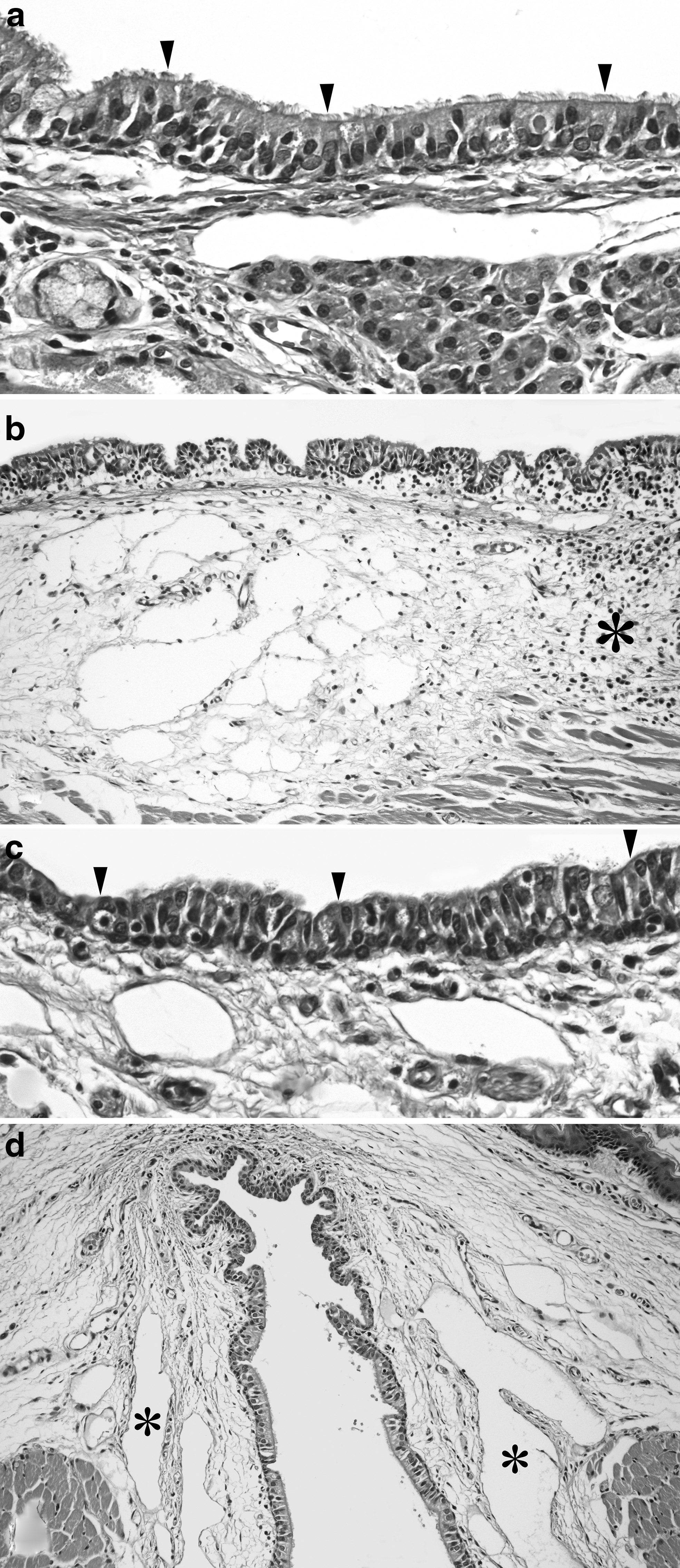
Comparison of ciliated cells and edema between the control and oleic acid-cisplatin groups.
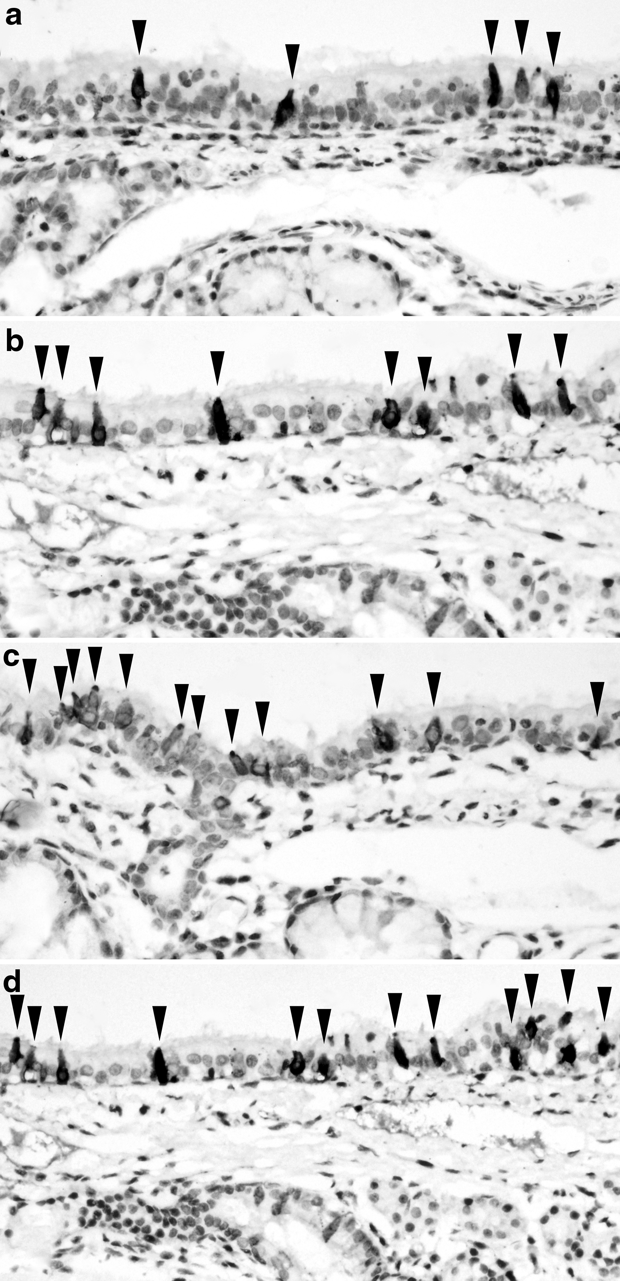
Comparison of Villin immunohistochemical expressions of groups.
Significant P Values of the Statistical Multiple Comparisons Between Groups (Kruskal–Wallis Test)
Statisticallay significant after Bonferroni correction.
Cis, cisplatin; Cox2, cyclooxygenase-2; HPF, A high-power field; Mel, melatonin; Ole, oleic acid; p-Cou, p-coumaric acid; Res, resveratrol; vitD, vitamin D.
Significant P Values of the Statistical Comparison Between Groups
Pearson chi-squared test.
Mann–Whitney U test.
Cis, cisplatin; vs, versus; C, control; p-Cou, p-coumaric acid; vitD, vitamin D; Mel, melatonin; Res, resveratrol; Ole, oleic acid; Cox2, cyclooxygenase-2; HPF, A high-power field.
The edema, number of Cox2-positive epithelial cells (Fig. 2c), and stromal expression of Cox-2 (Fig. 2d) in the vitamin D group were considerably higher than in the control group. The stromal expression of Cox-2 (Fig. 2d) in the vitamin D group was considerably higher than in the only cisplatin group. Only the edema in the melatonin group was considerably higher than in the control group and only the Ki67 index in suprabasal columnar epithelium was considerably higher than in the only cisplatin group. Villin-positive cells in the resveratrol group were higher than both in the control group and only cisplatin group (Fig. 3d). Interestingly number of Cox2-positive epithelial cells in resveratrol group (Fig. 2b) was considerably lower than in the both only cisplatin group and control group (Fig. 2a). The edema, vasodilation (Fig. 1d), cilia loss (Fig. 1c), number of villin-positive (Fig. 3c) cells, and Ki67 index in suprabasal columnar epithelium in the oleic acid group (Fig. 4b) were considerably higher than in the control group. Ki67 index in suprabasal squamous epithelium in the oleic acid group also was considerably higher than in the only cisplatin group (Fig. 4d).
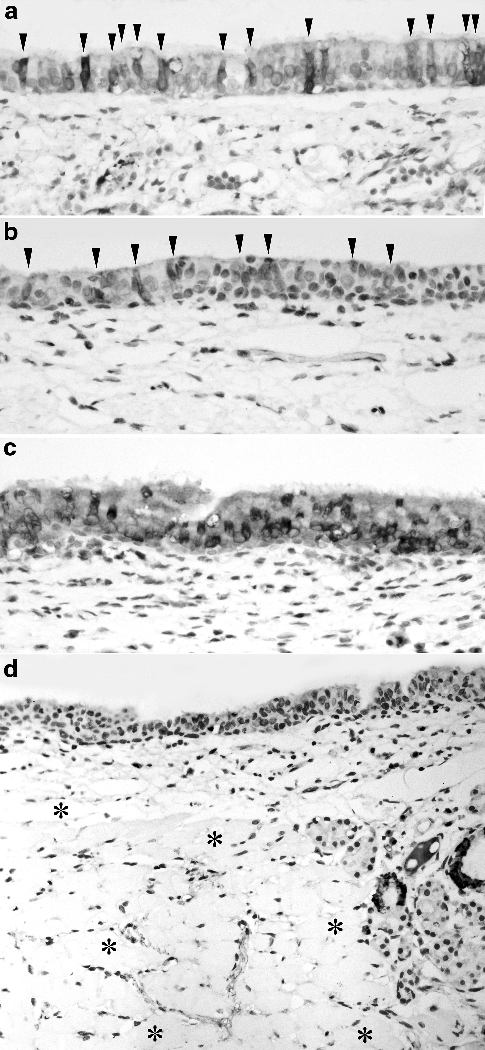
Comparison of count of Cox2-positive epithelial cells of groups.
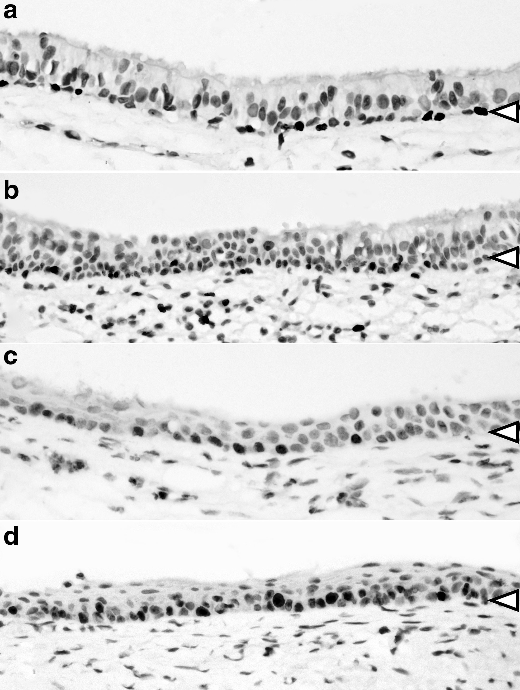
Comparison of Ki67-positive cells in surface columnar epithelial cells and surface squamous epithelial cells of groups. Black and white arrow heads sign basal cell layers of epithelium.
Discussion
Mucositis is a cisplatin-caused adverse effect related to the mucosal sites of the head and neck, giving rise to pain and an impairment of the quality of life. 5,6,11,15 In addition to mucositis, toxic effects on the salivary glands, pharynx, esophagus, and larynx were reported. 3,4,5,7,12 Side effects vary depending on the dose, duration, and combination of chemotherapy and radiotherapy. 10 Many agents containing antioxidants such as glutamine, vitamins, honey, zinc, aloe vera gel, ellagic acid, selenium, and so on were used in cisplatin-induced mucositis. 8,13,19,20 There were limited reports of the toxic effects of cisplatin on the larynx such as laryngitis, 5,10 laryngeal mucositis, 9 laryngeal mucosal ulcer, 33 and required tracheostomy. 10 Laryngeal mucosal ulceration has been reported during the cisplatin treatment of nasopharyngeal carcinoma. 33
The study showed that edema develops in the larynx with cisplatin. In our study, it was observed that cisplatin did not increase the vascular dilatation, the amount of inflammatory cells and the loss of cilia. The association of p-coumaric acid, vitamin D, melatonin, and oleic acid with cisplatin does not alter edema compared to cisplatin. This correlates with the increased edema effect of cisplatin, indicating that these antioxidants do not have any reducing or enhancing effect on edema. Khan et al. reported that cisplatin-caused distorted mucosal glandular architecture, villous atrophy, crypt ablation, distorted crypts, inflammatory infiltration in the mucosa and submucosa, and goblet cell disintegration in the jejunum of the rats. 7 Vitamin D affects T and B cells directly, plays a role in the adaptive immune response, modifies the activation response, inhibits the differentiation of plasma cells and immunoglobulin production, regulates macrophages and dendritic cells, 34 –36 and exhibits immunomodulating 37 and anti-inflammatory effects. 38 Rodrigues et al. proposed that oleic acid can modulate inflammatory and immune responses in skin lesions resulting from differential wound repair. 39
It was demonstrated that oleic acid has a proinflammatory effect, stimulating neutrophils to release VEGF-a, IL-1b, and CINC-2a/b, which results in speeding up the wound-healing process. 40 Although cisplatin alone does not increase the vascular dilatation in the larynx, it shows that oleic acid has an enhancing effect on vascular dilatation in the association with cisplatin. Alone cisplatin or in combination with antioxidants, it was observed that there was no change in inflammatory cells, Cox-2 expression in glandular epithelial cells, and goblet cell count in the laryngeal mucosa. The association of p-coumaric acid, resveratrol, and oleic acid with cisplatin increases the villin expression by control and only the cisplatin group. Vitamin D and melatonin do not cause a change in villin expression in the mucosa of the larynx. Athman et al. suggest a role for villin in cell motility, cell morphogenesis, and epithelial cell plasticity in response to cell injury. 25 Thus, when p-coumaric acid, resveratrol, and oleic acid are used in combination with cisplatin, they appear to be protected from the mucositis.
Even if multiple comparisons did not show significance, parameters showing significance in binary comparison were interpreted as follows. The fact that the loss of cilia in the coexistence of oleic acid and cisplatin was significantly higher than that of the control group and that there was no difference according to the cisplatin group, showed that the association of oleic acid with cisplatin increased the loss of cilia. It seems that combined use of oleic acid with cisplatin may have adverse effect on the mucociliary protective mechanism of the larynx. The association of vitamin D with cisplatin increases stromal Cox-2 expression compared to both control and cisplatin groups. The increase in Cox-2 expression correlates with mucositis severity, and Cox-2 plays amplifying role. 41 This study shows that the association of vitamin D with cisplatin also may reveal a side effect of increasing mucosal damage. For other antioxidants, inflammation does not differ significantly in comparison for each of the three groups. This suggests that cisplatin alone or in combination with antioxidants does not cause stromal Cox-2 change in the laryngeal mucosa. Vitamin D increases the number of Cox-2 expressed epithelial cells in the association of cisplatin, while the association of resveratrol cisplatin significantly decreases the number of epithelial cells showing Cox-2 expression (P = .01). Although there is no statistical significance with multiple comparisons, this suggests the efficacy of resveratrol in reducing the mucosal damage as a single component with the effect of increasing the number of villin-expressing cells. Increasing of villin expression could effect upper respiratory tract cilia motility, aid the mucosal defense system, protection against infections, and helps shorten healing periods.
The association of p-coumaric acid, vitamin D, melatonin, and resveratrol with cisplatin does not alter the Ki67 expression in the suprabasal layer of squamous epithelium, but increases the Ki67 proliferative index in the suprabasal layer of squamous epithelium when oleic acid is added to cisplatin (P = .007). The Ki67 proliferation index was not statistically significantly decreased in the cisplatin group, but the mean values were decreased. Cisplatin has been proved to alter cell proliferation in the rat intestinal epithelium, and supplementation of nutrient vitamins was shown to attenuate cisplatin-induced mucosal damage by increasing cell turnover. 16 The effect of oleic acid on increasing the Ki67 index will make laryngeal mucosa resist and will recover from the existing damage.
In the English literature, there were numerous articles about antioxidants, benefiting against side effects, enhancing the therapeutic effect of cisplatin, and protecting against cancer development, but there were no reports on reverse effect of antioxidants on therapeutic effect of cisplatin. It has been emphasized that almost all antioxidants are promoted to protect against all undesired side effects due to cisplatin and increase efficacy of cisplatin. Since the rats that are given cisplatin in the animal model in the study did not have tumors, it is not possible to obtain information about the antioxidant's effects on the therapeutic effect of cisplatin; this may be the subject of another further study.
Conclusion
Cisplatin-induced laryngeal toxicity decreases patient comfort in cancer treatment. In our study, the findings indicate that cisplatin causes evident laryngeal mucosal edema. Resveratrol, p-coumaric acid, and oleic acid might prevent cisplatin-induced mucositis in larynx by reducing Cox-2 expressed epithelial cells, increasing villin expressed cells, and increasing Ki-67 proliferation index of suprabasal squamous epithelial cells. However, oleic acid may have adverse effect on the mucociliary protective mechanism by increasing loss of cilia. Resveratrol and oleic acid were the most potent antiedema agents for the larynx in the study. On the contrary, when vitamin D is associated with cisplatin, it can be seen that increased stromal Cox-2 expression may have an effect on increasing mucosal damage.
Ethics Registration
Marmara University Animal Experiments Local Ethics Committee Project Approval Form Number:
Footnotes
Author Disclosure Statement
All named authors have read and agreed to the contents of the submitted version of the article. Authors have no such conflict of interest.
Funding Information
This research did not receive any specific grant from funding agencies in the public, commercial, or not-for-profit sectors.