
Abstract
Resveratrol (RES) (trans-3, 5,-4′-trihydroxystilebene) is a multi-biofunctional compound found in a variety of plants such as grapes and mulberries. Studies of nanoencapsulated resveratrol have indicated that this compound can inhibit the growth of cancer cells and free radicals. The aim of this study was to isolate resveratrol from Vitis vinifera, develop and evaluate resveratrol nanostructured lipid carriers (NLCs) and/or resveratrol encapsulated chitosan-coated nanostructured lipid carriers (CSNLCs) using low-viscous chitosan for anticancer therapy. In addition, our study was carried out to examine the prophylactic potential of RES, NLC, and CSNLC on paraquat-induced injury in rat hepatocytes. In this study we isolated resveratrol and encapsulated NLCs in phosphate-buffered saline solution using a phase inversion method. In addition, CSNLCs were prepared by ionic gelation method of NLCs using chitosan. NLCs and CSNLCs were then characterized for their particle size, zeta potential, morphology, and entrapment efficiency. Furthermore, NLCs and CSNLCs were evaluated for their cytotoxic effect on Hep-G2, human HCT-116 (colorectal cancer cell line), lymphoblastic leukemia (1301), and human MCF-7 (Michigan Cancer Foundation-7) cells as well as their effect on caspase-3 and death receptor (DR-4). In addition, incubation of hepatocytes with paraquat resulted in increased formation of TBARS (thiobarbituric acid reactive substances) with a parallel increase in lactate dehydrogenase (LDH) leakage at 1 h after incubation. Time-dependent depletion of cellular glutathione (GSH) was observed starting 2 h after incubation with paraquat. The mean particle size of NLC and CSNLC were 67.0 and 98.41 nm, zeta potential were (−) 24.8 and (+) 31.6 mV, entrapment efficiency were 74.15% and 85.46%, respectively, with the observed shapes of nanoparticle being spherical. The treatment of Hep-G2, human HCT-116, lymphoblastic leukemia (1301), and human MCF-7 cells with NLC led to high inhibition in the cell proliferation as concluded by the low IC50 values 27.7, 17.43, 35.39, and 47.66 μg/mL, respectively, whereas CSNLC had high cytotoxic effect on Hep-G2, human HCT-116, lymphoblastic leukemia (1301), and human MCF-7 cells with low IC50 values 13.29, 10.56, 16.79 and 22.60 μg/mL, respectively. Both NLC and CSNLC possess apoptotic properties through activation of the caspase-3 and death receptor (DR-4). In addition, incubation of hepatocytes with RES, NLC, and CSNLC markedly protected against paraquat-induced formation of TBARS, increase in LDH leakage, and prevented GSH depletion. The most effective doses for ethyl acetate, ethanolic, and aqueous extracts were 7.5, 10, and 12.5 μg, respectively. The results presented here may suggest that nanoencapsulated resveratrol isolated from the stems of V. vinifera to obtain NLC and CSNLC possess anticancer and apoptotic effects on cell proliferation, and therefore, can be used as new approach of pharmaceutical drugs. In addition, the results clearly suggest that the RES, NLC, and CSNLC exerted protective effect against cytotoxicity induced by paraquat. On the contrary, the effect decreased in order of CSNLC, NLC, and RES.
Introduction
Resveratrol (RES
Under the effect of oxidative enzymes in the presence of oxygen, light, and temperature, trans-resveratrol may be isomerized to cis-version, 11 reducing its activity. 12,13 It conjugates in the intestinal enterocytes and liver by glucuronidation and sulfate conjugation of the phenolic groups to give resveratrol-3-O-sulfate and resveratrol-3-O-glucuronid. 14,15 In addition, intestinal microbiota changes the aliphatic trans double bond of resveratrol by hydrogenation to give dihydroresveratrol. 11,15,16
Chitosan is a cationic polysaccharide, and it has been suggested that positively charged polymeric hydrogels could develop additional molecular attraction forces by electrostatic interactions with negatively charged mucosal surfaces. 17 The mechanism of chitosan nanoparticle formation is based on electrostatic interaction between amine group of chitosan and negatively charged group of polyanion such as hydroxide anion. 18
Studies have shown that the nanoparticle drug system has significant advantages over classic one, including increasing the solubility, enhancing absorption, and improving in vivo bioavailability of drugs in the body. 19 Drugs are controlled into specific target organs or cells upon using this system, which reduces the side effects of the drug. 20
Paraquat (1,1′-dimethyl-4,4′ bipyridinium dichloride), a nonselective herbicide, has toxic effects on various organs, including lungs, liver, and other organs. 21 It produces oxidative stress by redox cycling with a variety of cellular diaphorases and oxygen to produce superoxide radicals. 22
The objectives of this article were aimed to isolate resveratrol from Vitis vinifera stems, which is considered a waste product, and characterize nanostructured lipid carriers (NLCs) and chitosan-coated nanostructured lipid carriers (CSNLCs) formulas, evaluate their cytotoxic effect on Hep-G2, human HCT-116 (colorectal cancer cell line), lymphoblastic leukemia (1301), and human MCF-7 (Michigan Cancer Foundation-7) cells and their effect on caspase-3 and death receptor (DR-4) as well as to evaluate the possible hepatoprotective effect, in vitro, of RES, NLCs, and CSNLCs against cytotoxicity induced by paraquat.
Materials and Methods
Materials
Chitosan medium molecular weight, washing buffer, blocking buffer, diluents buffer, and substrate buffer (Catalog No. T7517-5G; Sigma-Aldrich Chemical Co.), triglyceride tripalmitate (Catalog No. T8127-100G; Sigma-Aldrich Chemical Co.), and soy lecithin (Catalog No. 206775; Avanti Polar Lipids), chloroform (HPLC grade) were used. MTT reagent was purchased from Merck (USA).
Hepatocellular carcinoma Hep-G2 (hepatoma G2) cells, breast adenocarcinoma MCF-7 cells, colon carcinoma HCT-116 cells, and lymphoblastic leukemia cells (1301) were purchased from ATCC (USA).
Isolation and identification of resveratrol
Freshly collected plant material of the stems of V. vinifera was collected from a private farm in Giza, Alexandrian Desert Road. The plant was identified and authenticated by Agriculture Research Center, Giza, Egypt. Voucher specimen number (12016) was deposited at the Department of Pharmacognosy, Faculty of Pharmacy, October 6 University.
The stems were air dried in shade and then powdered. The powder was extracted with ethyl acetate (6 L) under reduced pressure at 40°C, to give a residue of 25 g.
EtOAc-hexane (1:12) was used as eluting mobile phase (12 mg) to obtain resveratrol.
IR (KBr, cm−1) of resveratrol: 3252 cm−1 (OH), 2962 cm−1 (CH-arom.), 2827 cm−1 (CH-aliph.). It was confirmed by 1 H-NMR spectra; measured on Bruker 400 MHz, 1 H NMR (dimethyl sulfoxide, DMSO): δH 6.10 (m, 1H, H-4), 6.2 (d, 2H, J = 2 HZ, H-2,H-6), 6.70 (d, 2H, J = 8 Hz, H-3′,H-5′), 6.80 (d, 1H, J = 16 Hz, Ha), 6.96 (d, 1H, J = 16 Hz, Hb), 7.01 (d, 2H, J = 8 Hz, H-2′, H-6′), 9 (s, 2H, 3-OH and 5-OH), and 9.5 (s,1H,4′-OH).
13 C-NMR (DMSO, 100 MHz): δC 128.4 (C-1), 104.3 (C-2), 158.6 (C-3), 101.9 (C-4), 158.6 (C-5), 104.5 (C-6), 125.7 (C-a), 127.7 (C-b), 139.4 (C-1′), 127.9 (C-2′), 115.6 (C-3′), 157.3 (C-4′), 116 (C-5′), 127.9 (C-6′). NMR spectra were identical to those reported. 23
Methods
Preparation of nanoparticles
Resveratrol NLCs were prepared in 1 × phosphate-buffered saline solution using a phase inversion method. NLCs were made from a lipid mixture composed of the following lipids: isolated resveratrol, soy lecithin, glyceryl tridecanoate, glyceryl tripalmitate, vitamin E acetate, Kolliphor HS15, and an aqueous mixture containing 1% of NaCl in deionized water. Next, the lipid mixture and aqueous mixture were heated separately to 75°C, then the aqueous solution was mixed into the lipid solution (2:1) using a magnetic stirrer (Shimadzu, Japan) set at 375 rpm/min. The mixture was cooled to 35°C at a rate of 4°C/min. Then, the temperature of the mixture was increased to 75°C. When the mixture was cooled to 30°C, ice-cold deionized water (0°C) was added. Subsequently, magnetic stirring was applied to the suspension. Resveratrol encapsulated nanoparticles (NLCs) were coated with 0.2% (w/v) of chitosan (Sigma, St. Louis, MO, USA) using a magnetic stirrer for 1 h at 4°C to form resveratrol chitosan-coated NLC (CSNLC). All steps were performed under nitrogen to prevent resveratrol and lipid degradation. During all processes of encapsulation, precautions were taken to ensure that degradation does not take place.
High-performance liquid chromatography
Resveratrol, NLC, and CSNLC were detected using a high-performance liquid chromatography (HPLC) system (Shimadzu, Tokyo, Japan) with a C18 reverse-phase column and an ultraviolet (UV) detector and the flow rate was 1 mL/min. The mobile phase was made with 500 mL methanol, 2.5 mL acetic acid, and 500 mL deionized water. The HPLC was used to quantify the concentrations nonencapsulated and nanoencapsulated resveratrol. The external standardization method was used to calculate the peak area.
Morphology and NLC and CSNLC characterization
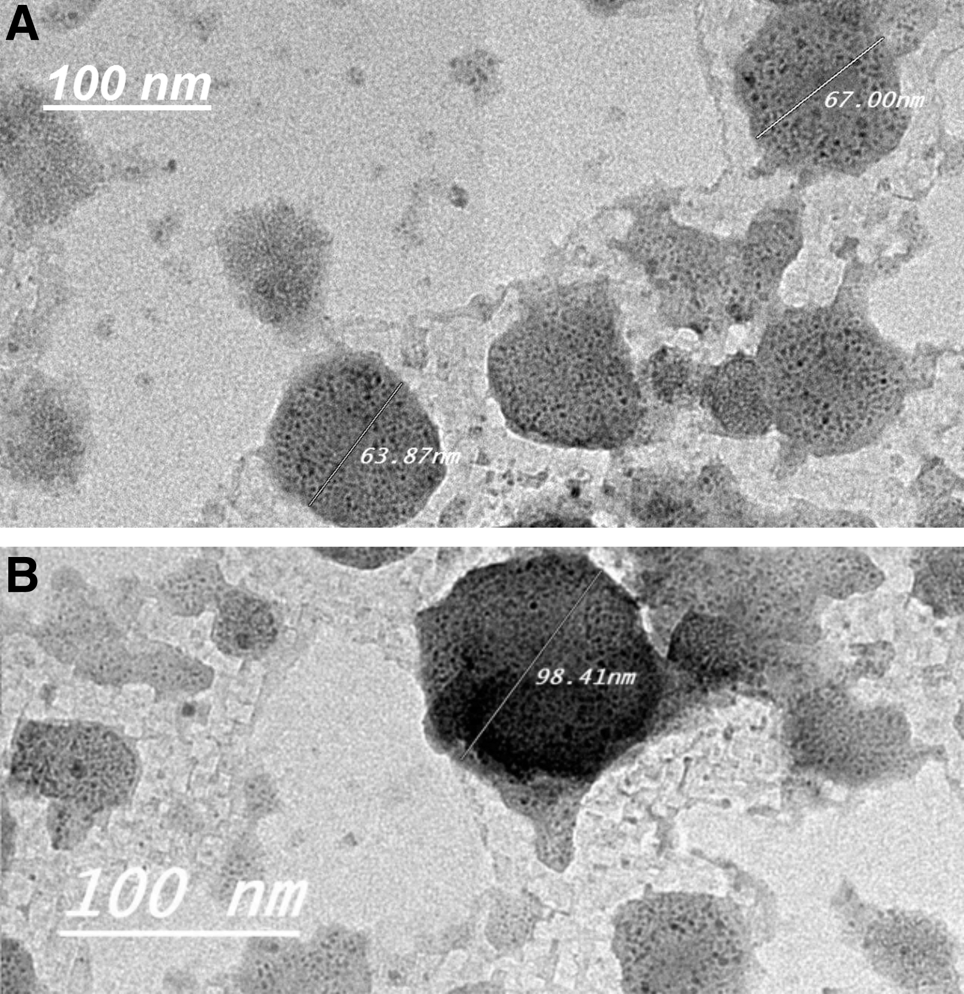
Morphological characteristics of resveratrol encapsulated NLC and/or resveratrol CSNLC were examined using scanning electron microscope (JSM-690; Jeol, Inc., Tokyo, Japan) and FETEM (field emission transmission electron microscopy, JSM-2100F; Jeol, Inc.) at 200 kV. In addition, their structures were proved using infrared (IR) spectral data (Shimadzu MR 470). In brief, a drop of nanoparticles was placed over carbon coated and allowed at room temperature for 24 h. The transmission electron microscopy image showed that the average diameter of the NLC and CSNLC was ∼67 and 98 nm, respectively, and had a round shape.
Entrapment efficiency
NLC and/or CSNLC of 5.0 mg were crushed and dispersed in a mixture of 5.0 mL distilled water and 5.0 mL of ethyl acetate. Ethyl acetate phase was separated after shaking the mixture carefully. UV-Vis spectrophotometer was used to determine free resveratrol content in ethyl acetate phase at 419 nm. The entrapment efficiency (%) of the drug was calculated using Equation (1).
Stability assay
The stability of the nanoparticles was evaluated in artificial gastric and/or intestinal fluids. About 5.0 mg of nanoparticles was dissolved in 5 mL artificial gastric and/or intestinal fluid. The mixture was shaken at 39°C and 100 rpm, using Mini-300 Orbital-Genie Ratcheting Clamps, USA. Ethyl acetate was then added at particular time intervals. The mixture was shaken and ethyl acetate phase was separated. UV-Vis spectrophotometer was used to estimate the amount of resveratrol released from the nanoparticles at 419 nm. The experiment was performed in triplicates.
Anticancer assay
Cell culture
Hep-G2, HCT-116, lymphoblastic leukemia (1301), and MCF-7 cells were routinely cultured in Dulbecco's modified Eagle's medium, 10% fetal bovine serum, 2 mM
Anticancer activity
MTT cell viability technique was used to assay the cytotoxicity of NLC and CSNLC against Hep-G2, HCT-116, lymphoblastic leukemia (1301), and human MCF-7 cells. In brief, MTT assay is based on the ability of active mitochondrial dehydrogenase enzyme of living cells to cleave the tetrazolium rings of the yellow (3-[4,5-dimethylthiazole-2-yl]-2,5-diphenyltetrazolium bromide) and form dark blue insoluble formazan crystals that is largely impermeable to cell membranes, resulting in its accumulation within healthy cells. In brief, in a flat bottom 96-well microplate, cells (0.5 × 10⁵ cells/well), in serum-free media, were plated and treated with different concentrations of NLC and/or NLC CSNLC (20 μL) for 48 h at 37°C, in a humidified 5% CO2 atmosphere. After incubation, media were removed and 40 μL MTT solution/well were added and incubated for an additional 4 h. MTT crystals were solubilized by adding 180 μL of acidified isopropanol/well and plate was shaken at room temperature, followed by photometric determination of the absorbance at 570 nm using microplate enzyme-linked immunosorbent assay (ELISA) reader.
24
Triplicate repeats were performed for each concentration and the average was calculated. Data were expressed as the percentage of relative viability compared with the untreated cells compared with the vehicle control, with cytotoxicity indicated by <100% relative viability. The percentage of relative viability was calculated using the following Equation (2):
The half maximal inhibitory concentration of cell viability (IC50) was calculated by the linear equation of sample concentration and cell viability %.
Caspase-3 and death receptor-4 estimation
Quantitative assay of Caspase-3 and death receptor (DR-4) levels were measured in cell lysate by ELISA technique. 25 In brief, in 96-well microplate, cell lysates (50 μL/well) was plated and then incubated for 1 h at 37°C and blocking buffer was added, incubated for 90 min at 37°C, washed and then antibodies (1 mg/mL Rabbit polyclonal to DR-4 and/or caspase-3; Abcam, Inc., Cambridge, MA, USA) were added. After 1-h incubation at 37°C, polyclonal goat anti-rabbit peroxidase conjugate (1:2000; Jackson Immunsearch Lab, USA) was added. The wells were washed after 1 h, and the substrate buffer was added. The color development was stopped by adding 1 M HCl and the intensity of yellow color was measured at 450 nm. The DR-4 level was compared with the activity of paclitaxel.
Prophylactic potential of RES, CSNLC, and NLC on paraquat-induced toxicity in rat hepatocytes
Experimental design
This experiment was carried out to examine the prophylactic potential of the extracts on paraquat-induced injury in isolated suspended rat hepatocytes. The rats were killed by cervical decapitation and the hepatocytes were performed using the collagenase perfusion method, as described by Moldėus et al. 26 The obtained hepatocytes were suspended in Krebs–Henseleit buffer at a concentration of 5 × 10⁶ cells/mL. Isolated suspended hepatocytes were then divided into 18 rotating round-bottomed flasks, each containing 0.5 mL of the suspended hepatocytes and were incubated with the following:
After a 30-min incubation period, the hepatocyte suspensions in flasks 2–18 were subjected to 1 mL paraquat (5 mM). 27
Samples of cell suspensions were collected at different time intervals namely at 30 min before incubation and zero time incubation with CSNLC, NLC, and RES and before exposure to the paraquat, then at 60, 120, and 180 min after addition of hepatotoxicant. Each sample was divided into two parts (0.5 mL each), one aliquot was used for the determination of TBARS (thiobarbituric acid reactive substances) level according to the method of Uchiyama and Mihara, 28 and the other was centrifuged and the clear supernatant was used for the determination of lactate dehydrogenase (LDH) activity according to the method of Buhl and Jackson 29 while the residue was suspended in phosphate buffer for the estimation of glutathione (GSH) level according to the method modified by Ahmed et al. 30
Statistical analysis
The results are expressed as mean ± standard deviation. Comparisons between groups were performed using one-way analysis of variance. Differences between individual treatment groups were compared using Dunnett's test. Statistical significance was set at P < .05 and P < .01, and the statistical analyses were performed using SPSS software, version 15.0 (SPSS, Inc., Chicago, IL, USA).
RESULTS
Retention time, peak area, and concentration of RES, NLC, and CSNLC given in Table 1 showed that the HPLC profile of standard resveratrol showed at specific retention time. Resveratrol and resveratrol extracted from NLC and CSNLC also showed similar HPLC pattern at required retention time at 6.42, 6.41, and 6.42, respectively. In addition, resveratrol and resveratrol extracted from NLC and SNLC also showed similar HPLC peak area at 299340, 3559555, and 1610156, respectively.
Retention Time, Peak Area, and Concentration of Resveratrol, Resveratrol Nanostructured Lipid Carrier and Chitosan-Coated Nanostructured Lipid Carrier
CSNLCs, resveratrol encapsulated chitosan-coated nanostructured lipid carriers; NLC, resveratrol nanostructured lipid carrier.
Figure 1 and Table 2 shows entrapment efficiency (%), particle size, polydispersity index (PI), and zeta potential values of NLC and CSNLC. NLC had size of ∼67.0 ± 1.4 nm with negative zeta potential of −24.85. In addition, CSNLC had size of ∼98.41 ± 2.2 nm with positive zeta potential of +31.60.
Entrapment Efficiency (%), Particle Size, Polydispersity Index, and Zeta Potential Values
Table 3 provides the stability of NLC and CSNLC nanoparticles in artificial gastric and/or intestinal fluids. The medium pH values mentoring the release of resveratrol from the nanoparticles formula is given. In artificial gastric fluid with acidic pH, chitosan was soluble and then degraded leading to the release of resveratrol from the nanoparticles.
Stability of Resveratrol Nanostructured Lipid Carrier and Resveratrol Encapsulated Chitosan-Coated Nanostructured Lipid Carriers Nanoparticles in Artificial Gastric and Artificial Intestinal Fluids
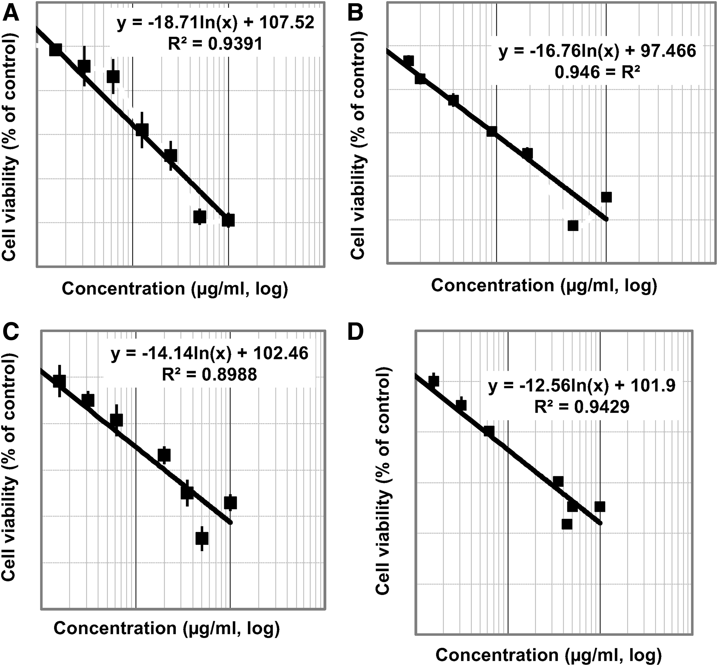
As given in Figures 2 and 3, the treatment of Hep-G2, human HCT-116, lymphoblastic leukemia (1301), and human MCF-7 cells with NLC led to a high inhibition in the cell proliferation as concluded by the low IC50 values (27.7, 17.43, 35.39, and 47.66 μg/mL), respectively, which revealed a high antitumor activity of the NLC against liver, colon, and blood carcinoma and moderate breast carcinoma, whereas CSNLC had high cytotoxic effect on Hep-G2, human HCT-116, lymphoblastic leukemia (1301), and human MCF-7 cells with low IC50 values (13.29, 10.56, 16.79, and 22.60 μg/mL), respectively, which revealed a high anti-tumor activity of the NLC against liver, colon, blood, and breast carcinoma.

Figures 4 and 5 show the effect of treatment with resveratrol encapsulated NLC and/or resveratrol chitosan-coated NLC (CSNLC) 50% of IC50 (21.83% and 11.3%), respectively, on DR-4 and caspase-3 levels in MCF-7 cells compared with paclitaxel activity.
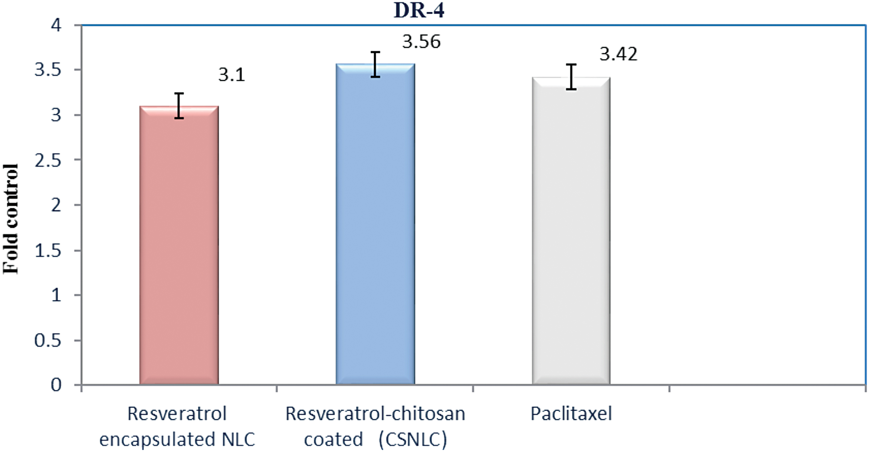
The effect of treatment with resveratrol encapsulated NLC and/or resveratrol CSNLC 50% of (IC50) 21.83% and 11.3%, respectively, on DR-4 levels in MCF-7 cells compared with paclitaxel activity. DR-4, death receptor. Color images are available online.
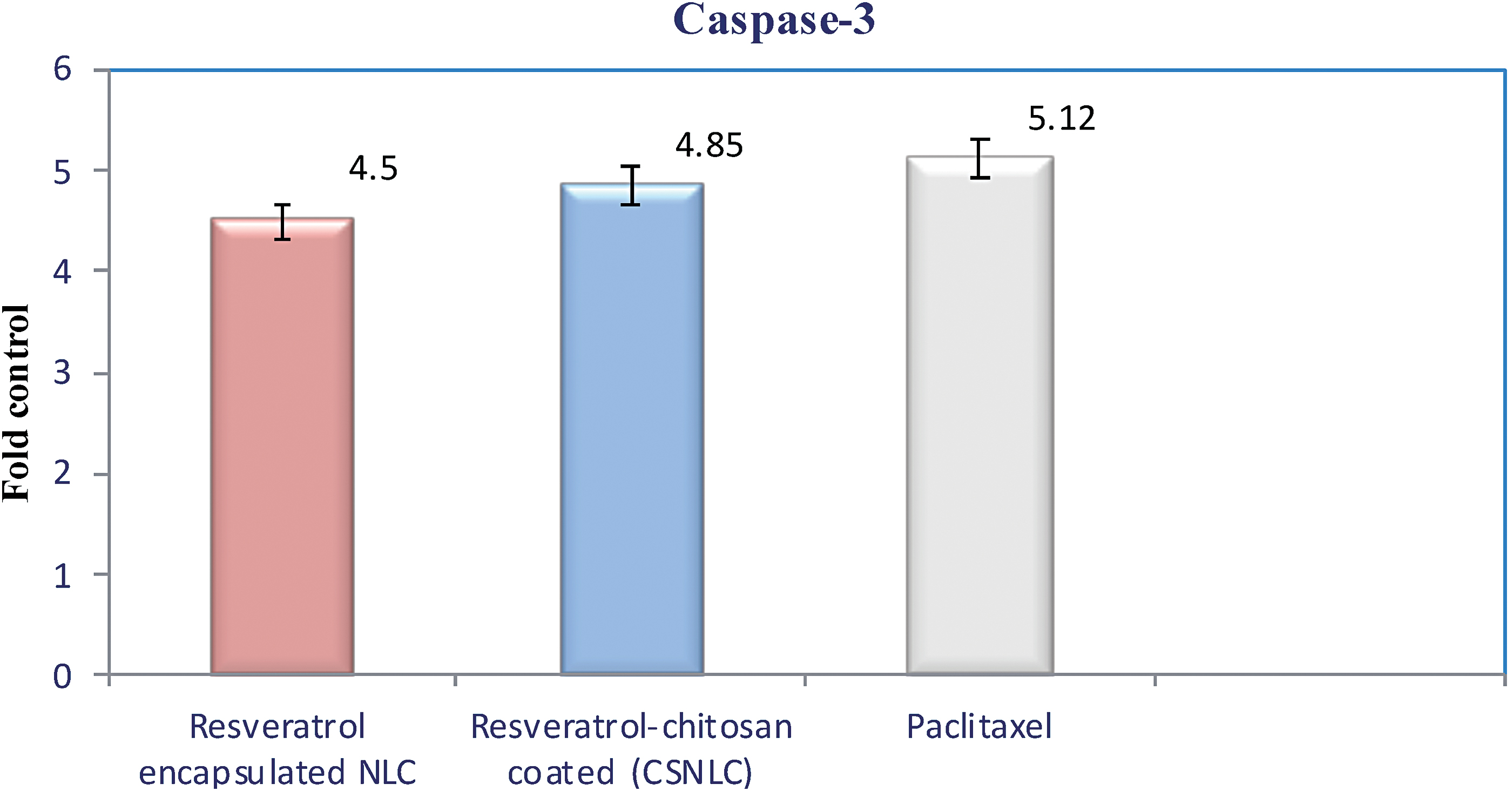
The effect of treatment with resveratrol encapsulated NLC and/or resveratrol CSNLC 50% of (IC50) 21.83% and 11.3%, respectively, on caspase-3 levels in MCF-7 cells compared with paclitaxel activity. Color images are available online.
Prophylactic potential of RES, CSNLC, and NLC on paraquat-induced toxicity in rat hepatocytes
The results in Table 4 show that the paraquat (5 mM) produced a marked depletion of GSH within 120–180 min of incubation period as evidenced by a 45.8–54.18% depletion of GSH compared with the respective normal values. Previous incubation of hepatocytes with CSNLC, NLC, and RES offered a significant protection against hepatotoxin-induced GSH depletion detected as early as 120 min after exposure to paraquat for CSNLC, NLC, and RES. The most effective doses for CSNLC, NLC, and RES were 7.5, 10, and 12.5 μg/mL, respectively. Table 5 revealed that the paraquat (5 mM) produced a marked increase in the rate of lipid peroxidation within 60–180 min of incubation period as evidenced by elevation in TBARS to two- to fourfold as compared with the respective normal values. Previous incubation of hepatocytes with CSNLC, NLC, and RES offered a significant protection against paraquat-induced lipid peroxidation as early as 60 min after exposure to paraquat; they produced a marked production starting from 120 min after exposure to hepatotoxin. The most effective doses for CSNLC, NLC, and RES were 7.5, 10 and 12.5 μg/mL, respectively. Table 6 showed that the paraquat (5 mM) produced a marked increase in the leakage of LDH within 60–180 min of incubation period as evidenced by elevation in leaked of LDH to two- to fourfold as compared with the respective normal values. Preincubation of the hepatocytes with CSNLC, NLC, and RES offered a significant protection against paraquat-induced LDH leakage as early as 60 min after exposure to paraquat. Preincubation of the hepatocytes with CSNLC (7.5 μg/mL), NLC (10 μg/mL), and RES (12.5 μg/mL) showed a protective effect against paraquat-induced LDH leakage after 180 min of exposure to paraquat by 66.8%, 48.22%, and 33.96%, respectively, as compared with the control group.
Effects of Different Concentrations of Chitosan-Coated Nanostructured Lipid Carrier, Nanostructured Lipid Carrier, and Resveratrol Treatment on Cellular Glutathione Leakage Induced by Paraquat Using Isolated Suspended Rat Hepatocytes
Paraquat (in DMSO) was added to the incubation media in all groups at a concentration of 5 mM, except in the normal group where only the vehicle DMSO was added. Paraquat control group were compared with normal control group. Experimental groups were compared with paraquat control group.
Significantly different from normal group at P < .05.
Significantly different from control group at P < .05.
DMSO, dimethyl sulphoxide; GSH, glutathione; RES, resveratrol.
Effects of Different Concentrations of Chitosan-Coated Nanostructured Lipid Carrier, Nanostructured Lipid Carrier, and Resveratrol Treatment on Thiobarbituric Acid Reactive Substances Formation Induced by Paraquat Using Isolated Suspended Rat Hepatocytes
Paraquat (in DMSO) was added to the incubation media in all groups at a concentration of 5 mM, except in the normal group where only the vehicle DMSO was added. Paraquat control group were compared with normal control group. Experimental groups were compared with paraquat control group.
Significantly different from normal group at P < .05.
Significantly different from control group at P < .05.
TBARS, thiobarbituric acid reactive substances.
Effects of Different Concentrations of Chitosan-Coated Nanostructured Lipid Carrier, Nanostructured Lipid Carrier and Resveratrol Treatment on Lactate Dehydrogenase Leakage Induced by Paraquat Using Isolated Suspended Rat Hepatocytes
Paraquat (in DMSO) was added to the incubation media in all groups at a concentration of 5 mM, except in the normal group where only the vehicle DMSO was added. Paraquat control group were compared with normal control group. Experimental groups were compared with paraquat control group.
Significantly different from normal group at P < .05.
Significantly different from control group at P < .05.
LDH, lactate dehydrogenase.
Discussion
In this study we isolated resveratrol from the stems of V. vinifera. The powdered stem was extracted with ethyl acetate to give a residue of 25 g. Hexane-EtOAc (2:1) was used as eluting mobile phase, and 12 mg of resveratrol was obtained and identified using spectral data.
Resveratrol is a promising natural compound for many diseases prevention and treatment. However, its low levels of stability and cellular bioavailability limit its anti-tumor activity. Two approaches have been used to increase its stability and bioactivities: (i) formation peracetate ester of resveratrol
31
or resveratrol docosapentaenoic acid ester
32
and (ii) using nanocarriers such as nanoliposomes
33
and NLCs.
34
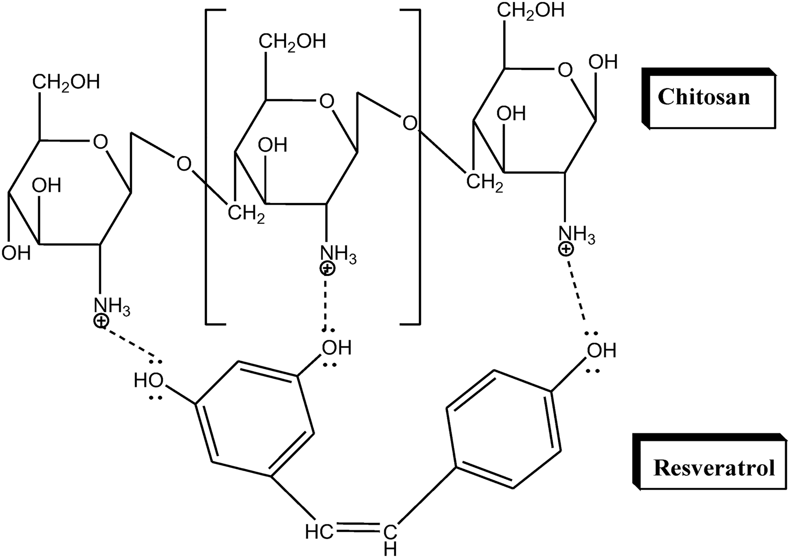
The chemical modification makes lipophilic resveratrol prodrugs, which requires chemical cleavage before releasing nonencapsulated resveratrol. Nanoliposomes are not stable, and encapsulated compound can be leaked out. NLCs do not have those problems and have been widely used in pharmaceutical and nutraceutical research. There are a number of characteristics necessary for chitosan to exhibit ionic interaction with resveratrol. These include the following: Strong hydrogen bonding groups (−OH, −COOH). High molecular weight Sufficient chain flexibility Surface energy properties favoring spreading onto mucus. In addition, the presence of lone pairs of electrons in hydroxyl groups of cis resveratrol facilities the formation of hydrogen bonds with amino groups of chitosan (Fig. 6). Proposal diagram for interaction between chitosan and resveratrol.
IR spectrogram of the resveratrol raw powder shows a phenol hydroxyl group absorption peak at 3252 cm−1 and benzene ring absorption peaks at 2827 and 2920 exists. IR spectrogram of resveratrol nanostructured lipid carrier (NLC) shows a hydroxyl group absorption peak at 3436 cm−1. In addition, the IR spectrum of resveratrol CSNLC shows that the characteristic absorption peak appeared at the same position, but the characteristic absorption peak of resveratrol did not appear, proving that resveratrol had been completely wrapped in chitosan.
Our results confirmed with the results of Grenha et al. 35 and Boonsongrit et al., 36 who showed that the addition of chitosan in the presence tripolyphosphate will lead to increase in particle diameters. In addition, PI was used to estimate the uniformity or dispersity homogeneity of the nanoparticles. In this study, NLC and CSNLC showed 0.732 and 0.623, respectively, of PI values. The present values indicate the heterogeneity dispersion of NLC and CSNLC. 31
Furthermore, in the presence of proteins, resveratrol would be more protected from trans-to-cis isomerization than when it is in the free form. 37,38
In this study, NLC and CSNLC size was characterized (Fig. 1).
The entrapment efficiency of resveratrol NLCs and resveratrol-CSNLC were 74.15% and 85.46%, respectively. The resveratrol entrapments in nanoparticles were in satisfaction as they were >70%.
At pH 7,
Antitumor activity
Using MTT assay, the effect of the resveratrol encapsulated NLC and resveratrol CSNLC on the proliferation of Hep-G2, human HCT-116, lymphoblastic leukemia (1301), and human MCF-7 cells was studied after 48 h of incubation.
This study demonstrated that resveratrol CSNLC and NLC decreased the viability of Hep-G2, human HCT-116, lymphoblastic leukemia (1301), and human MCF-7 cells than resveratrol encapsulated NLC. From research in the field of nanotechnology, it was noted that nanoparticles improved drug solubility, controlled drug release, enhanced bioavailability, increased stability, and improved long-term storage. 29 Resveratrol possesses antiproliferative effects through the induction of death in many different cell lines, including colon cancer. 39 In addition, it has low solubility, stability, and bioavailability. 40 In this study, we prepared NLC and then encapsulated it with chitosan to form CSNLC to prevent its active sites against photobleaching. 41 Nanoparticles were shown to improve the stability and enhance the compound's bioavailability. 41,42 The results of the MTT assay with nanoparticles indicate the anticancer activity of both resveratrol formulas. This may be owing to the resveratrol encapsulated nanoparticles' rate of bioavailability. Many studies indicate that nanoparticles improve bioavailability. 43 However, if the rate of absorption is too great, it may lead to toxicity on cancer cells. This could be the issue in this study.
The executioner caspases such as caspase-3, -6, and -7 induced apoptosis by activation of death receptor (extrinsic) and mitochondrial-mediated (intrinsic) pathways leading to nuclear shrinkage and DNA fragmentation. 44,45 In this study, DR-4 and caspase-3 levels were significantly increased in MCF-7 cells treated with the NLC and CSNLC compared with untreated cells and possessed a similar activity of paclitaxel. Many research studies have demonstrated that resveratrol can induce cancer cell death in cellular studies. 46 –49 This is a promising attribute in the field of cancer research. It is important for the area of nanoparticles to perfect precise targeting of cancer cells, and optimal time release.
Prophylactic potential of RES, CSNLC, and NLC on paraquat-induced toxicity in rat hepatocytes
The results presented here show strong antioxidative effect in vitro on paraquat-induced injury in isolated suspended rat hepatocytes. Depending on the test parameters (TBARS, LDH, and GSH), the magnitude of the effects varied considerably between RES, CSNLC, and NLC. The antioxidative effect decreased in the order CSNLC, NLC, and RES. The differences in potency between RES, CSNLC, and NLC were seen at the levels of TBARS, LDH, and GSH. The strong antioxidative influence of resveratrol is in agreement with other reports, particularly those describing effects of the polyphenols. The ability of RES to attenuate the hepatotoxic effect of paraquat could be the result of the antioxidant property of RES because paraquat is known to exert its toxicity through the induction of free radicals. 50 Polyphenols for example are known to be soluble chain breaking inhibitors of the peroxidation process, acting as scavengers of intermediate peroxyl and alkoxyl radicals, and chelating metal ions. 51 –53 Prevention of DNA oxidation is also achieved by these polyphenols mainly by quenching free radicals and modulating biometabolism enzymes. 54 This study demonstrated that resveratrol CSNLC and NLC had more potent hepatoprotective effect than RES because of the nanoparticles' improved drug solubility, controlled drug release, enhanced bioavailability, increased stability, and improved long-term storage. 37 The hepatoprotective activity of CSNLC being more pronounced than NLC may be because of the presence of chitosan that exhibits ionic interaction with resveratrol and increases its free radical scavenging activity.
In conclusion, in this study, we used the low viscous chitosan to prepare resveratrol nanoparticles. In addition, characterization of obtained spherical nanoparticles (NLC and CSNLC) was evaluated using different procedures. NLC and CSNLC were stable in artificial gastric and/or intestinal fluids. NLC and CSNLC showed significant anticancer activity against Hep-G2, human HCT-116, lymphoblastic leukemia cells (1301), and human MCF-7 cell lines. Both NLC and CSNLC possess significant apoptotic properties through activation of the caspase-3 and DR-4. The anticancer and apoptotic effect was more pronounced in CSNLC compared with NLC. This could be owing to the methods used. When preparing nanoparticles, to increase stability, chitosan is added to the encapsulation of resveratrol. Further studies need to be carried out to investigate all aspects of nanoencapsulated resveratrol. In addition, this study showed that CSNLC, NLC, and RES possess potent in vitro antioxidant activity against liver damage induced by paraquat. Overall, our results suggested that NLC and CSNLC could be a promising new approach for anticancer and hepatoprotective therapies.
Declarations
Ethics approval and consent to participate
Ethical approval for the data collection was granted by the Research Ethics Committee at the Faculty of Applied Medical Sciences, October 6 University, Egypt (No. 20180902). No laboratory animals or humans were used in this research; in vitro experiments only were used.
Consent for publication
The authors gave consent for their data to be used in the article.
Availability of data and Materials
Supporting data will be made available as it contains spectroscopic and analytical data to prove the structure of obtained nanoparticles and their in vitro antitumor and antiapoptotic activities.
Footnotes
Authors' Contributions
Experimental design of this study was carried out by all authors. Phytochemical studies (extraction, isolation, and structure elucidation of resveratrol) were carried out by Heba A. Elgizawy. Preparation of NLCs and CSNLCs and their physiochemical studies as well as antitumor and antiapoptotic activity was carried out by Ali A. Ali and Mohammed A. Hussein. Wrote the protocol, wrote the first draft of the article was carried out by M.A.H., managed the analyses of the study was carried out by H.A.E., managed the literature searches carried out by A.A.A. All authors read and approved the final version of the article.
Author Disclosure Statement
No competing financial interests exist.
Funding Information
No funding was received for this article.