




Abstract
The Asteraceae family is widely known for its therapeutic, aromatic, and nutritional properties. Chaptalia nutans (C. nutans), a member of the family, is widely used in folk medicine in southern Brazil. In this study, we aim to assess compounds present in root extracts of C. nutans, and evaluate their antioxidant capacity and toxicity. To determine the chemical composition of the extract, was performed through Liquid Chromatography coupled with Mass Spectroscopy. Antioxidant capacity, toxicity (Artemia salina biosassay), cytotoxicity, genotoxicity (Allium cepa test), and neurotoxicity (Drosophila melanogaster model) were evaluated. A large number of bioactive phytoconstituents were determined to be present, such as alkaloids, coumarins, flavonoids, terpenes, and especially phenolic compounds, which may explain the antioxidant capacity of the extract. Extracts had the capacity to protect cells from protein and lipid damage, and inhibit the formation of oxygen radicals. The A. salina bioassay revealed that extracts were only slightly toxic. In A. cepa, cells exposed to 1.5 mg/mL extract were protected against chromosomal damage caused by glyphosate, and had mitotic index values that were reduced by 49%. A concentration of 10 mg/mL extract did not kill flies, and when coadministered with paraquat (PQ) (52.5%) produced a mortality rate of only 18.75%. These findings indicated that the extract had the potential to protect against PQ-induced neurotoxicity. Taken together, these data reveal for the first time that the root extract of C. nutans is a rich source of natural antioxidants. The extract may be useful in the food and pharmaceutical industries.
Introduction
The Asteraceae family is widely known for its therapeutic, cosmetic, aromatic, and nutritional properties. It is the largest family in terms of the number of species, and contains nearly 1500 genera and 25,000 species. 1 Among these, Chaptalia nutans is popularly known as Língua-de-vaca or Arnica-do-campo. The species is widely distributed throughout Latin America, with the exception of Chile and can be found in grassland, forest, and wet soil habitats, along with areas with intense anthropic influence. 2,3 Folk medicine of southern Brazil has used its leaves for creating topical (heated) preparations to treat bruising, injuries, bleeding, and headaches. Leaves of the plant have also been used to make as tea. Roots of the species have been used to treat fevers, lung problems, constipation, skin diseases, and syphilis. 3 –5
Few studies exist in the literature that have assessed the effects of the species with respect to its nutritional and therapeutic properties. Pharmacological trials have confirmed the anti-inflammatory, cholinergic, and antimicrobial activities of its leaf extracts. 6,7 Coumarin (7-O-β-D-glucopyranosylnutranocoumarin) was isolated from crude root extract, and was responsible for antibacterial activity against Bacillus subtilis and Staphylococcus aureus. 8,9 The authors of the study suggested that the effectiveness of the treating of contaminated wounds could be attributed to coumarins.
Although studies have confirmed the antibacterial and anti-inflammatory activity of C. nutans extracts, there are no reports that have assessed its antioxidant activity and toxicity. The assessment of its toxicity is especially important since the plant is consumed as tea. However, it has been well established that most of the pharmacological properties of plant species are derived from antioxidants and polyphenols. 10 According to Ulewicz-Magulska and Wesolowski 11 and Taşkin et al., 12 natural polyphenols are commonly found in plants and represent the majority of antioxidants consumed as a part of the human diet. The use of natural products as supplements and medicines is widespread. Furthermore, although synthetic drugs are available and highly effective, many believe that natural ingredients are less harmful than synthetically produced compounds. 13
The perception that plant natural products are safe, and the consumption of plant-derived compounds is devoid of adverse effects, is false. In fact, several studies have shown that medicinal plants are capable of producing a wide range of adverse effects. 14,15 The indiscriminate use of plants and the lack of phytochemical, pharmacological, and especially toxicological knowledge is concerning from a public health standpoint. 12 Thus, knowledge of the bioactive compounds contained within widely used remedies and their modes of action within living organisms is of great importance.
The present study aimed to determine the chemical composition of C. nutans root extracts using chromatographic analysis, and evaluate the in vitro antioxidant activities of identified compounds in vivo. Furthermore, the cytotoxic, genotoxic, and neurotoxic effects of the extracts were assayed (Artemia salina, Allium cepa, and Drosophila melanogaster.
Materials and Methods
Plant materials
Plant material (C. nutans) was collected from São Francisco de Assis/RS (Brazil) (29°33'01”S, 55°07'52”W). The botanical characterization (family, genus, and species) of the plant was performed by Patrícia de Oliveira Neves, a botanist from the Federal University of Pampa (UNIPAMPA). The exsiccate was filed in the Unipampa herbarium (HBEI 1203). Plant roots (8.83 g) were dried, ground, and macerated in a hydromethanolic solution (30/70) at a concentration of 30 g/100 mL (material/solvent) for 4 weeks. The solvent used for the extraction was renewed every 7 days and was filtered and concentrated using a rotary evaporator (40°C) to obtain crude extract.
Phytochemical analysis
Phytochemical screening of C. nutans roots was based on classical methodologies described by Simões et al. 16 and Matos. 17 The screen was designed to identify the presence of cyanogenic glycosides, phenols, tannins, anthocyanins, proanthocyanidins, flavonoids, catechins, steroids, triterpenoids, saponins, resins, alkaloids, and quaternary bases.
Determination of chemical composition by HPLC-DAD-MS
The identification of the metabolites in the hydrolyzed crude extract of C.nutans roots in the present study follow the Vieira et al. 18 methodology with minor modifications. The technique was performed using a high-performance liquid chromatography (HPLC) coupled to mass spectrometry (HPLC-DAD-MS) and a diode detector, Shimadzu (Kyoto, Japan). Analyses were carried out using a C-18 column (4.6 × 250 mm, Merck, Germany) packed with 5 μm diameter particles within the C-18 precolumn (RP 18 5 μm, Merck).
The first mobile phase, phase A, comprised acetic acid (2%) and was set to pH 4.2, and the second mobile phase, phase B, comprised methanol, acetic acid, and distilled water at a ratio of 18:1:1, respectively. The elution gradient was as follows: 0 min; 20% B; 0–25 min: 50% B, 25 min: 20% B, 30 min: 20% B (end of run), and compounds were eluted at the flow rate of 0.8 mL/min. Peaks were obtained and compared with the retention times and mass spectra of compounds from the software library and external standards.
Analyses were performed using external standards, including an 40% isoflavone pool (Daidzin: 3.1%, Glycitin: 1.56%, Genistin: 0.98%, Daidzein: 35.49%, Glycitein: 0.1%, and Genistein: 0.03% - Dongming Huiren Biological Products, Shandong, China), 0.156–2.34 mg/mL glycitin, 3.5–53.2 mg/mL of daidzein, 3–4.5 μg/mL, and 1.5 and 24 μg/mL genistein, caffeic acid, gallic acid, chlorogenic acid, catechin, luteolin, coumarin, quercetin, and rutin (Sigma-Aldrich, St Louis, MO, USA). Samples of C. nutans and standards were tested in triplicate. Results were presented as mean ± standard deviation (SD).
Dosing of total polyphenols, flavonoids, and condensed tannins
Total polyphenol content of C. nutans root extract was determined using the Folin–Ciocalteau method, as described by Chandra and Mejia, 19 with small modifications as indicated below. The extract was diluted to a concentration of 150 μg/mL in ethanol and 1.0 mL of the solution was added to 0.5 mL 2N Folin–Ciocalteau reagent. After 5 min, 2 mL 20% sodium carbonate solution was added. Gallic acid was used as standard (0.001–0.03 mg/mL gallic acid), and were assessed at 730 nm (Y = 30.767x–0.0087, r = 0.9992). Results were measured at 420 nm and expressed as mg of gallic acid equivalents per g dry plant weight (mg GA/g dry plant).
Total flavonoid content was determined according to a method described by Woisky and Salatino. 20 Total flavonoid levels within extracts were measured at 420 nm and expressed as mg of quercetin equivalents per mL extract (mg QUE/mL EXT). The following equation was obtained using a quercetin standard (0.002–0.018 mg/mL; Y = 40.175x + 0.001, r = 0.9998). Condensed tannins were assessed using a method described by Morrison et al. 21 To calculate levels of condensed tannins, a catechin curve was used as a standard (0.0025–0.2 mg/mL; Y = 0.0015x–0.0005, r = 0.9968), and samples were measured at 500 nm. The results were expressed in mg catechin equivalents per g dry plant (mg CAT/g dry plant). All experiments were performed in triplicate using a Uv-Vis spectrophotometer (Spectroquant™ Pharo 10-Merck/Brazil).
In vitro antioxidant activity
2,2-diphenyl-1-picrilhydrazyl radical scavenging activity
The antioxidant activity of C. nutans root extract was assessed through a 2,2-diphenyl-1-picrilhydrazyl (DPPH) radical absorbance assay, using a procedure described by Choi et al.,
22
at concentrations of 500, 250, 125, 62.5, 31.25, 15.6, and 7.8 μg/mL. Absorbance was read at 518 nm using a spectrophotometer after the reaction had proceeded 30 min. Ascorbic acid was used as positive control (PC). The assay was performed in triplicate. The percentage of free radical inhibition of DPPH was calculated using the following equation:
where Abssample, Absblank, and Abscontrol indicate the absorbance of the sample, blank, and negative control (NC), respectively. The inhibitory concentration IC50 was calculated through the interpolation from a linear regression.
Ferric-reducing/antioxidant power assay
The antioxidant power activity of C. nutans root was assessed using a ferric-reducing/antioxidant power (FRAP) assay according to Rufino et al. 23 Briefly, samples were prepared at a concentration of 1000 μg/mL and diluted with distilled water. The test was performed in triplicate by adding 200 μL sample and 1800 μL FRAP reagent. Subsequently, the samples were stored in a 37°C oven for 4 min. Samples were assessed using UV-visible spectrometry (UV-VIS) at 593 nm. To calculate iron reduction ability, a standard ferrous sulfate curve was used at concentrations of 1000 mM to 62.5 mM (Y = 0.000049x + 0.036181, R 2 = 0.9922).
Lipid peroxidation assay (TBARS)
To evaluate the capacity of C. nutans root extracts to inhibit lipid peroxidation, thiobarbituric acid-reactive substances (TBARS) was determined, as described by Okhawa et al. 24 Briefly, samples were mixed with 1 mL 10% trichloroacetic acid (TCA), 1 mL 0.67% thiobarbituric acid, and heated using a boiling water bath for 30 min. TBARS levels were determined by measuring absorption at 535 nm. Results were expressed as malondialdehyde (MDA) equivalents per milligram protein (Eq. MDA/mg protein).
TBARS induced by ferrous sulfate
Another assay used to assess lipid peroxidation is TBARS with induction of free radical generators on a lipid-rich substrate (egg yolk), which was performed in accordance with procedures described by Vyncke, 25 with some modifications. Egg yolk homogenate was diluted 1% (v/v) in 100 mM TRIS HCl buffer, pH 7.4, mixed with TCA (10%), and centrifuged at 1200 rpm (10 min). The concentrations of C. nutans root extract used were 10, 100, and 1000 μg/mL (w/v). Next, 13.9 μg/mL aqueous ferrous sulfate was added to induce lipoperoxidation. Tubes were incubated at room temperature for 15 min and thiobarbituric acid (0.67%) was then added and incubated at 100°C for 30 min. Absorbance of 532 nanometers was determined using a spectrophotometer. The buffer solution was used as NC. Analyses were performed in quadruplicate and results were expressed as equivalents of MDA/mL substrate.
Determining the protein content through detecting carbonyl groups
Oxidized protein content (carbonyl grouping) was determined by detecting a reaction between carbonyl groups ad 2,4-dinitrophenylhydrazine (DNPH), as previously described by Levine et al. 26 Briefly, plasma (100 μL) in either the absence or presence of different dilutions of crude extract was incubated with 1 mL 0.2% DNPH in HCl (2 M) for 1 h at room temperature. Proteins were precipitated from the solution using 20% TCA. The protein pellet was washed three times with 10% TCA, ethanol, and ethyl acetate (1:1, v:v), and resuspended in 1 mL, 6 M guanidine and TCA. The concentration of carbonyl groups was calculated by determining the absorbance of the suspension at 370 nm using 22 L/mM/cm as the molar extinction coefficient, and results were expressed as nmol carbonyl per mg protein (mmol carbonyl/mg protein).
Assessment of dichlorodihydrofluorescein diacetate oxidation
The intracellular formation of reactive oxygen and nitrogen species (RONS) in C. nutans roots was measured using a dichlorodihydrofluorescein diacetate (DCFH-DA) substrate, a general index of oxidative stress according to Myrhe et al. 27 To perform the assay, a reaction mixture, consisting of 150 μL 0.1 μM potassium phosphate buffer (pH 7.4), 40 μL distilled water, 5 μL DCFH-DA (200 μM, final concentration 5 μM), and 5 μL sample (dilution 1:10) was combined. The fluorescence emission of 2′-7′dichlorofluorescein (DCF), which resulted from the oxidation of DCFH, was monitored for 10 min, at 30 s intervals, at 488 and 525 nm, excitation, and emission wavelengths, respectively. Hydrogen peroxide (H2O2) was used as a PC. The rate of DCF formation was expressed as a percentage of the control value (% of control group).
Toxicity assay
Toxicity to the nauplii of Artemia salina
Toxicity of C. nutans root extracts was evaluated using Artemia salina (Leach) according to methodology described by Silva et al. 28 A. salina cysts were incubated at 30°C in artificial saline (23 g/L sea salt and 0.7 g/L sodium bicarbonate in distilled water) and kept under conditions of constant aeration and agitation for 48 h to promote hatching. Ten nauplii were transferred to test tubes containing artificial seawater and three different concentrations of root extract (10, 100, and 1000 μg/mL). The assay was performed in triplicate. The number of dead and live nauplii was counted after 24 h. Treatment with artificial sea water was used as NC and treatment with sodium lauryl sulfate served as a PC. After determining numbers of living and dead nauplii, the LC50 and its confidence interval were calculated.
Evaluation of the genotoxicity of A. cepa
To evaluate the cytotoxic and genotoxic effects of treatment with C. nutans root extracts, the A. cepa (onion) toxicity test was used according to the methodology of Tedesco and Laughinghouse. 29 Briefly, eight groups of five bulbs were placed in distilled water for 48–72 h and were allowed to root. Next, bulbs were treated with different concentrations of crude extracts (0.1, 0.5, 1.5 mg/mL) for 24 h. Distilled water was used as NC, and 2% glyphosate was used as a PC.
To determine the capacity of the extract to protect against glyphosate damage, bulbs were placed in contact with 2% glyphosate solution for 24 h. Afterward, the bulbs were left in extract solutions for the same period. Subsequently, roots were collected and fixed in ethanol/acetic acid (3:1) (24 h). Radicles were removed from the fixative and packed using 70% alcohol.
To evaluate morphological and structural (abnormality index) abnormalities within cells, and to determine mitotic indexes (MIs), roots were soaked in 1 N hydrochloric acid for 5 mi after washing in distilled water and stained with 2% acetic orcein. Lamina was prepared using the crush technique
30
and phases of the cell cycle (interphase, prophase, metaphase, anaphase, and telophase) were examined using an optical microscope (40 × ). In the assays, 1000 cells per bulb were analyzed (5000 cells per treatment), and the average number of cells in each phase of the A. cepa cell cycle were calculated. MI was determined according to Equation 2, as follows:
The percentage of abnormalities (AN %) was determined according to Equation 3, as follows:
Fly behavior assay
Drosophila stock
Wild D. melanogaster was obtained from the National Stock Center (Bowling Green, OH, USA). Flies were kept inside 200-mL glass tubes under a 12-h light/12-h dark cycle at a constant temperature (22°C ± 1°C) and humidity (60%). Adult flies and larvae were fed standard nutrient media, as described by Paula et al. 31
Survival rates
To determine appropriate dosages and durations of exposure, adult flies were exposed for 7 days to a range of extract concentrations (15, 20, 25, 30, and 35 mg/mL). Throughout the process, the flies were fed a standard diet and the extract was provided every 24 h. The number of live and dead flies was counted on a daily basis. For each group assessed, 80 flies were tested. Each test was performed in quadruplicate. The results were analyzed and the percentage of surviving flies relative to the control (considered 100%) were plotted.
Treatment with C. nutans root extract and Paraquat®
A paraquat (PQ) assay was performed using 1- to 4-day-old adult flies. Twenty flies were used for each treatment, and the assay was performed in quintuplicate. For the assay, a liquid diet was provided, in accordance with protocols outlined in Soares et al. 32 Briefly, 300 μL diet solution was administered every 24 h. Experimental groups received the following solutions: EC, Extraction Control (10 mg/mL leaf extracts); PC (3 mM PQ); T1, 3 mM PQ +1 mg/mL extract; T2, 3 mM PQ +5 mg/mL extract; T3, 3 mM PQ +10 mg/mL extract. Flies were treated 4 days and kept in an incubator at 22°C ± 1°C at 60% relative humidity and exposed to a 12-h light/12-h dark cycle. Concentrations of PQ (3 mM) and C. nutans root extracts were determined based on survival curves and corresponded to the minimum time and concentration required to induce significant locomotor deficits and toxicity in flies. Surviving flies were used for behavioral trials.
Negative geotaxis assay
The locomotor activity of flies was determined using a negative geotaxis behavioral test, as described by Jahromi et al., 33 with some modifications. Flies were subjected to cryoesthesia and placed in plastic bottles (15 cm long, 2 cm diameter, 10 flies per bottle). After flies had recovered from exposure to cold (∼10 min), we tapped the bottom of each bottle gently to stimulate the climbing. The number of flies that rose to the 15 cm mark in 8 s was recorded. The assay was repeated six times per group and data were expressed as an average of six trials per repetition. Control flies were scored in the same manner.
Statistical analyses
Phytochemical analyses and in vitro antioxidant activity results are reported as mean ± SD. In vivo results were expressed as mean ± SEM. All experiments were performed in triplicate. Comparisons between more than two groups of data were performed using one-way ANOVA followed by Tukey's test or Newman–Keuls Multiple Comparison Test and differences were considered significant when P < .05, .01, or .001, according to the test used. Statistical analyses were performed using GraphPad Prism 5 software.
Results
Phytochemical analysis
A qualitative analysis of the secondary metabolite classes present in C. nutans root crude extract revealed the presence of condensed tannins, flavones, flavonoids, xanthones, saponins, coumarins, alkaloids, and quaternary bases. Results indicated that the extracts may be pharmacologically useful, after additional studies are performed.
Polyphenol, flavonoid, and condensed tannin content
Table 1 includes quantitative results in which levels of phenols, tannins, and flavonoids are shown. The results show that C. nutans root crude extract possess the highest levels of polyphenols, followed by condensed tannins, and that flavonoids are present at the lowest levels compared with the other two classes of compounds.
C. nutans Root Crude Extract Antioxidant Analysis
ND, not detected
HPLC-DAD-MS assay
HPLC-DAD-MS analysis of C. nutans root crude extract revealed the likely presence of four compounds: isoferulic acid, iosmarinic acid, ribitol, and arbutin (Table 2). Peaks were identified by comparing results obtained with peak retention times provided in a software library, mass spectra, and external standards.
The Phenolic Compounds Identified in Extracts Roots of Chaptalia nutans by HPLC-DAD-MS
Rt, retention time; [M-H]− (m/z), molecular ion peak in negative mode.
In vitro antioxidant activity
DPPH and FRAP analysis
Antioxidant activity obtained by DPPH and FRAP methods (Table 1B) using C. nutans root extracts suggest that they possess low levels of antioxidant activity. Using the DPPH method, the IC50 of the extract was 1011.53 ± 50.76 μg/mL. For the FRAP method, no antioxidant activity was detected.
TBARS, carbonyl protein, and DCFH-DA assays
Figure 1 shows results obtained using TBARS, carbonyl protein, and DCFH-DA assays. The findings indicate that crude extracts of C. nutans roots have the capacity to reduce levels of TBARS, protein oxidation, and peroxyl radicals. MDA is a byproduct of lipid peroxidation. Addition of all three concentrations (0.1, 1.0, and 10 mg/mL) of the extract reduced levels of MDA formation compared with basal controls (Fig. 1A), indicating that crude extracts of C. nutans roots inhibit formation of MDA. Regarding protein oxidation, the addition of root extract protected the cell from damage when all three concentrations of the extract were added. Concentrations of 1 and 10 mg/mL produced similar results (Fig. 1B), and both provided the maximal level of protein tissue protection, which was statistically higher than treatment with the control.
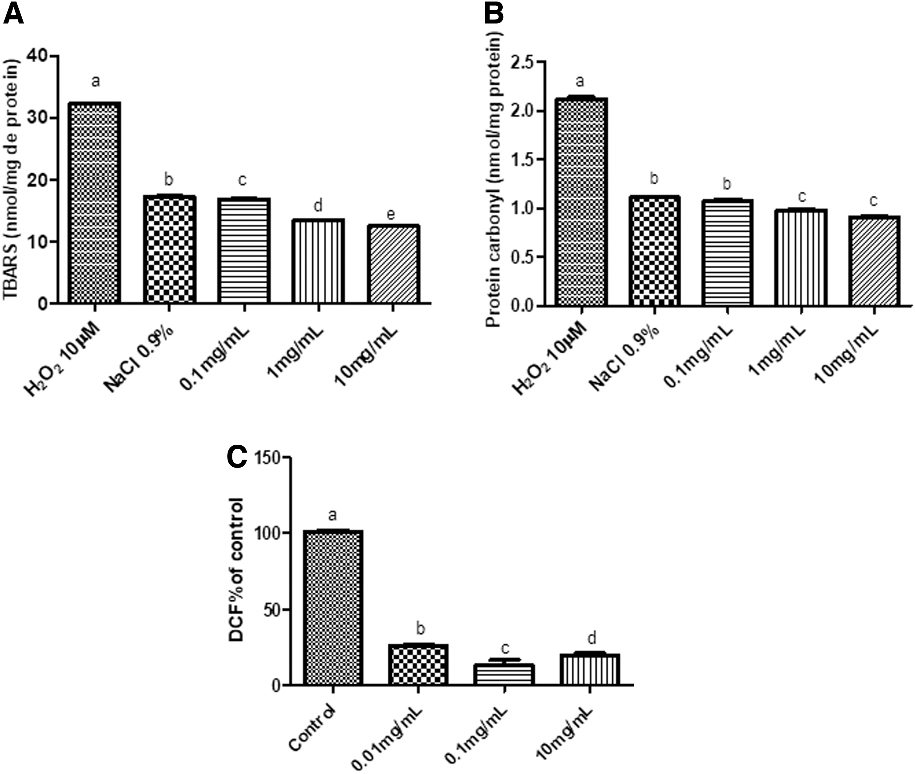
Figure 1C shows data that indicates that C. nutans root extract has the capacity to inhibit DCFH oxidation to DCF, which is indicated by a decrease in fluorescence intensity. The three concentrations of root extracts tested differed statistically from each other, and also differed from control levels. We determined that the 0.1 mg/mL concentration of root extract possessed the greatest potential to neutralize oxygen radicals. These results demonstrate the significant capacity of the extract to neutralize a variety of ROS sources.
TBARS induced by ferrous sulfate
Previous results obtained using TBARS method to test the effects of C. nutans crude root extract (Fig. 1) only considered baseline controls. To evaluate the response to the induction of free radical generators, lipid peroxidation with ferrous sulfate was induced and the capacity of the extract to protect against damage was verified. Figure 2 shows results obtained through TBARS analysis. The data are similar to that which was produced by performing a TBARS assay previously (Fig. 1), in which extract controls (ECs) were assessed at concentrations of 10, 100, and 1000 μg/mL, were statistically equal to the NC, which confirmed that the extract did not increase MDA levels.
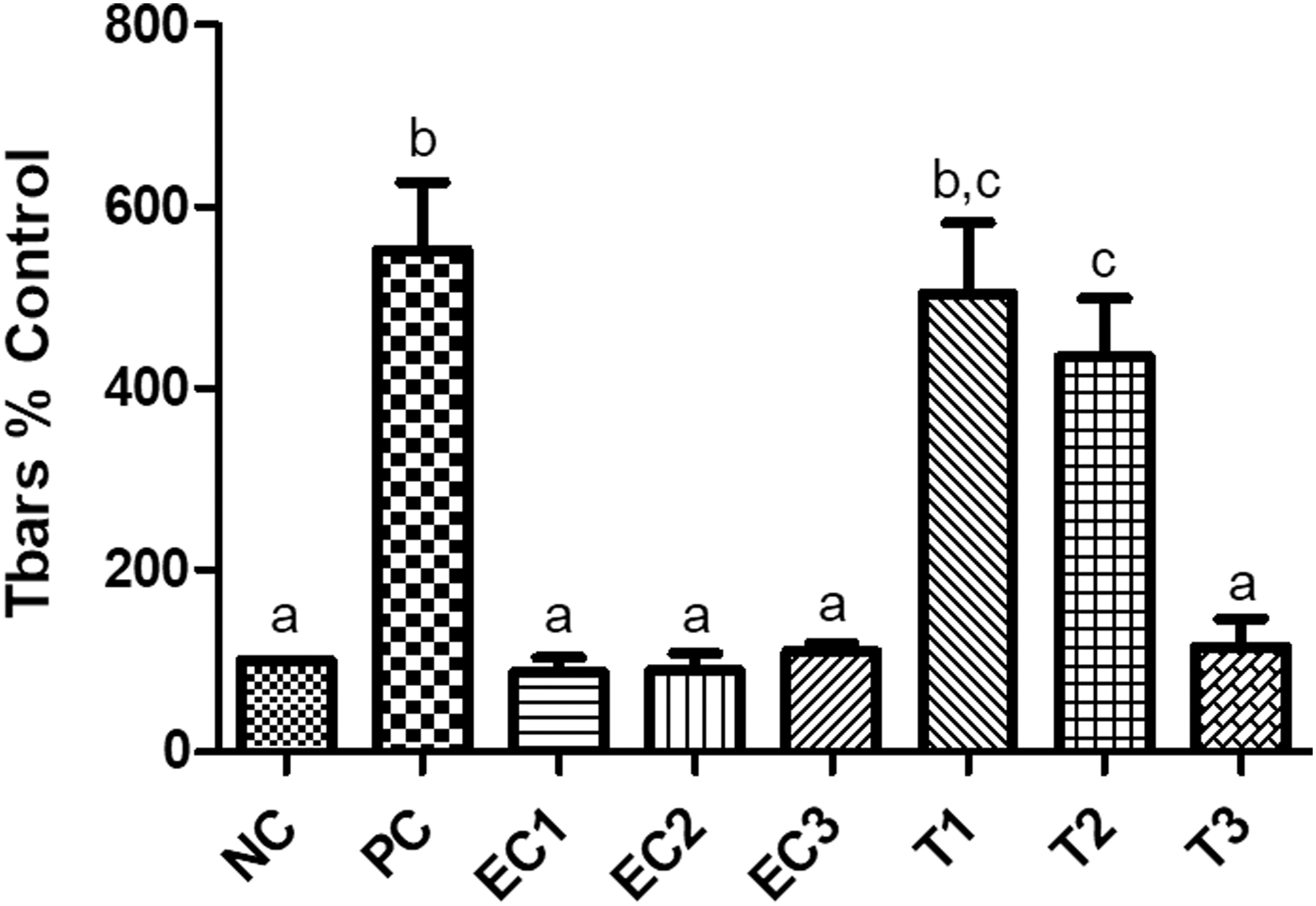
Analysis of the antioxidant activity of the crude extract of C. nutans root by lipoperoxidation assay (TBARS) induced for FS NC (buffer solution); PC (FS 13.9 μg/mL); EC (EC1: Extract Control 10 μg/mL; EC2: Extract Control 100 μg/mL; EC3: Extract Control 1000 μg/mL); Treatment (T1: FS 13.9 μg/mL+ extract 10 μg/mL; T2: FS 13.9 μg/mL + extract 100 μg/mL; T3: FS 13.9 μg/mL + extract 1000 μg/mL). Different letters represent statistical differences according to the Tukey's test (P < .001). FS, ferrous sulfate; NC, negative control; PC, positive control; EC, extract control.
Regarding the potential of the extract to protect against SF-induced lipid peroxidation, the lowest concentration of extract had no effect, and did not differ statistically from the PC. The 100 μg/mL concentration of extract did not differ statistically from the 10 μg/mL concentration; however, both were statistically different from the PC, and reduced MDA levels by 20% relative to controls. The highest concentration (1000 μg/mL), on the other hand, had the greatest capacity to inhibit formation of MDA, and was statistically equal to NC, and protected ∼80% of the cells assessed from damage. Thus, the assay demonstrated that 1000 μg/mL C. nutans root crude extract can decrease the lipid peroxidation caused by ferrous sulfate.
Toxicity and genotoxicity
The acute toxicity of crude extracts of C. nutans roots against A. salina nauplii is shown Table 3. The dose required to kill 50% of the nauplii over a 24-h period was 642.91 μg/mL, a value that was 11-fold greater compared with the PC (57.80 μg/mL). This result indicated that crude extracts of C. nutans roots are mildly toxic. Concentrations used in this assay were standardized for the A. cepa test.
Lethal Concentration for 50% of Artemia salina Nauplii (LC50) Obtained for C. nutans Roots Crude Extract, Positive Control, and Its Confidence Interval
The antiproliferative and genotoxic effects of C. nutans root crude extract against A. cepa are shown in Figure 3. The results were obtained by analyzing the number of cells in interphase and the different stages of cell division (prophase, metaphase, anaphase, and telophase) after they were treated with extracts. We observed that none of the three extract concentrations prevented cell proliferation (Fig. 3A), and levels were comparable to the PC (2% Gly). Treatment with extracts differed statistically from each other; 0.1 and 0.5 mg/mL concentrations of extract affected MI values least, and were statistically equal to the NC (H2O). The 1.5 mg/mL concentration of extract differed from both the positive and NC, and possessed a MI that was 49% lower compared with the NC. Therefore, it can be inferred that high concentrations of extract possess antiproliferative activity.
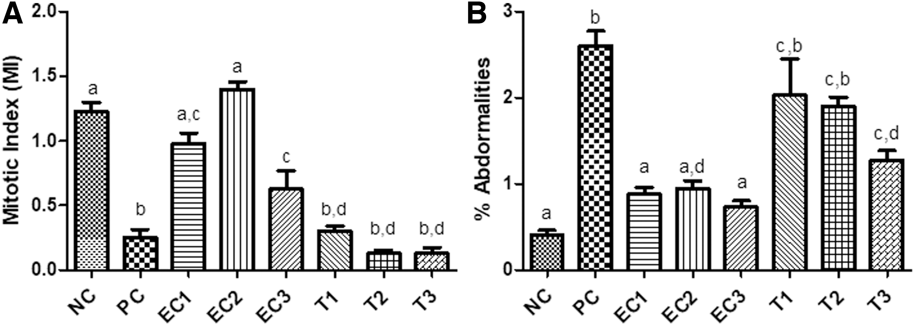
However, the three concentrations tested did not adequately prevent glyphosate damage, and were statistically equal to the PC. The C. nutans root crude extract at the three concentrations tested did not cause chromosomal changes in A. cepa cells, did not differ from each other, were statistically equal to the NC, and differed statistically from the PC (Fig. 3B). This indicated that the extract is not genotoxic. Regarding the prevention of chromosomal abnormalities caused by glyphosate, the concentration 1.5 mg/mL showed the potential to protect against chromosomal damage because it differed statistically from the PC, and protected against 50% of the damage that incurred within the nuclei of cells.
D. melanogaster assays
Survival
The concentration of C. nutans root extract capable of killing 50% of the D. melanogaster assessed (LC50) is the benchmark concentration reported in toxicity studies. In this study, the LC50 of C. nutans root extract was determined to be 26.05 mg/mL (R 2 = 0.7592) 5 days postexposure at the concentrations tested (Fig. 4). For this test, better results could not be obtained because it was only possible to solubilize the root extracts up to 30 mg/mL. These findings demonstrated that the root extract has a low degree of toxicity, and potential for use in future pharmacological studies is promising.

Survival of adult flies exposed to different concentrations of C. nutans root extract for 7 days. LC50 (26.05 mg/mL) of the extract response. The results expressed in percentage of survivor flies presented, correspond to the concentrations of the 15, 20, 25, 30, and 35 mg/mL. The test was effectuated in quadruplicate. R 2 = 0.7592
Treatment with C. nutans root extract and exposure to PQ
D. melanogaster was subjected to PQ poisoning and treated with 1, 5, and 10 mg/mL concentrations of C. nutans root extract. Results of the assay are shown in Figure 5. Treatment with 10 mg/mL extract produced a result that were statistically equal to the NC. It was observed that PQ caused about death of 52.5% of the flies assessed (PC), and this effect reversed by 35.7% posttreatment with 10 mg/mL extract, a value that differed statistically from the PC. Treatment with 1 mg/mL extract did not reduce levels of PQ damage observed, and values did not significantly differ from the PC. Results showed that treatment with 5 mg/mL and 10 mg/mL concentrations of extracts reversed effects of damage, especially since treatment with the highest concentration (10 mg/mL) of extract did not cause the death of the flies. Administration of the extract concomitantly with PQ has the potential to reverse damage caused by PQ.

PQ poisoning control trial represented by mortality (number of dead individuals) per group. NC (1% sucrose); EC (10 mg/mL extract roots); PC (3 mM PQ); Treatment: (T1 = 3 mM PQ +1 mg/mL extract; T2 = 3 mM PQ +5 mg/mL extract; T3 = 3 mM PQ +10 mg/mL extract). Different letters represent statistical differences according to Tukey's test (P < .001). PQ, paraquat.
Negative geotaxis assay
Negative geotaxis was assessed by determining the number of flies capable of reaching the top of a plastic bottle. Figure 6 shows that PQ exposure caused a decrease in the locomotor activity of flies, which resulted in 50% of the flies remaining at the bottom of the flask. Simultaneous treatment of flies with varying concentrations of C. nutans root extract (1, 5, and 10 mg/mL) and PQ reversed locomotor deficits observed by 30%. In these assays 80% of the flies treated with root extracts and PQ were able to reach the top of the bottle. There was no statistical difference between the three concentrations of root extracts used. The effect of treatment with extracts were observed in relation to the EC, and differed from the NC. This finding led researchers to believe that the extract has a stimulating effect on flies.
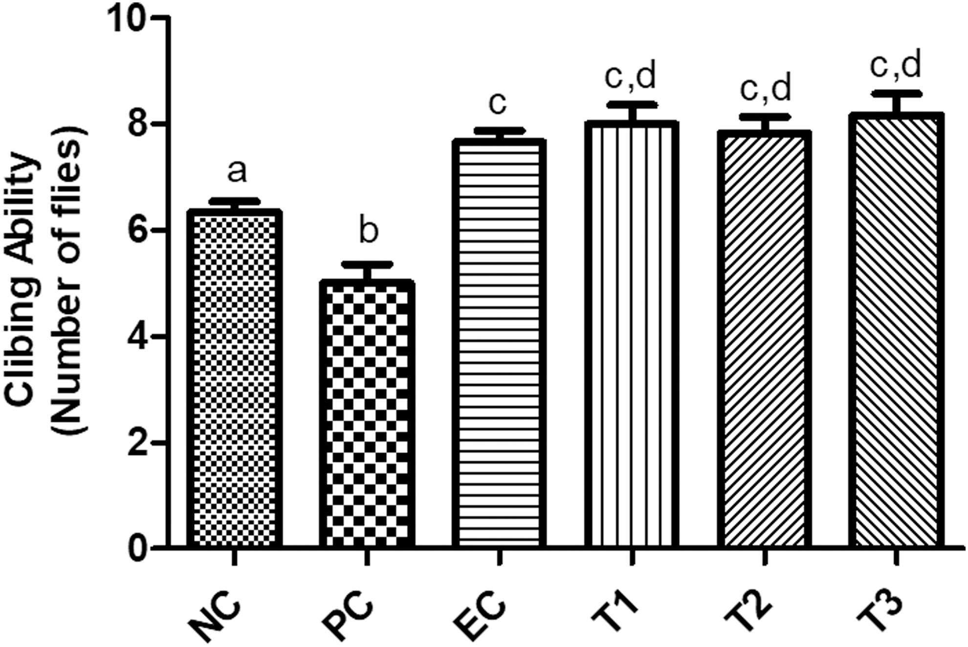
Effect of C. nutans extract on negative geotaxis (10 flies per replicate) in flies exposed to PQ for 4 days (n = 3). NC (1% sucrose); EC (10 mg/mL extract roots); PC (3 mM PQ); Treatment: (T1 = 3 mM PQ +1 mg/mL extract; T2 = 3 mM PQ +5 mg/mL extract; T3 = 3 mM PQ +10 mg/mL extract). Different letters represent statistical differences according to Tukey's test (P < .0001).
Discussion
Currently, there is a great interest in the investigation of medicinal or food extracts with antioxidant activity. The antioxidant capacity of the hydromethanolic extract of C. nutans roots was evaluated. A DPPH assay showed that C. nutans root extracts had very high IC50 and were unable to sequester the DPPH radicals. FRAP assay results were also negative (Table 1B). However, TBARS, carbonyl protein, and DCF assays showed that C. nutans root extracts had a significant degree of antioxidant capacity. They demonstrated that 0.1 and 10 mg/mL concentrations of extract were capable of protecting cells against from protein and lipid damage, and could also inhibit the formation of oxygen radicals (Fig. 1).
The TBARS assay, which used a lipid peroxidation-inducing agent, revealed that all three concentrations of extracts tested did not increase levels of MDA. Furthermore, 1000 μg/mL extract protected against 80% of the SF-induced lipid damage (Fig. 2). No studies have previously reported the presence of antioxidant activity in C. nutans, nor has antioxidant activity been reported for members of the genus Chaptalia. A study investigating a species that is a member of the same family as C. nutans produced similar results. Fabri et al. 41 and Paula et al. 42 also observed antioxidant activities in other species of Asteraceae, which confirmed the usefulness of the family for treating pathologies related to enhanced ROS production.
Although no substantial antioxidant activity was observed for C. nutans root extracts using either DPPH or FRAP methods, significant levels of antioxidant activity were observed when levels of lipid peroxidation, protein oxidation, and ability to eliminate ROS, through DCFH-DA method, were assessed. Our results confirm observations of Choi et al., 22 who stated that due to the complexity of chemicals present in crude extracts, it is necessary to evaluate plant antioxidant capacity using at least two methods.
Promising findings determined using the lipid peroxidation assay, protein oxidation assay, and the DCFH-DA method can be attributed to the phytochemical composition of the C. nutans root extract. The presence of a large amount of active phytoconstituents, such as phenols, alkaloids, coumarins, terpenes, etc. explain the antioxidant capacity of the extract. Several studies have confirmed that medicinal plants produce important pharmacological benefits as a result of the presence of bioactive constituents produced through secondary metabolic pathways. 43 Flavonoids, for example, prevent the growth (or kill) of many bacterial strains, inhibit specific enzymes, sequester free radicals, and stimulate some hormones and neurotransmitters. Coumarins are capable of functioning as antioxidants because they are capable of chelating iron ions and prevent lipid peroxidation. 44,45
The only studies found in the literature that have identified phytochemical constituents of C. nutans were reported by Truiti & Sarragioto 8 and Truit et al., 9 and these researchers reported the presence of a coumarin compound in both the leaves and roots of the species, which was isolated and its antibacterial activity was assessed.
Levels of tannins, flavonoids, and polyphenols in C. nutans species (Table 1A) may justify their popular consumption as laxatives and antitussives (internally). Furthermore, it may explain their effectiveness as topical treatments of injury and hemorrhage, 4,3 and their excellent antioxidant potential. 16 Flavonoids, tannins, and phenolic substances are plant constituents with potential antioxidant activity, mainly as a result of their capacity to scavenge free radicals. Chromatographic analysis through HPLC-DAD-MS revealed that the composition of C. nutans root extract includes phenolic compounds (Table 2), which could explain the high degree of antioxidant activity possessed by the extract.
The presence of isofeluric acid, a phenolic compound with significant antioxidant activity in lipid and aqueous media, 46 may explain results observed in the species. Wang et al. 46 and Li et al. 47 assessed Rhizoma cimicifugae species, and claimed that the ability to protect cells from oxidative damage was a result of the presence of isofeluric acid. Additionally, the presence of rosmarinic acid in C. nutans roots confirms its antioxidant activity, since as rosmarinic acid, a phenolic acid, is known to possess antioxidant activity and is found in many medicinal and crop plants capable of eliminating free radicals. 48,49
Another phenolic compound found in C. nutans root extract included arbutin, a well-known, plant-based compound with a great degree of value in both the medicinal and cosmetic industries as a result of its antioxidant and antifungal activities, which have been effectively used to treat urinary tract infections. 39,50 Ribitol found in C. nutans extract may be a result of bacterial contamination of plant roots during the collection and/or preparation of material, since it is a teichoic acid that functions to strengthen the cell wall of most Gram-positive bacteria. 51
In addition to the phytochemical study of C. nutans root extract, which indicated the presence of natural components that may be useful for preventing oxidative damage, its toxicity against A. salina, A. cepa, and D. melanogaster was assessed. As a preliminary toxicity test, we used the A. salina model, which is widely used in natural product laboratories, to perform a bioassay that monitored the toxicity of the plant extracts. 52,53 In this assay, we observed that the extract was slightly toxic. It had an LC50 value of 642.91 μg/mL, which was 11-fold greater compared with the PC (Table 3).
According to Nguta et al., 54 both organic and aqueous extracts with LC50 values between 500 and 1000 μg/mL are considered to possess low levels of toxicity, and extracts with LC50 values above 1000 μg/mL are considered to be nontoxic. Likewise, extracts of Senecio westermanii, a species from the same family as C. nutans, was determined to possess excellent antioxidant activity through multiple mechanisms, and its extract was determined to lack toxicity against A. salina. 55
Assays using A. cepa are generally employed to preliminarily evaluate antiproliferative and genotoxic effects of medicinal plant extracts because of its high degree of sensitivity and comparability with mammalian systems. 15,29,56 Results of assays evaluating the effects of C. nutans root extracts at a concentration of 1.5 mg/mL were promising. The addition of extract reduced MI values by 49% (Fig. 3A), demonstrating its antiproliferative potential. With respect to protecting cells from glyphosate damage, the addition of extract was ineffective.
Regarding genotoxicity, none of the three concentrations of extract tested caused chromosomal abnormalities in A. cepa cells (Fig. 3B). Dalla Nora et al. 57 also reported that the extracts lacked genotoxic effects against Mikania glomerata, suggesting that the chemical composition of the extract may enhance preservation of nuclear structures. Yuet Ping et al. 58 reported that the low MI of the Euphorbia hirta extract correlated with its genotoxic effects, which is in accordance with our results showing that C. nutans root extract is neither antiproliferative nor genotoxic. Additionally, 1.5 mg/mL extract protected against 50% of the chromosomal damage was caused by glyphosate.
This effect can be attributed to the phytochemical composition of the extract, since its components have a high antioxidant capacity. Phytochemicals, such as phenolic compounds, with these properties are generally nontoxic. Therefore, both the A. salina toxicity bioassay and the A. cepa genotoxicity assay effectively produced preliminary results showing that C. nutans extracts were not highly toxic.
D. melanogaster (fruit flies) functioned as a more complex experimental model, and were used to assess the biological properties of C. nutans root extract, and determine their toxicity. 59 The properties make D. melanogaster a great model for studying neurodegenerative diseases and neurotoxicity. 60 –62
Our findings confirmed that C. nutans root extract lacked toxicity. In fact, a very high concentration of the extract was needed to reach its LC50 (Fig. 4), concentrations that were much higher than those needed to produce sufficient antioxidant potential. Additionally, when compared with PQ values that caused the death of 52.5% of flies tested, C. nutans 10 mg/mL root extract did not cause the death of the flies, and levels of survival were statistically the same as the NC (Fig. 5). PQ is widely used to test intoxication and behavioral attributes in the D. melanogaster model, because it causes significant levels of brain damage, and will kill individuals after acute exposure. It is also useful for evaluating neuroprotective effects of compounds. Development of movement disorders and neurodegeneration in D. melanogaster were assessed in this case. 33,63
Treatment with C. nutans root extract, when coadministered to the PQ, produced a mortality rate of 18.75%. This indicated that the extract was able to protect 35.7% of the damage that occurred as a result of PQ treatment (Fig. 5). The locomotor performance of flies exposed to PQ and C. nutans root extract was evaluated using the negative geotaxis test, which is a common assay that assesses locomotor behavior in which flies move vertically (climbing) when placed in a bottle. 33 Results showed that C. nutans root extract protected from damage caused by PQ. When treated with PQ alone, 50% of the flies considered remained at the bottom of the bottle. When PQ was coadministered with root extracts, 80% of the flies assessed reached the top of the bottle (Fig. 6).
Similar results were found for the root extract of Decalepis hamiltonii and Bougainvillea glabra, which were able to protect flies from both mortality and locomotor impairment induced by PQ. 33,59 Opposite results were determined for Croton campestris hydroalcholic extract 64 ; it had an LC50 of 26.51 mg/mL after 3 days of treatment, while our study reached approximately this concentration after the sixth day of treatment. The authors of the work stated that the extract was toxic when administered concomitantly with PQ, which increased D. melanogaster mortality rates, and altered fly locomotor behavior.
Notably, these results that C. nutans root extracts function as antioxidants. PQ, which has been widely used as a model of oxidative stress in D. melanogaster, causes dopaminergic neurons to die as a result of increased levels of ROS production and decreased levels of dopamine, which leads to movement disorders similar to those of patients with Parkinson's disease. 33,65 The chemical composition of C. nutans root extract and its ability to neutralize different sources of ROS in vitro may affect its potential to protect against PQ-induced neurotoxicity. Soares et al. 59 suggested that the capacity of B. glabra extract to suppress oxidative stress induced by PQ was related to free radical elimination, since the extract showed antioxidant activity in vitro and in vivo. The results suggested that the antioxidant capacity of C. nutans root extract was sufficient to prevent PQ-induced neurotoxicity.
Our study demonstrates that the roots of C. nutans are rich sources of natural antioxidants, which are useful for both the food and pharmaceutical industry. Furthermore, they are not cytotoxic, genotoxic, or neurotoxic. Further studies are needed to confirm these unprecedented results.
Conclusion
In this study, we revealed for the first time that C. nutans root extract is a source of several natural antioxidants, which significantly prevent oxidative stress-induced damage. In addition, synergism between the antioxidant capacity and neuroprotective effects of the extract, along with its lack of cyto- and genotoxicity, is likely.
Footnotes
Author Disclosure Statement
The authors declare that there are no conflicts of interest regarding the publication of this article.
Funding Information
The authors are grateful to CNPq, CAPES, and FINEP agencies.