
Abstract
Polysaccharide from Ma-chi-xian (Portulacae oleracea L., POLP) was prepared and the therapeutic effect on dextran sodium sulfate-induced colitis mice was investigated in this study. The results of clinical activity score and H&E staining confirmed the therapeutic effect of POLP. POLP could diminished the symptoms of colitis and improve colon histopathological structure of the colitis mice. The expression levels of four cytokines were determined. The concentrations of PGE2 and IL-6 were downregulated by POLP treatment. The COX-2 protein expression levels and the STAT3 phosphorylation levels were detected. The results showed that these two protein levels were all increased in colitis and decreased after POLP treatment, indicating that these two proteins were closely related with the protective effect of POLP. Because the synthesis of PGE2 is catalyzed by COX-2 and phosphorylation of STAT3 can induce the expression of COX-2, it was concluded that STAT3 was a key protein related to the POLP exerting its activity in colitis.
Introduction
Ma-chi-xian (Portulaca oleracea L. [POL]) is a widely used traditional Chinese medicine (TCM) and the main function is recorded as clearing heat toxicity. 1 Its Chinese name is “ma chi xian” and first recorded as a cold medicine in Ben Cao Jing Ji Zhu. According to the principle of treating heat syndrome with cold-natured drugs, cold POL is used to treat damp-heat dysentery. 2 More evidence exhibited that there are large amounts of polysaccharide, alkaloids, and various kinds of flavonoids in POL. 3 The main pharmacological effects of POL include antibacterial, anti-inflammatory, and wound healing. 4,5 Therefore, it is widely used in clinical trials, especially for colitis.
Colitis is defined as an inflammatory bowel disease (IBD) and can result in bloody diarrhea that can be life threatening. And it can affect any part of the gastrointestinal tract. 6 Although there is much research about colitis, the concrete mechanisms involved in its etiology remain elusive. Emerging evidence indicates that the occurrence and progression of these disease are caused by many factors, including immunoregulatory, genetic, and environmental factors. 7,8 Furthermore, it is considered that the occurrence and progression of IBD are closely related with the imbalance of inflammatory cytokines. 9 Especially, the equilibrium between anti-inflammatory cytokines and proinflammatory cytokines are supposed to be key in maintaining health. Moreover, accumulating experiments suggest that almost all of the cytokines are connected with cellular signaling proteins to modulate the function of the target cell. 10,11
To explore the pharmacodynamic material basis of POL, the total alkaloids, flavones, and polysaccharide were extracted from POL, and the protective effects were investigated in our previous study. 12 The results showed that polysaccharide from POL (POLP) could ameliorate the health conditions of colitis mice and the expression levels cytokines IL-6 and PGE2 are greatly changed after treatment with POLP. These results implied that IL-6 and PGE2 were closely related with the protective effect of POLP.
IL-6 is an important proinflammatory cytokines and plays significant roles in inflammation. It has been proved that IL-6 could activate STAT3 to exacerbate the progression of inflammation. 13 The translocation of phosphorylated STAT3 into nucleus gives rise to gene expressions of migration, apoptosis, and so on. 14,15 Literatures suggest that IL-6 has the ability to activate STAT3, which is an important part in IBD pathogenesis. 16 PGE2 is a significant growth factor related to cell growth. It is reported that PGE2 is the principal metabolic product of COX-2, which is an important cellular factor related with cell growth and can be modulated by NF-κB and STAT3. 17 In the past decades, number of colitis model have been established and used to decipher the underlying mechanisms as well as to evaluate number potential therapeutics. Among these models, dextran sulfate sodium (DSS)-induced colitis model is widely used for its rapidity, controllability, simplicity, reproducibility, and especially many similarities with human intestine colitis. 18
For the advantages of DSS-induced colitis model, this model was employed in our experiments to investigate the roles of POLP and the underlying mechanisms of these protective effects.
Materials and Methods
Extraction and purification of POLP
POLP was extracted and purified from P. oleracea L. through traditional process combined with membrane separation technology reported in our previous study. 12 P. oleracea L. was purchased from the market in Xi'an, Shaanxi Province, China. The plant was identified as P. oleracea L. by senior engineer Jitao, Wang, department of pharmacology, Shaanxi University of Chinese Medicine. A voucher specimen has been deposited in Shaanxi Collaborative Innovation Center of Chinese Medicinal Resources Industrialization, Shaanxi University of Chinese Medicine (20151129). The plant (1 kg) was ground and extracted with 10 × of petroleum ether two times (2 h/time). The solvent was filtered and the powder was collected and dried. The dried powder was extracted with water at 100°C for 3°h and repeated four times. The extraction was filtered and the solvent was collected. Then fourfold of 80% ethanol was added into the extraction solvent for 24 h. The precipitate was filtered and diluted in absolute ethyl alcohol and acetone. The proteins in the crude extraction were removed by repeating the Sevage method three times. The crude polysaccharide was freeze–dried and 91.2 g of product with an extraction yield of 9.12% was obtained. Finally, the crude product was separated with membrane separation technology through 0.8, 0.2, and 0.05 μm of membrane successively. And many of the polysaccharides were collected from the penetrated liquid filtered of 0.8 μm membrane. The penetrated liquid was evaporated and the polysaccharide was freeze–dried and about 60.8 g product was received. The concentration of polysaccharide in crude product was 92.7% determined by phenol-sulfuric acid method.
Animal models of colitis induced by DSS
Kun Ming mice weighing 20 ± 2 g (Animal Center of Health Science Center, Xi'an-Jiao-Tong-University) were used in this study. All animal experiments followed protocols confirmed by Institutional Ethical Committee of Shaanxi University of Chinese Medicine. Permits for the experiments were obtained with the specific approval signed by Institutional Ethical Committee of Shaanxi University of Chinese Medicine.
The mice weighing 20 ± 2 g were maintained in standard laboratory conditions, with ultrafiltered water and standard chow. DSS with average molecule weight of 40,000 Da was obtained (MP Biomedicals, USA) in powder form. Mice for control group were provided distilled water. The colitis mice were prepared by freely drinking 5% DSS solutions for 7 days. Then, colitis animals were randomly divided into four groups: the positive control group (9 mg/mL sulfasalazine) and three POLP groups (0.75, 0.5, and 0.25 g/mL POLP). Drugs were given in a 0.5 mL volume saline by intragastrical administration, 9 and therapeutic period were 7 days after colitis induction.
Clinical activity score
The previously reported evaluation method 19 for DSS colitis was adopted involving daily rectal bleeding, body weight, and stool consistency data. In this method, the clinical activity score (CAS) ranged from 0.0 (healthy) to 6.0 (the most severity colitis).
Cytokine determination
Inflammatory cytokines, including IL-1β, IL-6, IL-10, and PGE2, were tested in our study. ELISA kits of IL-β, IL-6, and IL-10 were all purchased from Neobioscience (Batch Code: M171109-604a, M171109-605a, and M171109-104a, respectively). The PGE2 kits were provided by Shanghai Zeye Biological Technology Co. (Batch Code, ZY-PGE2-Ge).
Histopathological evaluation
Mice colon tissues were dissected and spread onto plastic sheets, followed by 48-h-fixation with 10% neutral-buffered formalin and embedding in a paraffin block. The embedded tissues were stained with H&E method and observed under microscope by pathologists to give histological scores. Scoring standard consisted of inflammatory infiltration (0 for normal, 1 for the lamina propria inflammatory cells increasing, 2 for submucosa inflammatory cells confluence, and 3 for transmural extension of the infiltrate) and tissue damage (0 for normal, 1 for discrete lymphoepithelial lesions, 2 for surface mucosal erosion, and 3 for severe mucosal damage of deeper structures of the bowel wall). The combined histological score ranged from 0 (no changes) to 6 (extensive cell infiltration and tissue damage).
Immunohistochemistry
The earlier colon tissues sections were also used for immunohistochemistry study. The expressions of COX-2 (CST, No. 12282) and p-STAT3 (CST, No. 9145S) were detected with commercially available kits (KGOS60; KeyGEN). In brief, tissue slides were deparaffinized and incubated with 10 mM sodium citrate buffer (0.1% Tween 20) in boiled water bath, to unmask antigens. Then, primary antibodies were incubated with the slides and horseradish peroxidase polymer was added and incubated for 30 min. After imaging, slices were observed and the counting and quantification of labeled proteins were conducted with Image Pro +6.0 software (Media Cybernetics, USA).
Western blot analysis
Mice colon tissues were ground to homogenate in homogenizer and washed with cold PBS. Then the washed tissues were lysed by RIPA buffer (Heart Biological Technology Co., China) assisted by a homogenizer. The supernatants were piped out and protein concentrations were detected by BCA kits (Pierce, USA). SDS-PAGE was used to separate the prepared proteins. The separated proteins were then transferred onto PVDF membranes (Millipore, USA). Membranes were blocked with 5% defatted milk and incubated with primary antibodies of COX-2 (CST, No. 12282), STAT3 (CST, No. 12460S), and p-STAT3 (CST, No. 9145S). Then, secondary antibodies (1:3000) were incubated after visualizing using ECL Western blotting detection reagent (Millipore, USA). Images were gathered by the Multi Image Light Cabinet Filter Positions (Alpha Innotech, USA) system.
Statistical analysis
All results were presented as the mean ± standard deviation. All studies were replicated three times. P < .05 were considered statistically significant.
Results
POLP attenuated DSS-induced colitis
The CAS in Figure 1 exhibits the improved role of POLP on colitis. The colitis group with CAS was almost 3 on the 7th day, indicating that the colitis model was successfully established compared with control. After treatment for 7 days, the health conditions of colitis in positive control and POLP groups were all greatly improved. The CAS of POLP groups and positive control group were almost equal, especially 0.75 g/mL of POLP. All results and CAS indicated that POLP could ameliorate health conditions of colitis mice.
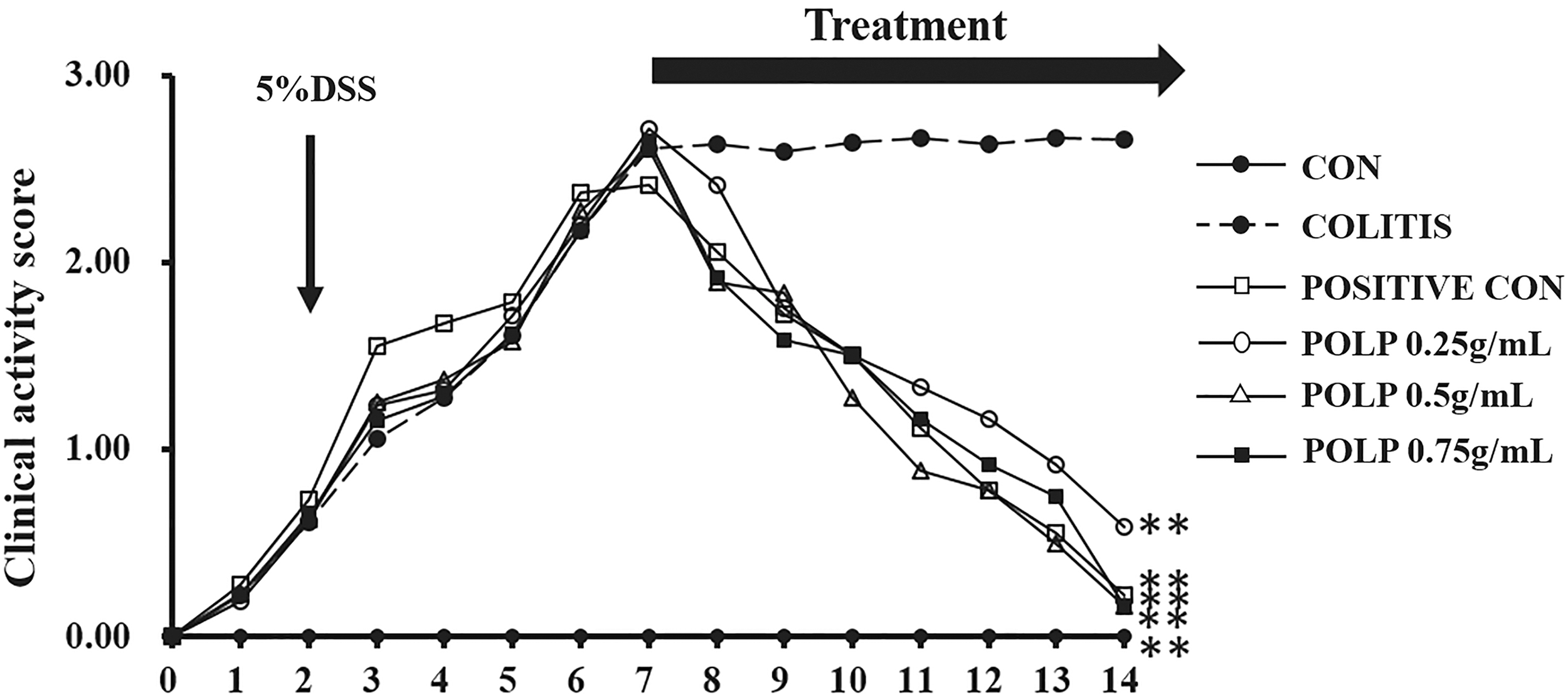
CAS of mice. After colitis induction, the animals were treated for 7 days with distilled water, DSS, sulfasalazine (“positive con”), and three concentrations of POLP. The untreated showed no significant improvement after 14 days, whereas treatment with sulfasalazine and three concentrations of POLP has significantly improved the CAS of colitis mice. n = 6, **P < .01 compared with colitis. CAS, clinical activity score; DSS, dextran sulfate sodium; POLP, polysaccharide from Portulaca oleracea L.
POLP decreased IL-6 and PGE2 levels
Because of the important roles of inflammatory cytokines in the pathology of colitis, the concentrations of four cytokines, IL-10, IL-1β, IL-6, and PGE2 were detected by ELISA kits. The results in Figure 2 showed that all the five cytokines were significantly changed in the colitis group compared with negative control. These results implied that these four inflammatory cytokines were closely related with colitis. After 7 days treatment with POLP, the concentrations of IL-6 and PGE2 were all greatly downregulated in the colitis group compared with model group. These observations indicated that IL-6 and PGE2 were closely related to the protective effect of POLP.
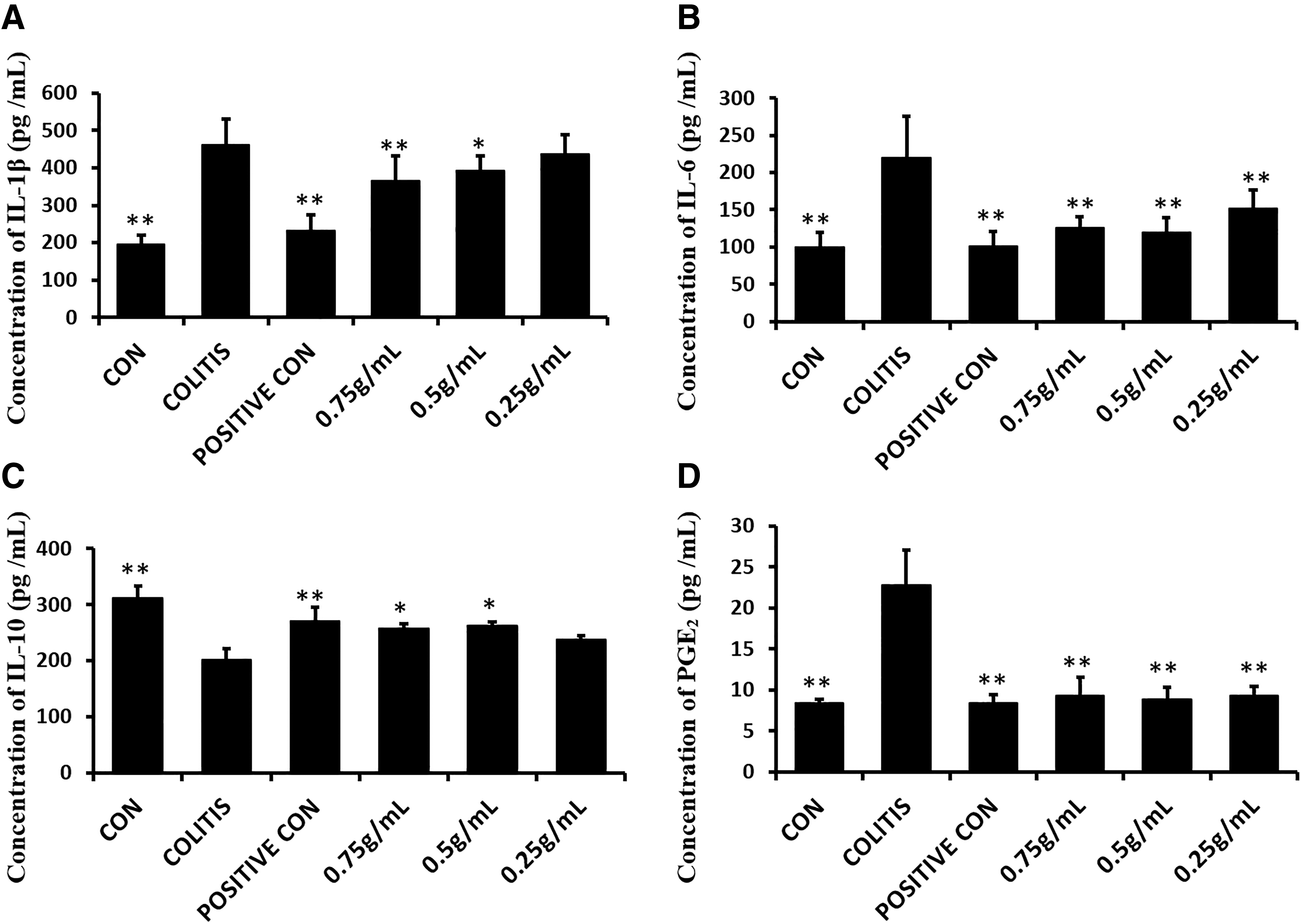
Levels of cytokines IL-1β, IL-10, IL-6, and PGE2 in each group.
POLP improved the histological alterations of colitis mice
To evaluate the protective effect of POLP, H&E staining was used in our study. Figure 3 shows histopathological sections of each group. As shown by the normal colon (Fig. 3A), the structure was clear, the crypts and goblet cells were regularly arranged, whereas in colitis (Fig. 3B), the colon crypts and goblet cells were rarely seen and many massive inflammatory cells were observed. The pathological section of Figure 3C shows a certain degree of protective effects of the positive drug, sulfasalazine. The intact colon structure and some inflammatory infiltration were observed. Similarly, POLP also ameliorate colitis with 0.5 g/mL concentration (Fig. 3D). The tissue damages caused by inflammation were inhibited by POLP, colon structures were still intact. The histopathological scores of each group are shown in Figure 3E; the column heights of positive control and POLP were considerably lower than that of colitis group.
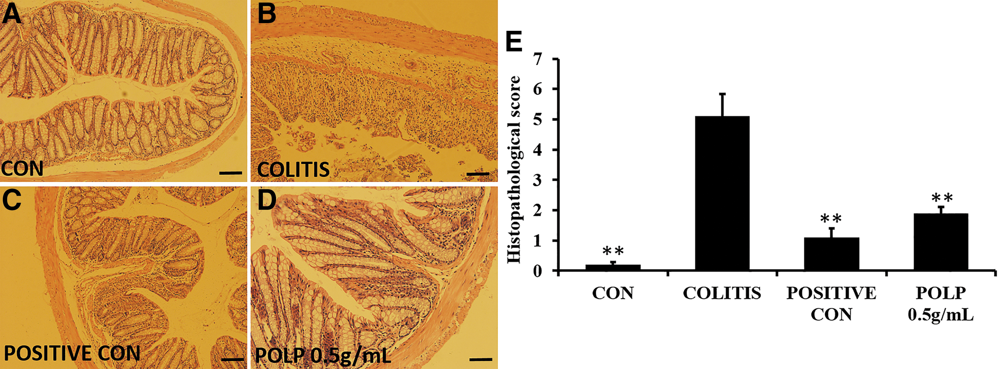
Histological characteristics of each group, magnification × 100.
POLP inhibited the phosphorylation of p-STAT3 and the protein level of COX-2
The results of Figure 4 exhibit the phosphorylation level of p-STAT3 in colon tissue. The protein level of p-STAT3 was considerably increased in colitis group, whereas the phosphorylation levels were all decreased by positive drug and POLP (0.5 g/mL).
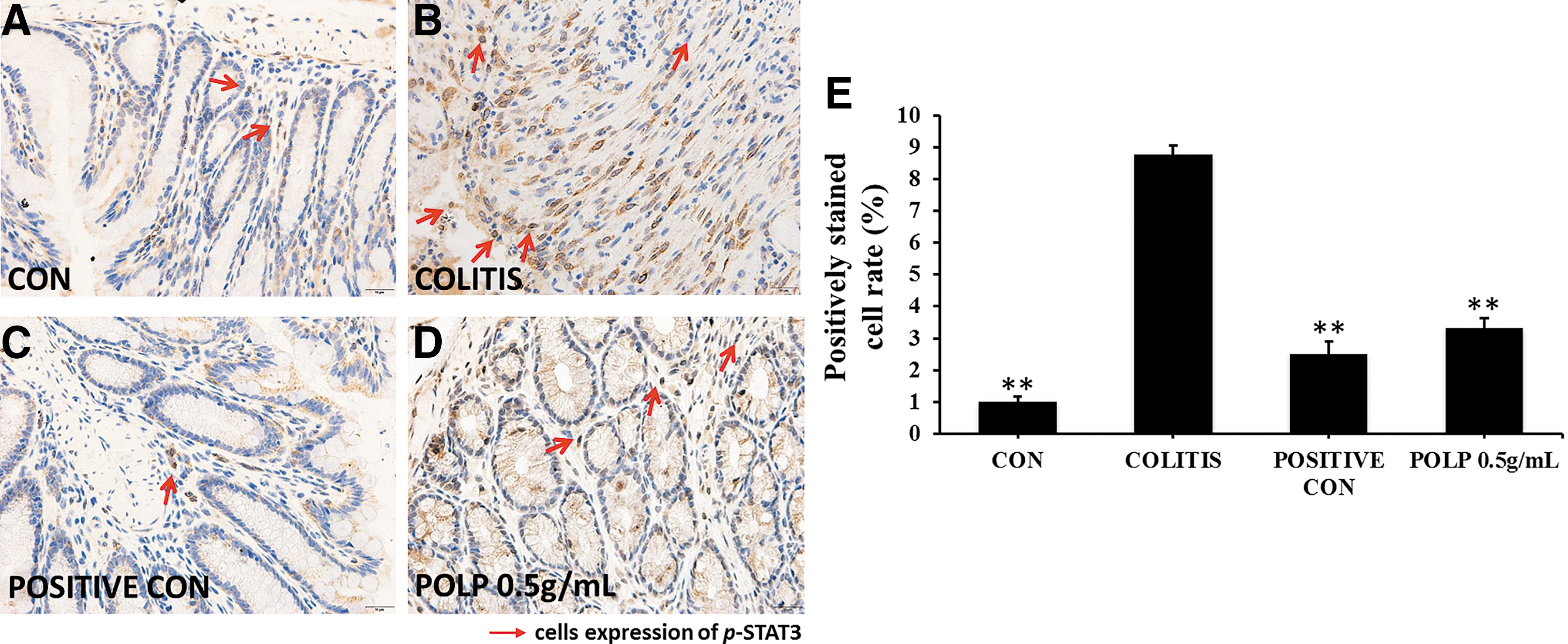
Treatment with POLP suppressed the expression of p-STAT3, magnification × 400.
PGE2 is an important growth factor that could inhibit the progression of inflammation. The synthesis of PGE2 is greatly regulated by COX-2, which can be modulated by STAT3. 20 Therefore, the protein concentrations of COX-2 in colon tissues were also investigated in our study. The results in Figure 5 exhibited that the variation curve of COX-2 was similar to that of p-STAT3. The protein expression level in colitis was greatly higher than that in control. The opposite results were observed after treatment with POLP.

POLP inhibited the expression of COX-2, magnification × 400.
To evaluate the concrete phosphorylation level of STAT3 and the protein level of COX-2, western blotting analysis was enrolled in our study. The results in Figure 6 displayed that the phosphorylation level of STAT3 was upregulated in colitis, indicating that STAT3 played a key role in colitis occurrence. After treatment with POLP, the phosphorylation level sharply decreased. These results suggested that POLP regulated the phosphorylation level of STAT3 to exert its protective effect. Furthermore, the protein concentration of COX-2 was also investigated, which was regulated by STAT3 and could catalyze the synthesis of PGE2. The variation curve was similar to that of p-STAT3, indicating the close relationship of STAT3 and COX-2.
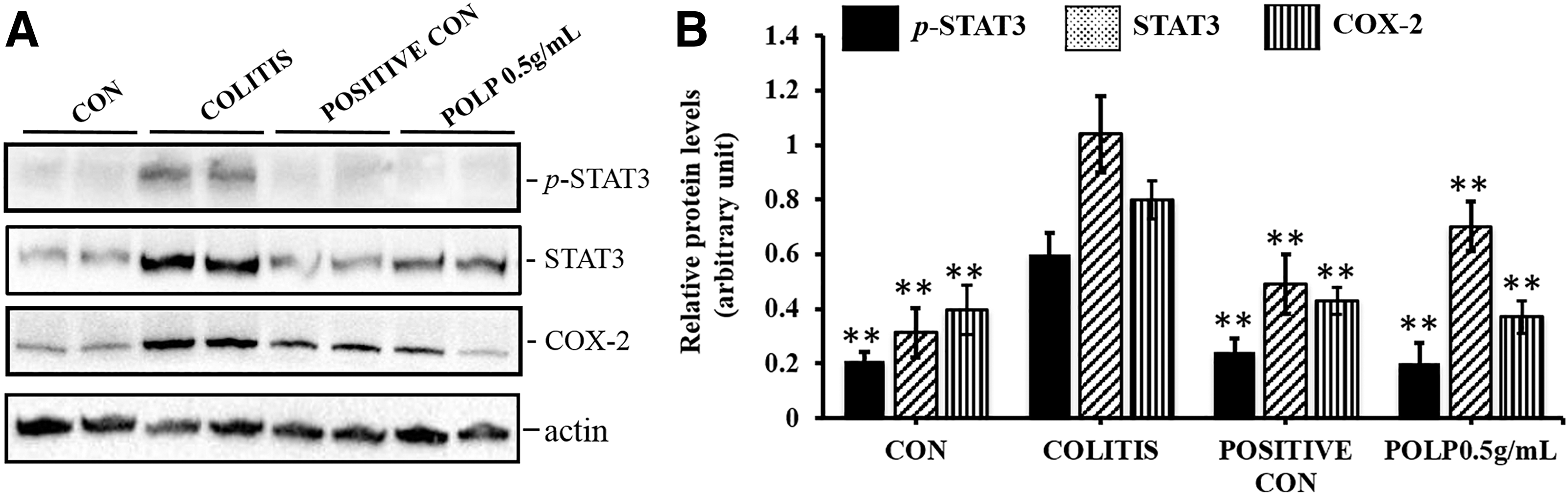
Effects of POLP on the protein levels of STAT3 and COX-2.
Discussion
P. oleracea L. is a cold TCM. Although it has been clinically used to treat colitis for 1000 years, the concrete mechanisms and pharmacodynamic characteristics remain unclear. In this study, polysaccharide was extracted from P. oleracea L. and the protective activity against colitis was evaluated.
The results of CAS implied that POLP could protect against colitis induced by DSS. The H&E experiment demonstrated that the activity of POLP on ameliorating health conditions was mainly associated with protecting the morphological structure of the colons of mice with colitis. After treatment with 0.5 g/mL POLP, the intact morphological structure was observed and the inflammatory cells had almost disappeared. These results definitely demonstrated that POLP could improve colitis syndrome and keep the colon intact.
To elucidate the mechanisms of this protective effect of POLP, four cytokines were detected in our study. The results indicated that IL-10, IL-1β, IL-6, and PGE2 were all related with the occurrence and progression of colitis. However, only IL-6 and PGE2 decreased after treatment with POLP implying that the therapeutic effect of POLP was closely related with these two cytokines. It is reported that IL-6 could activate the phosphorylation of STAT3, which is an important transcription factor related with cell proliferation and apoptosis. Activation of STAT3 by IL-6 plays a crucial role in inflammation-induced disease pathogenesis. 21,22
PGE2 is a significant growth factor and could bind to a G protein-coupled receptor to modulate the inflammation. The synthesis of PGE2 is catalyzed by COX-2 using arachidonic acid as materials. 23 It is an important inflammation cytokine and plays a significant role in occurrence of colitis. It is reported that IL-6 could activate COX-2 through its trans-signaling pathway, and the IL-6-STAT3–COX-2 axis was shown to be important in inflammation-induced disease. 24 Therefore, the expression level of COX-2 was also detected in our study. The results of immunohistochemistry and western blotting analysis revealed that the phosphorylation level of STAT3 and the protein expression level of COX-2 were all increased significantly in the colitis group, and these two protein levels were all decreased after treatment with POLP. These results suggested that the protective effect of POLP was closely related with STAT3 and COX-2. Referring to the previous reports, it was concluded that POLP exerted its protective effect through regulating the IL-6/STAT3/COX-2 pathway.
Ethical Statements
The protocols of animal were approved by Institutional Ethical Committee of Shaanxi University of Chinese Medicine. And the experiment procedures followed were in accordance with the Helsinki Declaration of 1975, as revised in 2008.
Footnotes
Author Disclosure Statement
Z.W., Y.L., D.Z., X.W., J.Y., Z.Z., Y.L., C.S., Z.T., L.L. designed and conducted the study, and analyzed the data. Z.W., Y.L., D.Z., and X.W. wrote the article. J.Y., Z.W., C.S., Z.T., and L.L. are the principal investigators, and revised and edited the article.
Funding Information
This study was supported by the Program for the National Natural Science Foundation of China (Nos. 81703924 and 81803951), The Youth Innovation Team of Shaanxi Universities, The Key Research and Development Program of Shaanxi Province (2018ZDCXL-SF-01-02-02), and Science and Technology Young Nova Program of Shaanxi Province (2019KJXX-025).