




Abstract
To reduce microbial loads in medicinal herbs, Cnidii Rhizoma and Alismatis Rhizoma were subjected to electron-beam (e-beam) irradiation at doses (≤10 kGy) as permitted by the Korean Food Code. The effects of e-beam irradiation on the microbial load, stability of the active components, and anti-inflammatory activity of medicinal herbs were determined. We observed that the total aerobic bacteria (TAB; 4.0–7.0 log CFU/g), yeasts and molds (Y&M; 3.3–6.8 log CFU/g), and coliform counts (CC; 3.2–3.8 log CFU/g) in both herb samples were effectively reduced in a dose-dependent manner, resulting in acceptable levels of <3.0 log CFU/g in TAB and Y&M and negative in CC at 10 kGy irradiation. The concentration of the active components (0.87–4.22 mg/g) of Cnidii Rhizoma, including z-ligustilide, chlorogenic acid, senkyunolide A, and ferulic acid, in order of prevalence and those (0.86–2.76 mg/g) of Alismatis Rhizoma, including Alisol B acetate and Alisol B, were not changed at irradiation doses of ≤10 kGy. The extracts of e-beam irradiated Cnidii Rhizoma and Alismatis Rhizoma showed a reduced production of inflammation-related factors, such as nitric oxide, prostaglandin E2, interleukin (IL)-1β, and IL-6, in a concentration-dependent manner, which was induced by lipopolysaccharide in RAW 264.7 cell. However, there was no significant difference observed at e-beam irradiation doses of 0, 1, 5, and 10 kGy. Thus, we confirm that e-beam irradiation up to 10 kGy was effective for the control of microbial load in Cnidii Rhizoma and Alismatis Rhizoma without causing considerable changes in their major active components and anti-inflammatory activity. The results show the potential of e-beam application for sanitization of medicinal herbs.
Introduction
The medicinal herb industry has been gradually increasing at a large scale, as there is an increased interest in the use of natural products in medicine and medical supplies. 1 Due to the growing demand for medicinal herbs, at least 70% of the herbs that are currently used in Korea are being imported. As imported medicinal herbs might be damaged by insects or contaminated by microorganisms or mycotoxins due to long shipping periods and unsterilized manufacturing processes, the need to control microbial growth has become a major priority in the medicinal herb industry. 2,3
Cnidii Rhizoma and Alismatis Rhizoma are examples of medicinal herbs that are generally sprayed with large amounts of pesticides to prevent insects and microbial contamination. 4 Cnidii Rhizoma is a medicinal herb used extensively globally for treating anemia, sinusitis, irregular menstruation, and gynecological conditions because it has antioxidant as well as anti-inflammatory activities. It also inhibits active microglial cell production. 5 –7
Cnidii Rhizoma has been reported to contain large amounts of phthalides and terpene compounds such as senkyunolide A and z-ligustilide. 8,9 Yan et al. reported the active components in Cnidii Rhizoma using high-performance liquid chromatography (HPLC). 10 Alismatis Rhizoma is also a medicinal herb that has anti-inflammatory, 11,12 anti-cancer, 13,14 and anti-diabetic effects, 15,16 and alisol B acetate is its major component. 17
Recently, gamma rays, electron-beams (e-beams), and X-rays have been used for the removal of pathogenic microorganisms and pests, and to delay germination and control maturation. 18,19 An e-beam is classified as a non-heat treatment method as they produce ionizing radiation energy. They have been of interest lately because they can be continuously applied and require a short penetration time, and unlike gamma rays, they do not produce radioactive waste. 20
Studies have reported the effects of e-beam irradiation on the color, nutrient composition such as sugars, organic acids, tocopherols, fatty acids, and antioxidant properties of dried plants, dried mushrooms, Portuguese chestnuts, and Matricaria chamomilla L. 21 –24 Irradiation with ionizing radiation is another method for disinfecting dried plant materials, which is an economically viable alternative to chemical fumigation. Irradiation of dried spices is widely recognized and is now legally accepted in more than 60 countries with a maximum overall average dose of 10 kGy. 25 In some countries, such as Australia, New Zealand, and the United States, a dose up to 30 kGy is permitted. 26
Studies on irradiation of medicinal herbs have been conducted in which herbs such as Angelica gigas root, licorice, Saposhnikovia root, Angelica dahurica root, Astragalus root, and Ephedra herb were irradiated with gamma rays. These studies reported the effects of gamma rays on the microbial contamination, antimicrobial and antioxidant activities, as well as the active components of these herbs. 27 –29 However, there have been a few reports on the application of e-beam irradiation for decontamination of medicinal herbs and on its effects on their anti-inflammatory activity, which is the major function of medicinal herbs.
Therefore, in this study, we intended to determine the effect of e-beam irradiation up to a dose of 10 kGy on microbial load and the stability of active components in the medicinal herbs, Cnidii Rhizoma and Alismatis Rhizoma. In addition, we also assessed the effects of e-beam irradiation on the anti-inflammatory activity of both herbal extracts, which is the main biological activity. This was done by investigating the production of inflammation mediators such as nitric oxide (NO), interleukin-1β (IL-1β), interleukin-6 (IL-6), and prostaglandin E2 (PGE2) in lipopolysaccharide (LPS)-induced RAW 264.7 cells to evaluate the applicability of e-beam for the sanitization of medicinal herbs.
Materials and Methods
Plant materials and e-beam irradiation
Cnidii Rhizoma and Alismatis Rhizoma were purchased from a Good Manufacturing Practice certified herbal medicine company in Yeongcheon-si, Gyeongsangbuk-do, Korea. The origin of Cnidii Rhizoma is Cnidium officinale Makino, which was cultivated in Bonghwa-gun, Gyeongsangbuk-do, Korea and Alismatis Rhizoma is Alisma orientale Juzepzuk, which was cultivated in Suncheon-si, Jeollanam-do. Both of these cultivars were produced as herbal medicines in 2015. These medicinal herbs were originally packed in polyethylene bags (600 g pack) and stored at 4°C until further usage.
We carried out the e-beam irradiation by using an electron accelerator (High Energy Linear Accelerator, 10 MeV; EB-Tech, Korea) at doses of 0, 1, 5, and 10 kGy. All of the irradiated samples were ground into a powder that could pass through a 20-mm mesh sieve and stored at below 4°C until further use.
Chemicals
Z-ligustilide, alisol B, and alisol B acetate standards were purchased from FUJIFILM Wako Pure Chemical Corporation (Osaka, Japan). The senkyunolide A standard was purchased from Chemfaces CN (Wuhan, China). Chlorogenic acid and ferulic acid were purchased from Sigma-Aldrich Co. (St. Louis, MO, USA). All of these had a purity of more than 98%.
Methanol and acetonitrile were of analytical grade, whereas HPLC solvents were of HPLC grade and were purchased from Merck Chemicals GmbH (Darmstadt, Germany). Acetic acid was purchased from FUJIFILM Wako Pure Chemical Corporation.
HPLC analyses were performed by using a Waters HPLC system (Waters Corporation, Milford, MA, USA) equipped with an Alliance photo diode array (PDA) detector.
Dimethyl sulfoxide (DMSO), 3-(4,5-dimethylthiazol-2-yl)-2,5-diphenyltetrazolium bromide (MTT), and Griess reagent were purchased from Sigma-Aldrich Co. (St. Louis), Dulbecco's modified Eagle's medium (DMEM) was purchased from Gibco BRL (Grand Island, NY, USA), and 10% fetal bovine serum (FBS) was purchased from Invitrogen Co. (Carlsbad, CA, USA). To measure the amount of PGE2, IL-1β, and IL-6 in the cell culture medium, a commercial competitive enzyme immunoassay kit was purchased from R&D Systems (Minneapolis, MN, USA). A water-jacketed 3010 (Thermo Electron Corp., Waltham, MA, USA) CO2 incubator and an enzyme-linked immunosorbent assay (ELISA) reader from Tecan Group Ltd. (Mἄnnedorf, Switzerland) were used.
Microbial analysis
Microbial analysis was carried out by following the Korean Food Standards Codex. 18 Ten grams of each sample was homogenized for 2 min in a sterile stomacher bag containing 90 mL of sterile 0.1% peptone water by using a stomacher (Bagmixer® 400; Interscience Co., Saint Nom, France). Serial decimal dilutions were prepared by using the same diluent and analyzed for total aerobic bacteria (TAB), yeasts and molds (Y&M), and coliforms counts (CC). Enumeration media for TAB, Y&M, and CC were prepared with plate count agar (Merck KGaA, Darmstadt, Germany), potato dextrose agar (Merck), and a desoxycholate agar (Merck), respectively. For the culture, the plates of TAB and CC were incubated at 32°C for 2 days, whereas those of Y&M were incubated at 25°C for 7 days. The colony forming units (CFU) were counted on the plate ranging from 30 to 300 CFU, and based on this, the CFU per gram of herbal medicine were calculated.
Analysis of the active compounds in e-beam irradiated Cnidii Rhizoma
The contents of the active ingredients in e-beam irradiated Cnidii Rhizoma were determined by a method described by Baek et al. 30 One gram of powdered Cnidii Rhizoma was added to 70% ethanol (50 mL), extracted by using sonication treatment (1 h), and filtered by using a 0.45-μm syringe filter, and this was used as the test solution. Then, we dissolved 10 mg each of chlorogenic acid, ferulic acid, senkyunolide A, and z-ligustilide standards separately in methanol to make exactly 100 mL, which was used as the standard solution. The sample and standard solutions were analyzed by using a Waters HPLC system (Waters Corporation) equipped with an Alliance and PDA detector and C18 column (Waters Atlantis dC18 4.6 × 150 mm, 5 μm) at 35°C. A (0.1% acetic acid in water) and B (0.1% acetic acid in acetonitrile) were used as the mobile phase by using a gradient condition (0 min, 20% B; 2 min, 20% B; 4 min, 55% B; 20 min, 85% B) to analyze the samples. The flow rate was 1.0 mL min−1, injection volume was 10 μL, and peaks were detected under UV at 280 nm.
Analysis of active compounds in e-beam irradiated Alismatis Rhizoma
The contents of the active ingredient in e-beam irradiated Alismatis Rhizoma were determined by the method of Baek et al. 31 For this, 0.5 g of powdered Alismatis Rhizoma was added to acetonitrile (10 mL) and extracted by using sonication (1 h) at 50°C, and the rest of the protocol was followed as previously described. The filtered solvent was removed by using a vacuum concentrator; acetonitrile was added to the residue to make exactly 2 mL and filtered by using a 0.45-μm syringe filter to make the test solution. Then, 10 mg each of alisol B and alisol B acetate standards was dissolved separately in acetonitrile to make exactly 100 mL, which was used as the standard solution. The sample and standard solutions were analyzed by using a Waters HPLC system (Waters Corporation) equipped with an Alliance and PDA detector and C18 column (Waters Nova-Pak 4.6 × 150 mm, 3.9 μm) at 35°C. A (water) and B (acetonitrile) were used as the mobile phase by using a gradient condition (0 min, 45% B; 20 min, 50% B; 30 min, 50% B) to analyze samples. The flow rate was 1.0 mL min−1, injection volume was 10 μL, and peaks were detected under UV at 207 nm.
Determination of anti-inflammatory activity
Preparation of extracts
Fifty grams each of irradiated Cnidii Rhizoma and Alismatis Rhizoma powder was extracted with 500 mL of 70% ethanol for 24 h at room temperature with stirring, and the extracted solution was filtered through filter paper and evaporated. The extracts were freeze-dried to obtain 17.4–18.1 g of Cnidii Rhizoma extract and 15.5–15.8 g of Alismatis Rhizoma extract, which were then stored at −20°C until further use. Ten milligrams of each extract was dissolved in 1 mL of ethanol and used as the sample solution.
Cell culture
RAW 264.7 cell murine macrophage cell line was purchased from the Korean Cell Line Bank (Korea). The cells were cultured in DMEM supplemented with 10% FBS and penicillin/streptomycin (100 U/mL), and sub-cultured at 37°C in a 5% CO2 incubator. Various concentrations of the CRE and ARE were dissolved in DMSO and added to the cells along with LPS. As the final concentration of DMSO used was less than 0.05%, cells were treated with only 0.05% DMSO as vehicle control.
Evaluation of cell viability using MTT assay
RAW 264.7 cells were seeded at a density of 5 × 10⁴ cells/well in a 96-well plate, and 0.02 mL of each extract was added to each well. The cells were cultured in a 5% CO2 incubator at 37°C for 24 h. In the control group, the same amount of distilled water as that of the sample was added to the cells and cultured under the same conditions. After adding 0.002 mL of 5 mg/mL MTT solution and incubating for 4 h, the culture solution was removed, and 0.15 mL of DMSO was added to each well. The reaction was allowed to proceed at room temperature for 30 min, and then absorbance was measured at 540 nm by using ELISA assay. Cytotoxicity measurements were carried out based on the rate of decrease in absorbance of the treated and untreated sample cells.
Inhibition of NO production
NO measurement was performed by measuring the amount of NO present in the cell supernatant in the form of nitrite and Griess. Griess reagent, a safe chemical that detects the reduction of nitrate to nitrite, was used. In a 96-well plate, 5 × 10⁵ cells were seeded into each well and incubated for 12 h up to 80% confluency. These cells were washed twice with phosphate buffered saline (PBS), and 1 μg/mL of LPS was added to all the wells except the control group for stimulation. After treating the cells with LPS for 1 h, they were cultured for 24 h. The supernatant was collected and incubated with Griess reagent for 10 min. NO production was measured by reading the absorbance at 540 nm. The amount of NO produced in the group supplemented with LPS alone was regarded as 100% and based on this, the absorbance of the samples was measured.
Evaluation of PGE2 production using ELISA
RAW 264.7 cells were seeded onto each well of a 24-well culture plate at a density of 5 × 10⁵ cells/mL and incubated overnight at 37°C. The cells were pre-incubated with different concentrations of the extracts of e-beam irradiated Cnidii Rhizoma and Alismatis Rhizoma for 1 h and then incubated for 24 h with or without LPS (1 μg/mL). The levels of PGE2 in the culture supernatant were quantified to determine the inhibitory activities of extracts by using PGE2 enzyme-immunoassay (EIA) kits according to the manufacturer's instructions.
Evaluation of IL-1β and IL-6 production using ELISA
RAW 264.7 cells were plated in a six-well culture dish at a density of 2 × 10⁶ cells/mL and then incubated with extracts of e-beam irradiated Cnidii Rhizoma and Alismatis Rhizoma in the presence or absence of LPS for 18 h. The supernatants of cell cultures were used to measure the IL-1β and IL-6 levels by using enzyme-linked immunosorbent assay (ELISA) kits according to the manufacturer's instructions. The cytokine concentrations in the samples were calculated from a standard curve developed by using a known concentration of recombinant IL-1β and IL-6.
Statistical analyses
Experimental data were presented as the mean and standard deviation of the mean. The data were analyzed by using Duncan's multiple-range test. The level of significance was set at P < .05. Statistical analyses were performed by using SAS 9.4 software (SAS, Inc., Cary, NC, USA).
Results
Effects of e-beam irradiation on microbial reduction in medicinal herbs
As shown in Figure 1, the two medicinal herb samples showed different microbial populations. Cnidii Rhizoma was contaminated with TAB of 7.0 log CFU/g, Y&M of 6.8 log CFU/g, and CC of 3.8 log CFU/g; whereas Alismatis Rhizoma was contaminated with TAB of 4.0 log CFU/g, Y&M of 3.3 log CFU/g, and CC of 3.2 log CFU/g, respectively. E-beam irradiation (0, 1, 5, and 10 kGy) induced a linear reduction in microbial loads in a dose-dependent manner. Irradiation at 5 kGy brought about a 3 to 4 log reduction in contaminated TAB and Y&M loads and negative for CC in both herb samples, respectively. At 10 kGy of irradiation, both medicinal herb samples showed reduced microbial populations from 3.0 log CFU/g to non-detectable levels of TAB and Y&M, which is considered as acceptable microbial loads in dried spices and herbs. Microbiological Specifications for Foods (ICMSF, 1974) state that spices are of unacceptable quality when the total bacterial count exceeds 6 log CFU/g and Y&M populations are greater than 4 log CFU/g. 32

Effect of different doses of e-beam on microbial reduction and production rate of No, PGE2, IL-1β, and IL-6 at 200 μg/mL in Cnidii Rhizoma
Waje et al. reported that gamma-irradiation at 10 kGy is sufficient to eliminate microorganisms, causing negligible changes in the physicochemical properties of black pepper, which supports the effectiveness of irradiation as a microbial decontamination method rather than the traditional method of steam treatment for disinfecting ground black pepper. 33
Stability of active components in e-beam irradiated medicinal herbs
To verify the stability of the active components in Cnidii Rhizoma and Alismatis Rhizoma irradiated with e-beam, the contents of all the active components of Cnidii Rhizoma, chlorogenic acid, ferulic acid, senkyunolide A, and z-ligustilide and the contents of alisol B and alisol B acetate, which are the active components Alismatis Rhizoma, were quantitated by using HPLC-PDA. The peaks corresponding to each component were well separated without interference. The peaks of chlorogenic acid, ferulic acid, senkyunolide A, and z-ligustilide were detected at 3.6, 5.9, 10.0, and 11.5 min, respectively, in Cnidii Rhizoma (Fig. 2A, B) and the peaks of alisol B and alisol B acetate were detected at 14.3 and 20.3 min, respectively, in Alismatis Rhizoma (Fig. 2C, D). Among the active components of Cnidii Rhizoma, the amount of z-ligustilide was highest, followed by chlorogenic acid, senkyunolide A, and ferulic acid. The content of z-ligustilide ranged from 4.06 to 4.22 mg/g, chlorogenic acid ranged from 2.15 to 2.18 mg/g, ferulic acid ranged from 0.87 to 0.89 mg/g, and senkyunolide A ranged from 0.89 to 0.91 mg/g (Table 1). These results were similar to those of Baek et al., 30 who reported the contents of four active components of Cnidii Rhizoma. The contents of four active components of the Cnidii Rhizoma were the highest at 10 kGy, but the difference was not significant. Alisol B acetate content was higher than alisol B content in Alismatis Rhizoma. The content of alisol B acetate ranged from 2.54 to 2.76 mg/g, and alisol B ranged from 0.86 to 0.92 mg/g (Table 2); these results were similar to those of Baek et al., 31 who reported the contents and stability of alisol B and alisol B acetate in Alismatis Rhizoma. The contents of alisol B acetate and alisol B were the highest at 1 kGy, unlike Cnidii Rhizoma, but the difference was not significant. As shown in Tables 1 and 2, the contents of active components were not significantly different by dose.
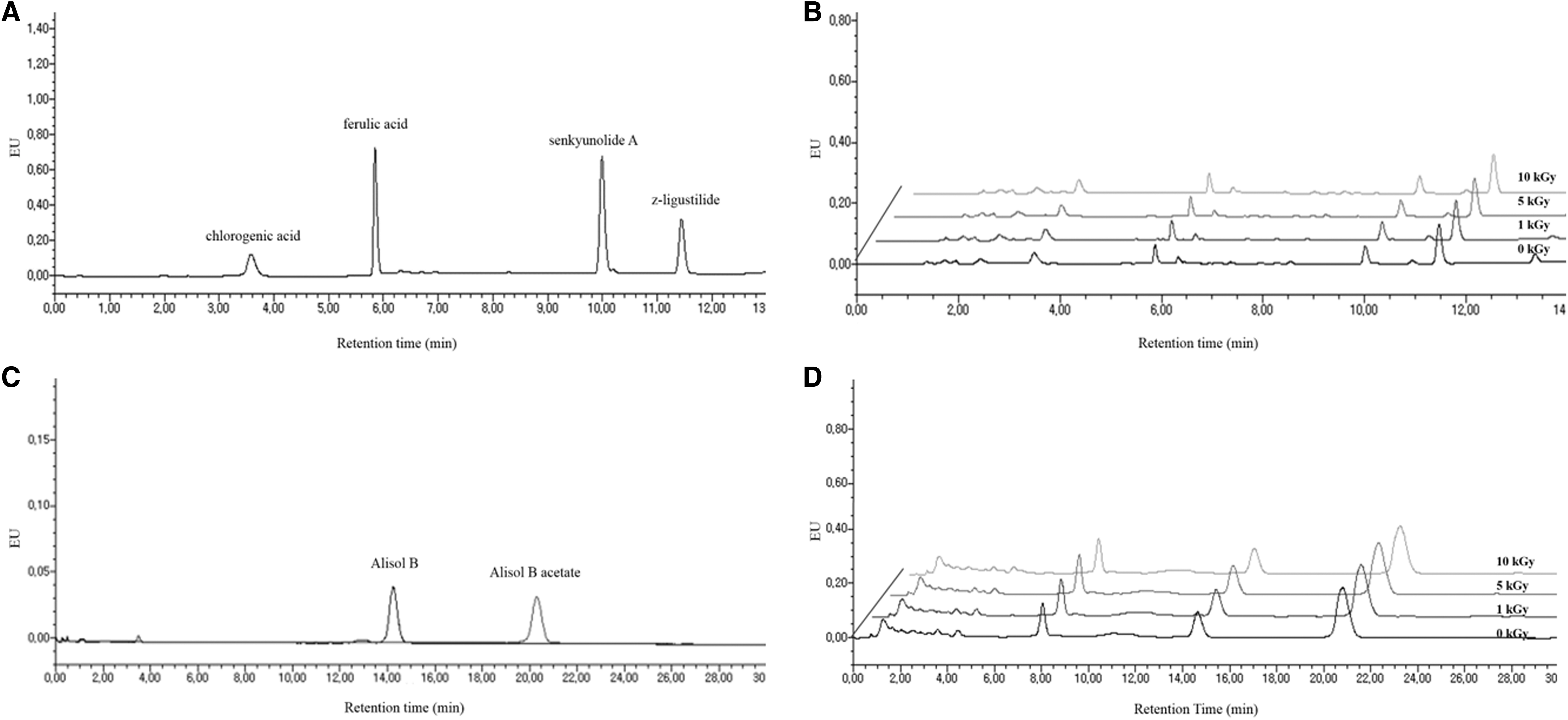
HPLC chromatograms of standard solution and active components in e-beam irradiated Cnidii Rhizoma and Alismatis Rhizoma
Concentration of Active Components in Electron-Beam Irradiated Cnidii Rhizoma
Data are presented as mean ± standard deviation (n = 3).
Means with different superscripts letters (a, b) in the same column are significantly different at P < .05 by Duncan's multiple-range test.
Concentration of Active Components in Electron-Beam Irradiated Alismatis Rhizoma
Data are presented as mean ± standard deviation (n = 3).
Means with superscript letters (a) in the same column are not significantly different at P < .05 by Ducan's multiple-range test.
Effect of e-beam irradiation on anti-inflammatory activity
To investigate the effect of e-beam irradiation on the anti-inflammatory activity of the medicinal herbs, RAW 264.7 cells were stimulated with LPS and the production of NO, PGE2, IL-1β, and IL-6 was investigated. First, MTT assays were performed to determine the cytotoxicity of the samples across a range of concentrations and as shown in Figure 3, the extracts of Cnidii Rhizoma and Alismatis Rhizoma irradiated with e-beams were not cytotoxic at a concentration of 200 μg/mL or less. Based on the results of the MTT assays, LPS-induced RAW 264.7 cells were treated with the extracts of Cnidii Rhizoma and Alismatis Rhizoma at concentrations of 50, 100, and 200 μg/mL and the production of NO, PGE2, IL-1β, and IL-6 was measured. The extracts of Cnidii Rhizoma reduced the rate of production by NO by 79.3–80.5% and the extracts of Alismatis Rhizoma by 77.6–79.4%. NO production was suppressed significantly by the extracts of Cnidii Rhizoma and Alismatis Rhizoma, and the highest level of inhibition of NO production was seen at 200 μg/mL (Fig. 4A, B). PGE2 production was suppressed significantly by the extracts of Cnidii Rhizoma and Alismatis Rhizoma in a concentration-dependent manner. The highest level of inhibition of PGE2 production was seen at 200 μg/mL, with a reduction of ∼69.2–72.1% in the extracts of Cnidii Rhizoma and by 64.8–71.8% in the extracts of Alismatis Rhizoma (Fig. 4C, D). The extracts of Cnidii Rhizoma and Alismatis Rhizoma showed a lower inhibitory effect on IL-1β production than NO and PGE2, but these extracts also elicited the lowest production rates of IL-1β at 200 μg/mL (Fig. 5A, B). The extract of 5 kGy irradiated Cnidii Rhizoma and 0 kGy irradiated (non-irradiated) Alismatis Rhizoma showed maximal inhibition of IL-6 production at 200 μg/mL (Fig. 5C, D). Overall, the higher the concentration of the extracts of Cnidii Rhizoma and Alismatis Rhizoma used for treatment, the more the inhibition of the production of NO, PGE2, IL-1β, and IL-6 was observed and the same tendency was seen for all irradiation doses (0, 1, 5, and 10 kGy). An et al. screened for the anti-inflammatory activity of these medicinal herbs extracts by measuring the production of NO, IL-1β, IL-6, and tumor necrosis factor-alpha (TNF-α). 34 In this article, the production rates of NO, IL-1β, and IL-6 after treatment with the extracts of Cnidii Rhizoma were 76.6%, 74.2%, and 65.2% and the production rates of NO, IL-1β, and IL-6 in the extracts of Alismatis Rhizoma were 76.8%, 62.9%, and 72.4%, respectively. These results were similar to ours.
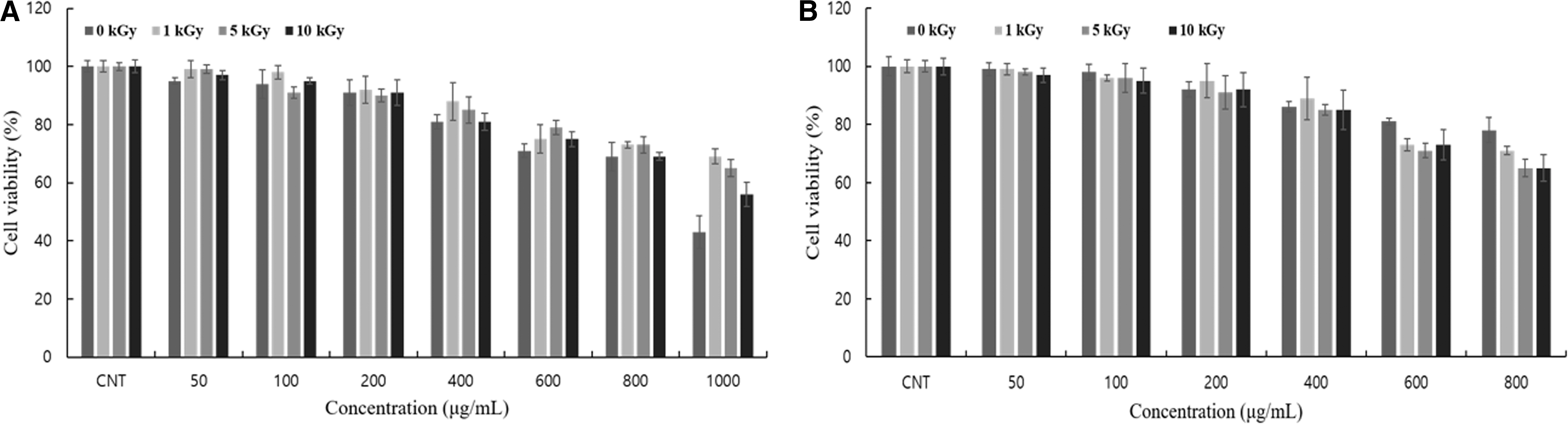
Measurement of cytotoxic effect of 70% ethanol extract from irradiated Cnidii Rhizoma
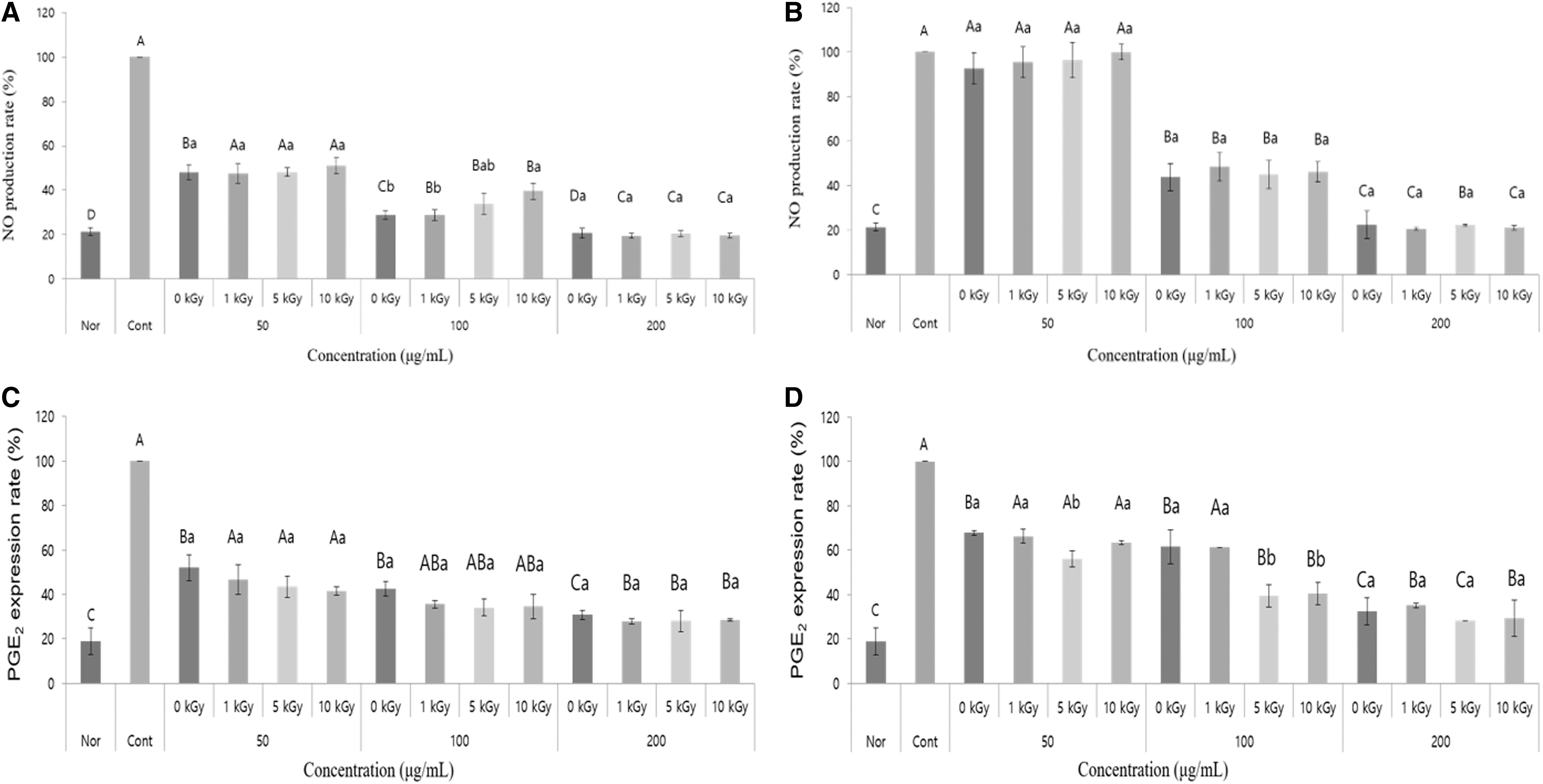
Comparison of NO production of 70% ethanol extract from irradiated Cnidii Rhizoma
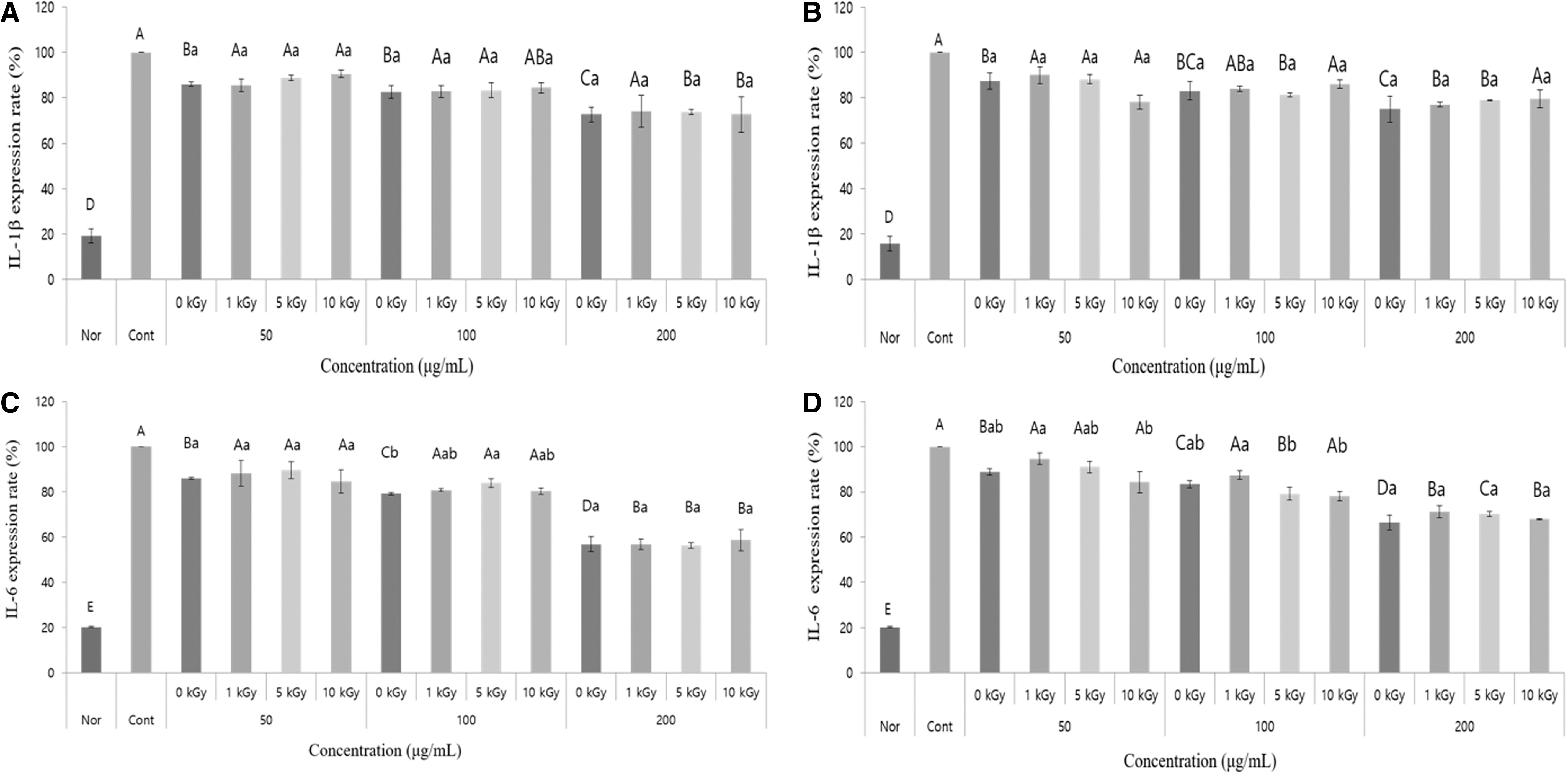
Comparison of IL-1β expression of 70% ethanol extract from irradiated Cnidii Rhizoma
Discussion
In this study, the effects of e-beam irradiation on the microbial population, stability of the active components, and anti-inflammatory activity of the medicinal herbs were examined.
With increases in the irradiation dose, the microbial loads in Cnidii Rhizoma and Alismatis Rhizoma reduced significantly (P < .05), and the 5 kGy e-beam irradiation reduced 3–4 log CFU/g of the microbial contamination level in both herb samples. Lung et al. reported that e-beam irradiation increases the energy in the microorganisms, breaking down the cellular homeostasis and damaging the DNA, thus killing the organisms. 35 This study confirmed that e-beam irradiation is an effective method for controlling microbial loads of medicinal herbs.
The content of chlorogenic acid, ferulic acid, senkyunolide A, and z-ligustilide of e-beam irradiated Cnidii Rhizoma and the content of alisol B and alisol B acetate of e-beam irradiated Alismatis Rhizoma were determined by using HPLC and no significant difference was observed over a range of irradiation doses. These results are consistent with previous reports that state that irradiation does not affect the active ingredients of medicinal herbs. Kim et al. reported that the content of gamma ray irradiated glycyrrhizin in licorice, berberine in Coptis Rhizoma, and amygdalin in apricot kernel determined by using HPLC was not significantly different than that found in non-irradiated samples. 28 Yu and Jo reported that the contents of decursin and decursinol angelate, which are the active components of Angelica gigas root, did not change after Angelica gigas was irradiated with gamma rays. The components of dried samples did not change because the production of free radicals from water molecules was suppressed in the dried sample. 27
Many studies have been conducted to explore the effects of irradiation on antioxidant and biological activities of medicinal herbs. Carocho et al. and Huang and Mau reported that gamma rays and e-beam irradiation not only maintained but also enhanced the antioxidant properties to some extent. 23,36 Anti-inflammatory activity is one of the important biological activities of medicinal herbs, and there have been a few reports on the effects of irradiation on the anti-inflammatory activity of medicinal herbs. It has been reported that the extract of 10 kGy γ-ray irradiated persimmon leaves inhibited the production of NO, PGE2, IL-6, and TNF-α more than the control. 37 Although our study is different from this article, we also investigated the effect of different e-beam irradiation doses (0, 1, 5, and 10 kGy) on the anti-inflammatory activity of medicinal herbs. However, we found that e-beam irradiation did not affect the anti-inflammatory activity of Cnidii Rhizoma and Alismatis Rhizoma, which was inconsistent with the result of the previous study. The activity of the anti-inflammatory compounds senkyunolide A and z-ligustilide, which are the major active components in Cnidii Rhizoma, 38 and alisol B acetate, the major active component in Alismatis Rhizoma, 39 was not changed by e-beam irradiation. We suggest that the reason that the anti-inflammatory activities of Cnidii Rhizoma and Alismatis Rhizoma were not changed by e-beam irradiation is due to the stability of the major active components. The e-beam with a dose of 10 kGy or less is considered safe 25 and does not induce apparent changes in food components as it is non-heating in nature. 20
Taken together, our results show that as the irradiation dose increases, the number of TAB, Y&M, and CC decreases; however, the concentration of the active components and anti-inflammatory activities of Cnidii Rhizoma and Alismatis Rhizoma do not change (Fig. 1). This is the first study to investigate the effect of e-beam irradiation on the anti-inflammatory activities of medicinal herbs by using a range of irradiation doses, and the results of our study support the application of e-beam irradiation for sanitization of medicinal herbs.
Footnotes
Author Disclosure Statement
No competing financial interest exists.