
Abstract
Urushiols are amphipathic compounds found in Rhus verniciflua Stokes that exhibit various biological activities. However, their practical use is very restricted due to their contact dermatitis-inducing property. Therefore, we applied the ionization method to remove the allergenic properties of the urushiols and to increase their usability. One of the natural urushiols, 3-pentadecylcatechol (PDC), was heated for 30 min with a solution of H2O and sodium carbonate (Na2CO3). The reaction product was analyzed by electrospray ionization mass spectrometry (ESI-MS). Ionized PDC with an m/z value of 316.9 and complexed PDCs with Na+ of 1 − 3 atoms with m/z values of 340.8, 365.2, and 380.8 were detected. PDC and ionized PDC (3 μmol/3 mg of Vaseline) treatments were applied on the rear of left ear of Sprague-Dawley rats once daily for 10 days. Erythema and swelling were observed on the ear skin treated with PDC, but not in case of ionized PDC. Compared with control, contact hypersensitivity-related biomarkers (neutrophils, eosinophils, immunoglobulin E, and histamine) in the blood were significantly higher only in the PDC-treated group. In addition, Il-1b, Il-6, Tnfα, and Cox-2 mRNA expression levels were dramatically increased in the ear tissue of PDC-treated rats, but in the ionized PDC-treated group, they were similar to those in the control group. Overall, it was confirmed that the allergenic property of the urushiol PDC was removed by ionization. This method is expected to be useful for preventing allergy induction in cooking and food processing using R. verniciflua Stokes.
Introduction
R
In our previous study, 3-pentadecylcatechol (PDC, Fig. 1), which is a urushiol derivative, was chemically synthesized. 17 PDC showed a strong radical-scavenging activity and inhibited lipid peroxidation in various in vitro systems. 17 Therefore, PDC would be useful as a bioactive compound for a variety of applications. Nevertheless, as mentioned above, the use of urushiols is limited due to its allergy-inducing property. Therefore, we tried to develop a method to suppress this property, while maintaining the amphipathic basic skeleton of PDC. In this study, we ionized the natural urushiol PDC, and then tested it for contact dermatitis induction, and hematological evaluations and blood absorption were evaluated.
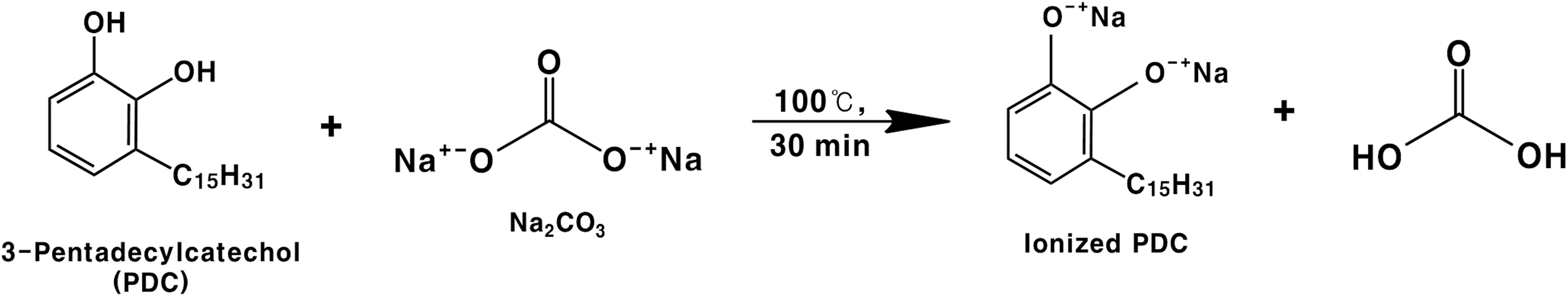
Figure for the ionization of PDC. PDC, 3-pentadecylcatechol.
Materials and Methods
Chemicals
The 3-pentadecylcatechol (PDC) was chemically synthesized according to the method described in our previous research. 17 Sodium carbonate (Na2CO3), β-glucuronidase type H-1 (from Helix pomatia, EC 3.2.1.31), and heparin sodium salt (from porcine intestinal mucosa) were obtained from Sigma-Aldrich Chemical Co. (St. Louis, MO, USA). All other chemicals and solvents were of analytical grade.
Ionization of PDC
PDC (1 g, 3.13 mmol) was mixed with 20 mL of Na2CO3 (1.66 g, 15.65 mmol) aqueous solution. The mixture was boiled at 100°C for 30 min. After cooling at room temperature, the reaction mixture was partitioned with methylene chloride (CH2Cl2, 20 mL, three times). The CH2Cl2 layer was evaporated in vacuo. Ionization was confirmed by thin-layer chromatography (TLC, silica gel 60 F254, 0.25 mm thickness; Merck), high performance liquid chromatograph-photodiode array (HPLC-PDA), and direct electrospray ionization mass spectrometry (DI-ESI-MS) analyses. The developing solvent for TLC was benzene/CHCl3/EtOAc [8:2:0.2 (v/v)]. The HPLC analysis was performed on a Shimadzu LC-6AD instrument with an SPD-M20A detector. Separation was performed on an ODS column (ODS-80Ts, 5 μm, 4.6 × 150 mm; Tosoh, Tokyo, Japan) maintained at 40°C, with a flow rate of 1 mL/min. The mobile phase was an isocratic solution of 90% MeCN. The elution pattern was monitored by measuring the absorbance at 275 nm. The Mass spectral data were obtained through DI-ESI-MS (EsquireHTC; Bruker Daltonics, Billerica, MA, USA).
Animals
All experimental procedures were approved by the Institutional Animal Care and Use Committee of Chonnam National University (no. CNU IACUC-YB-2012-26). Six-week-old Sprague-Dawley male rats weighing 180–200 g were supplied by Samtako Bio Korea (Osan, Korea). The animals were kept in an environmentally controlled animal facility operating on a 12-h dark/12-h light cycle at conditions of 20 ± 1°C and 55 ± 5% humidity, with free access to water and normal feed (Harlan Rodent diet, 2018S, by Samtako Bio Korea). The rats were acclimatized to the environment for 1 day before the commencement of the experiments. 17
Induction of contact dermatitis by application of PDC and ionized PDC on rat ears
PDC and ionized PDC (PDC, 960 μg; ionized PDC, 1093 μg) (3 μmol each) were mixed with 3 mg of Vaseline and applied onto the rear (1 cm2) of the left ear of rats (n = 6) once daily for 10 days. 17 The erythema visualized on the rat ears treated with the PDC and ionized PDC was reflective of contact dermatitis.
Hematological evaluation after induction of contact dermatitis by PDC and ionized PDC
After the PDC and ionized PDC treatments for 10 days, the rats (n = 6) were anesthetized with diethyl ether, and blood was collected from the abdominal aorta. 17,18 The blood was divided into two portions.
For determination of the white blood cell, eosinophil, and neutrophil levels, 1 mL of whole blood was placed into ethylenediaminetetraacetic acid-treated tubes, and analyzed using the Veterinary Multispecies Hematology System (Hemavet 850; CDC, USA) without freezing.
The remaining blood was stored at 4°C for 90 min. Blood serum for measuring the histamine and immunoglobulin E (IgE) levels was obtained by centrifugation (1026 g, 4°C, 15 min) and stored at −80°C until analysis. Serum histamine and IgE levels were measured using the histamine Enzyme-Linked Immunosorbent Assay (ELISA) Kit (Oxford Biomedical Research, Inc., Oxford, MI, USA) and the Rat IgE ELISA Complete Kit (Koma Biotech, Seoul, Korea), respectively, according to the manufacturer's instructions.
qPCR analysis of inflammatory response gene expression
The left ear tissues after the PDC and ionized PDC treatments were harvested for quantitative real-time reverse transcriptase polymerase chain reaction (qPCR) analysis. 19 Total RNA was isolated from ear tissue, using the GeneAll® Hybrid-R™ (GeneAll Biotechnology, Seoul, Korea). cDNA was synthesized using the ReverTra Ace® qPCR RT Kit (Toyobo, Osaka, Japan), and qPCR with amplification was accomplished using a Mx3000P qPCR System (Agilent Technologies). All reactions were performed in triplicate according to the manufacturer's recommended thermocycling conditions, and then subjected to melting curve analysis. The primer sequences for the transcript analysis are as follows: GAPDH, 5′-GTATTGGGCGCCTGGTCACC-3′ (forward), 5′-CGCTCCTGGAAGATGGTGATGG-3′ (reverse); Il-1b, 5′-CACCTCTCAAGCAGAGCACAG-3′ (forward), 5′-GGGTTCCATGGTGAAGTCAAC-3′ (reverse); Il-6, 5′-TCCTACCCCAACTTCCAATGCTC-3′ (forward), 5′-TTGGATGGTCTTGGTCCTTAGCC-3′ (reverse); Tnfα, 5′-AAATGGGCTCCCTCTCATCAGTTC-3′ (forward), 5′-TCTGCTTGGTGGTTTGCTACGAC-3′ (reverse); and Cox-2, 5′-TGTATGCTACCATCTGGCTTCGG-3′ (forward), 5′-GTTTGGAACAGTCGCTCGTCATC-3′ (reverse).
Oral administration of ionized PDC
All rats were fasted for 15 h and subjected to water deprivation for 3 h before ionized PDC administration. Ionized PDC was dissolved in 1 mL of propylene glycol and then orally administered (150 μmol [54.0 mg]/kg body weight) to rats. Blood was withdrawn from the abdominal aorta of rats under mild anesthesia (diethyl ether) at 0, 0.5, 1, 2, 4, and 8 h after the administration and placed into heparinized tubes. Plasma samples were obtained by centrifugation (2767 g, 4°C, 15 min) and stored at −80°C until use.
Determination of the presence or absence of ionized PDC in rat plasma
The presence or absence of ionized PDC in rat plasma after oral administration was determined by HPLC analysis as previously described. 20 Briefly, rat plasma (100 μL) was mixed with 200 μL of β-glucuronidase type H-1 (500 U, from Helix pomatia) solution in 0.1 M sodium acetate buffer (pH 5.0). The mixture was incubated at 37°C for 2 h. The reaction mixture was extracted using 1 mL of MeCN. The mixture was then vortexed for 30 sec, sonicated for 30 sec, and centrifuged at 2767 g for 15 min at 4°C. The supernatant was concentrated in vacuo and dissolved in 100 μL of MeOH. Twenty μL of the resulting solution was injected onto an ODS HPLC column (UG 120, 5 μm, 4.6 × 250 mm; Shiseido, Tokyo, Japan). The HPLC analysis was performed on a Shimadzu LC-6AD instrument with an SPD-M20A detector. The mobile phase was a solution of 15% MeOH containing 2% AcOH (Solvent A) and 100% MeOH (Solvent B). The gradient program for HPLC analysis was: A/B 100:0 to A/B 10:90 for 0–10 min, A/B 0:100 for 10–40 min, and isocratic A/B 0:100 for 15 min. The column temperature was maintained at 40°C and the flow rate was 1 mL/min. The elution pattern was monitored by measuring the absorbance at 275 nm.
Statistical analysis
Data are expressed as the mean ± standard deviation using the Statistical Package for Social Sciences (IBM, Armonk, NY, USA) 19.0 package program. Statistical differences were measured by one-way analysis of variance followed by Tukey–Kramer test. P < .05 was considered significant.
Results
Ionization of PDC
PDC was simply ionized by boiling at 100°C for 30 min after mixing with an aqueous solution of Na2CO3. The TLC analysis suggested that PDC (Rf 0.8) was converted to a relatively more polar compound (Rf 0.0) 30 min after the reaction (Fig. 2). In addition, the peak of PDC (tR 20.0 min) disappeared and a new peak (tR 14.0 min) was detected at a more polar elution site on the HPLC chromatogram using an ODS column (Fig. 3). The molecular weight of ionized PDC was confirmed by DI-ESI-MS (positive) analysis (Fig. 4). From the mass spectrum, the peaks corresponding to C21H34O2 2− (ionized PDC, m/z 316.9), C21H34O2Na− (m/z 340.8), C21H34O2Na2 (m/z 365.2), and C21H34O2Na3 + (m/z 380.8) were detected, but the peak of authentic PDC (C21H36O2, M.W. 320.8) was not observed. This result suggested that PDC was successfully ionized by boiling with Na2CO3 aqueous solution.
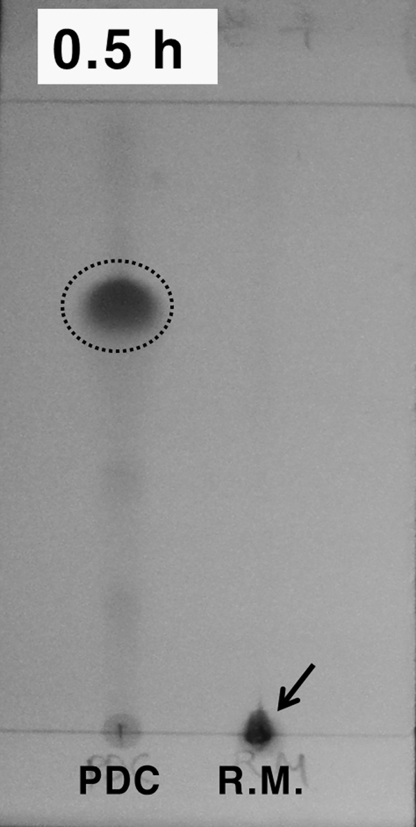
TLC analysis after ionization of PDC. TLC analysis was performed on silica gel 60 F254 (0.25 mm thickness). Developing solvent, benzene/CHCl3/EtOAc = 8:2:0.2 (v/v); detection, 254 nm. R.M., reaction mixture; TLC, thin layer chromatography.
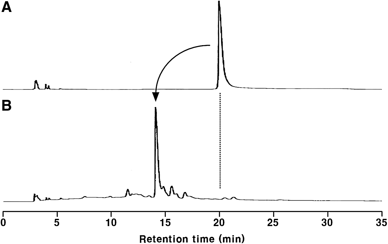
HPLC chromatogram of ionized PDC. HPLC conditions: column, ODS-80Ts (5 μm, 4.6 × 150 mm; Tosoh); mobile phase, isocratic solution of 90% MeCN; flow rate, 1 mL/min; detection, 275 nm.
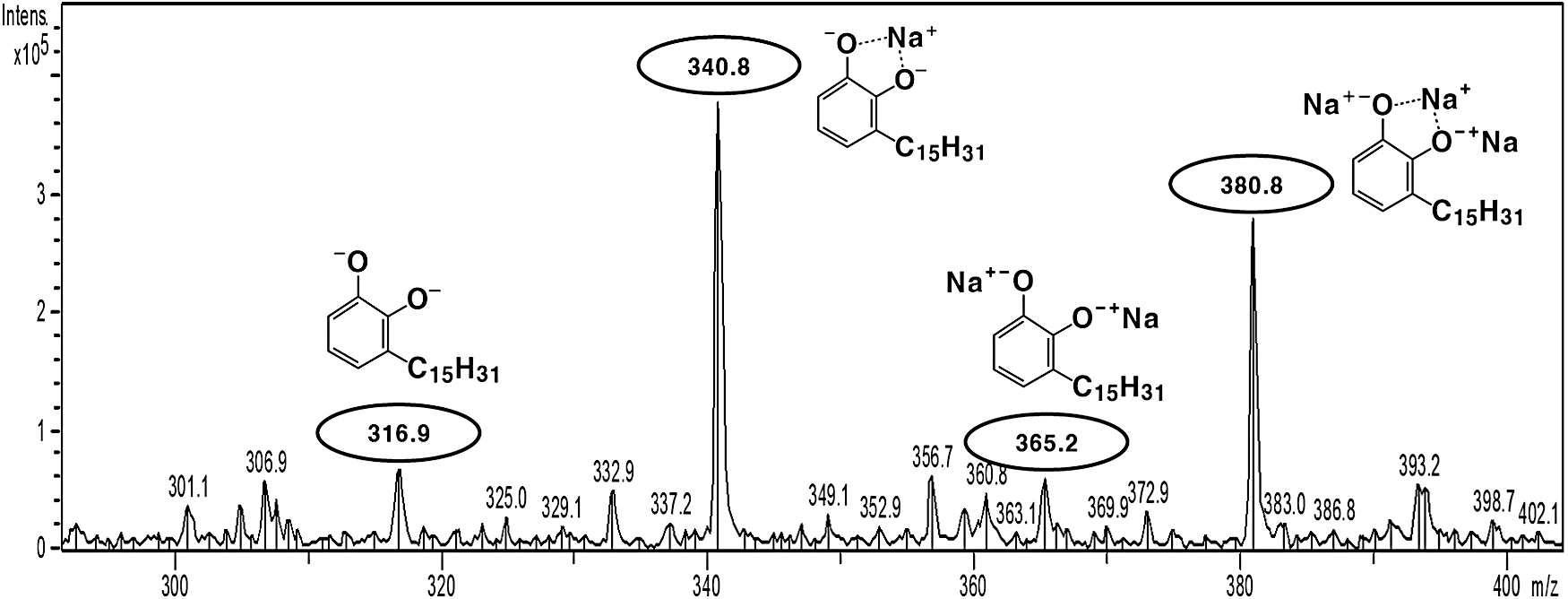
Direct electrospray ionization mass spectrometry (positive) spectrum of ionized PDC.
Contact dermatitis induction by PDC and ionized PDC administration on rat ears
PDC and ionized PDC were applied to the rear of the left ear of rats for 10 days. The morphological changes are shown in Figure 5. Serious inflammatory symptoms such as erythema and swelling were observed on the left ear skin of rats treated with PDC (Fig. 5B). In contrast, inflammatory symptoms were not observed in the left ear of the rats in the ionized PDC-treated group (Fig. 5C), like the case for those in the control group (Fig. 5A), which is a PDC-untreated group.

Induction of contact dermatitis by PDC and ionized PDC on rat ears (arrows) for 10 days (n = 6). PDC and ionized PDC (PDC, 960 μg; ionized PDC, 1093 μg) (3 μmol each) were mixed with 3 mg of Vaseline and applied to the rear of the rat ears.
Hematological evaluation in rat bloods after treatments with PDC and ionized PDC
White blood cell, eosinophils, neutrophils, histamine, and IgE levels in rat blood after PDC and ionized PDC treatments were determined (Fig. 6). The levels of eosinophil and neutrophil were more significantly increased (P < .05) in rat blood after PDC treatment than in the blood of rats in the control group. However, the levels of these parameters in the ionized PDC-treated group and the control group were similar. These phenomena were also observed for serum histamine and IgE levels.
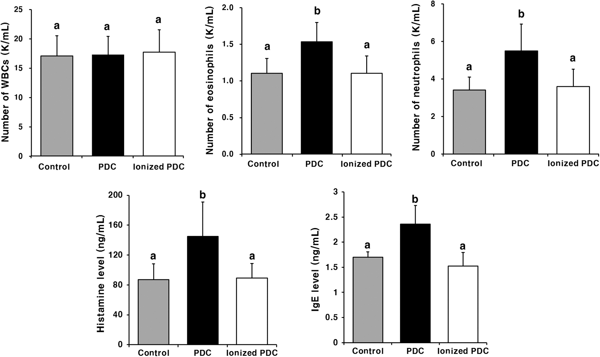
White blood cell, eosinophil, neutrophil, histamine, and IgE levels in rat blood after PDC and ionized PDC treatments of rat ears for 10 days (n = 6). The different letters indicate a significant difference (P < .05) by the Tukey–Kramer test. IgE, immunoglobulin E.
Relative expression levels of inflammatory factors in rat ear tissue after PDC and ionized PDC treatments
mRNA expression of the inflammatory genes, interleukin-1 beta (Il-1b), interleukin-6 (Il-6), tumor necrosis factor alpha (Tnfα), and cytochrome c oxidase subunit 2 (Cox-2), in the rat ear tissue after PDC and ionized PDC treatments for 10 days were assessed through qPCR. As shown in Figure 7, all inflammatory genes were dramatically upregulated in the PDC-treated group compared with the control group. Unlike the PDC-treated group, the expression levels of inflammatory factors in the ionized PDC-treated group were not increased at all compared with those of the control group. From the aforementioned results, it was clearly confirmed that the contact dermatitis induction property of PDC was eliminated by ionization.
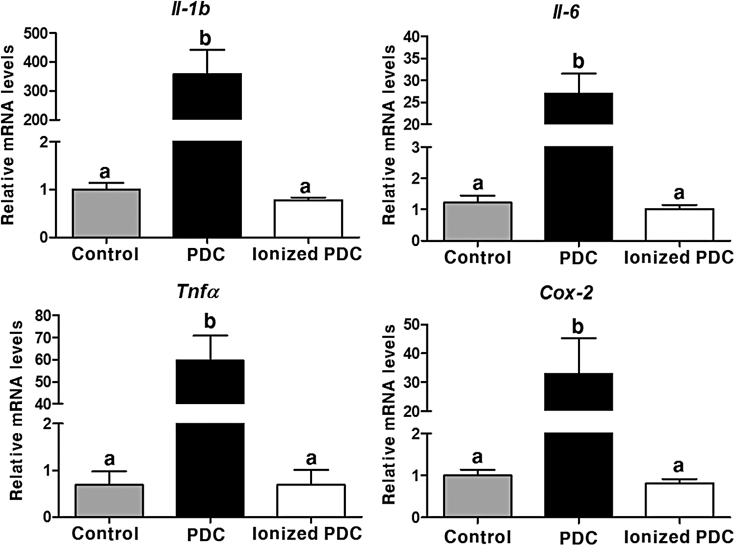
Relative expression levels of inflammatory response genes in rat ear tissue after PDC and ionized PDC treatments. PDC and ionized PDC were used to treat the left ear of rats for 10 days (n = 6). The different letters indicate a significant difference (P < .05) by the Tukey–Kramer test.
Presence or absence of ionized PDC in rat blood plasma after oral administration
Rat blood plasma after oral administration of ionized PDC was analyzed by HPLC-PDA (Figs. 8 and 9). All plasma samples before (Fig. 8) and after (Fig. 9) β-glucuronidase type H-1 treatment were analyzed. Significant peaks at the retention times of PDC (tR 47.2 min) and ionized PDC (tR 45.9 min) standards were not observed from the HPLC chromatograms of all plasma samples, regardless of β-glucuronidase type H-1 treatment. The detection limit for both PDC and ionized PDC was 15.0 pmol. Therefore, it was verified that neither ionized or intact PDC was absorbed into the blood stream, and was mostly excreted in feces after oral administration. 21
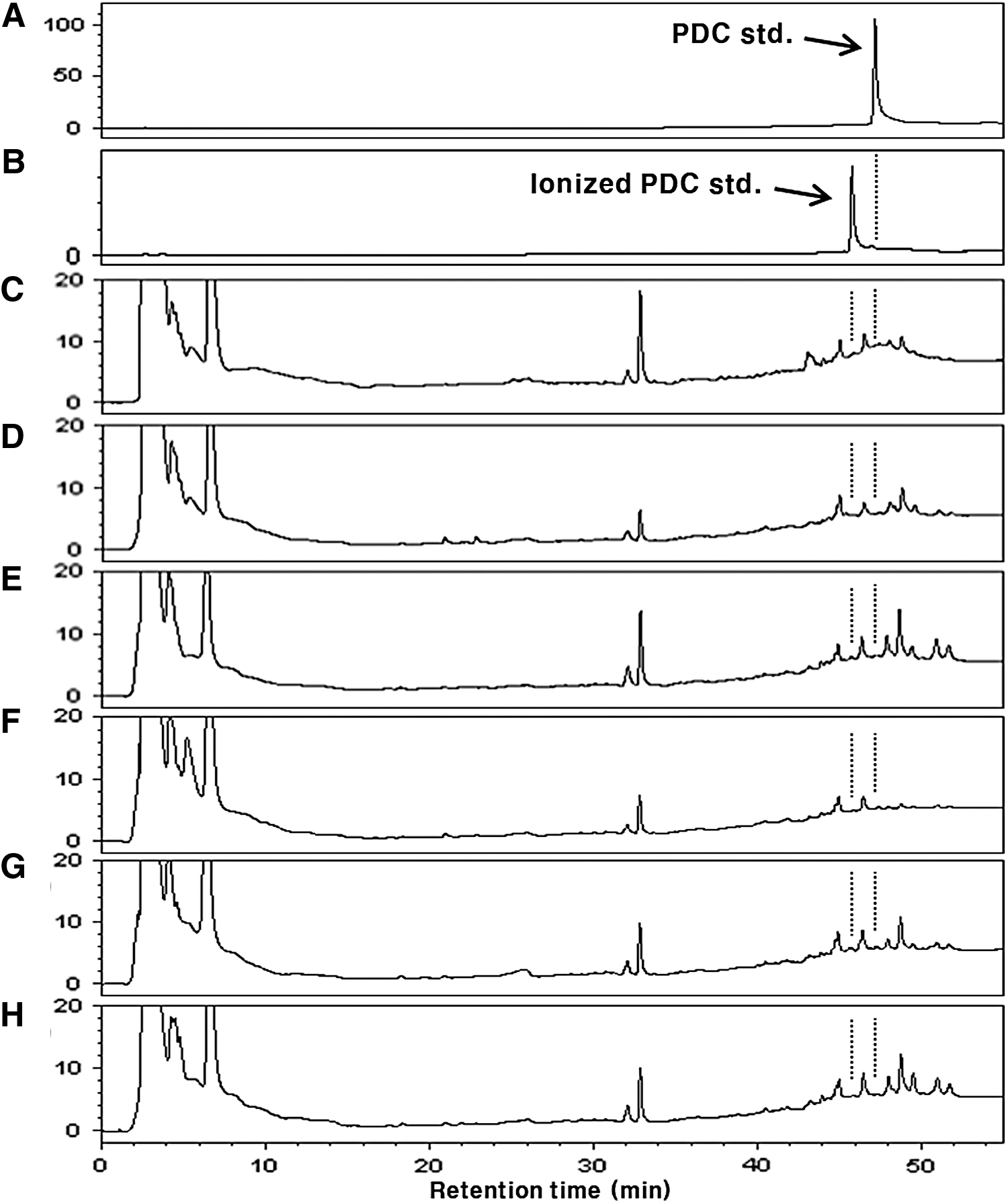
HPLC chromatograms of ionized PDC administered (54.0 mg/kg body weight) rat plasma extracts without β-glucuronidase H-1 treatment.

HPLC chromatograms of ionized PDC administered (54.0 mg/kg body weight) rat plasma extracts with β-glucuronidase H-1 treatment.
Discussion
Urushiols, major constituents of the lacquer tree sap, have been reported to exhibit various biological activities.4−9 However, their application is limited due to the contact dermatitis-causing property.10−12 Therefore, in this study, we simply ionized the catechol structure of PDC to remove the allergy induction property, and examined whether ionized PDC induces contact dermatitis.
The ionization of the PDC was carried out by a simple method of heating with an aqueous solution of sodium carbonate. The result of the TLC analysis suggests that PDC was converted to a relatively more polar compound after the reaction (Fig. 2). In addition, a similar phenomenon was observed in the result of HPLC analysis using an ODS column (Fig. 3). The DI-ESI-MS spectrum (Fig. 4) revealed the peaks corresponding to ionized PDC and ionized PDC complex-ion salt with 1 − 3 atoms of Na+. However, a significant peak corresponding to native PDC (M.W. 320.8) was not detected. Consequently, it was confirmed that PDC was successfully ionized by this method.
To confirm the elimination of the contact hypersensitivity property by ionization, PDC and ionized PDC were applied to the ears of rats for 10 days. The morphological changes are shown in Figure 5. The natural urushiol PDC, which is already known to cause contact dermatitis, 17 showed reproducibility in this experiment. That is, inflammation symptoms were observed on the ear skin of rats treated with PDC. Interestingly, ionized PDC treatment group did not show the inflammatory symptoms. In addition, eosinophil, neutrophil, histamine, and IgE levels in rat blood significantly increased after PDC treatment (Fig. 6). However, the ionized PDC treatment group showed similar levels of these parameters, compared with the control group. In addition to morphological changes and blood analysis, the expression levels of inflammatory factors in rat ear tissues treated with PDC and ionized PDC were analyzed by qPCR to more reliably confirm whether the ionized PDC causes contact dermatitis. In comparison with the control group, all of the markers analyzed (Il-1b, Il-6, Tnfα, and Cox-2) were significantly higher in the PDC-treated group, but not in the ionized PDC-treated group (Fig. 7). Therefore, it was clearly confirmed that the contact hypersensitivity property of PDC was eliminated by ionization.
The contact dermatitis induction mechanism of urushiol that has been proposed states that the catechol structure of urushiol is oxidized to O-quinone after it penetrates the skin, after which it binds to proteins. 10,22 This complex, which acts as an antigen, activates the Langerhans cells, which helps provide protective immune responses against skin infection. 10,22 Based on this, there are two mechanistic possibilities for allergenic property elimination of PDC by ionization. First, it is presumed that ionized PDC cannot be transformed to O-quinone structure due to the ionization of catechol, and thus, it cannot act as a hapten, which is a molecule that induces an immune response when it is bound to proteins. 10,22 Second, ionized PDC may not penetrate into the skin due to ionization, which would prevent complex formations.
In our previous study, 21 we have confirmed that PDC was not absorbed into the blood of rat after oral administration, and about 90% of the administered PDC was excreted through feces. We expected that PDC uptake into the blood would be increased by ionization. However, no significant peaks corresponding to PDC and ionized PDC were observed in the HPLC chromatograms of all the ionized PDC-administered rat plasma samples (Figs. 8 and 9). Therefore, it was certified that ionized PDC is not also absorbed by oral administration like PDC. 21 Accordingly, it may be difficult to expect an increase of useful in vivo biological activity of PDC by ionization. Therefore, it is considered that further studies on the absorption and metabolism of various urushiol derivatives, including ionized urushiols should be conducted. However, our findings suggested that the in vivo beneficial effects of lacquer-tree extract may be due to other phenolic compounds, 23 –25 such as fustin, fisetin, and sulfuretin, etc., rather than urushiol derivatives.
In this research, the contact hypersensitivity property of the natural urushiol PDC was removed by ionization. Various existing methods have also attempted to remove the urushiols and urushiol-induced contact dermatitis.13−16 However, the existing methods are hard to apply widely in general because of the complexity of the processes and their time-consuming nature. In comparison, our ionization method can very easily and quickly eliminate the contact hypersensitivity property of urushiols. Therefore, it is expected that ionization can be applied widely in terms of allergy elimination when using the R. verniciflua extract and its sap as foods, medicines, and similar products.
Footnotes
Acknowledgment
DI-ESI-MS spectral data were obtained from the Korea Basic Science Institute, Ochang.
Author Disclosure Statement
No competing financial interests exist.
Funding Information
This research was supported by Basic Science Research Program through the National Research Foundation of Korea (NRF) funded by the Ministry of Education, Science, and Technology (no. NRF-2017R1D1A1B03034520).