




Abstract
This study was designed to explore the impact of Lycium barbarum polysaccharide (LBP) on inflammation and gut microbiota in mice with allergic asthma. Mice were divided into four groups: control group, OVA (ovalbumin) group, Con+LBP group, OVA+LBP group. After 28 days of LBP intervention, mice were euthanized and associated indications were investigated. Histopathological examination demonstrated that LBP reduced lung injury. The results of our current study provide evidence that supplementation with LBP in asthmatic mice decreases TNF, IL-4, IL-6, MCP-1, and IL-17A in plasma and bronchoalveolar lavage fluid (BALF). Sequencing and analysis of gut microbiota indicated that compared with the OVA group, Lactobacillus and Bifidobacterium were increased, but Firmicutes, Actinobacteria, Alistipes, and Clostridiales were decreased in the OVA+LBP group. We also found that gut microbiota were related to inflammation-related factors. Therefore, we speculate that LBP may improve allergic asthma by altering gut microbiota and inhibiting inflammation in mice.
Introduction
Allergic asthma is one of the chronic inflammatory diseases that seriously threaten the public health. Its main characteristic is significant airway allergic reaction. 1 Statistics show that at least 300 million people around the world suffer from asthma. 2 Th1/Th2 imbalance is a major immune mechanism of asthma, as is Th17 dysregulation. When the body is exposed to allergens, inflammatory cells such as mast cells, eosinophils, and macrophages are activated. The inflammatory mediators (histamine, leukotriene, prostaglandin, etc.) released by the activated cells can stimulate the contraction of bronchi, thus eliciting inflammation in airway cells. Inflammation-related factors induce bronchospasm, airway smooth muscle contraction, increase of vasodilation and permeability, and so on, leading to allergic asthma. 3 –5 Current therapies include inhalation of glucocorticoids, leukotriene receptor modulators, bronchodilators, and injection desensitization. 6 Although these treatments can improve airway inflammation and control allergic reactions, they have many side effects. Therefore, the search for new and effective treatment has become a hot spot of asthma research.
It was found that the change of gut microbiota was related to the occurrence and development of allergic asthma. Probiotics such as Lactobacillus and Bifidobacterium may correct Th1/Th2 imbalance in the ovalbumin (OVA) allergic model by reducing airway hyperresponsiveness and Th2-mediated cascade reaction. 7 Other investigators have also found that Lactobacillus and Streptococcus can induce human peripheral blood mononuclear cells triggering secretion of IFN-γ and IL-12, which play a role in asthma development. 8 In addition, studies have shown that probiotics/prebiotics can promote the Th1 response and inhibit the Th2 response to alleviate asthma. 9,10
Lycium barbarum (goji berry) is a kind of Chinese herbal medicine that has many biological activities and pharmacological effects. It is mainly used to treat hyperlipidemia, 11 diabetes, 12 and cancer. 13 Lycium barbarum polysaccharide (LBP), the main active ingredient of L. barbarum, has anti-stress, anti-fatigue, anti-oxidation, blood lipid lowering, and immune system enhancement properties. 14 Recent studies have found that LBP can be a new “prebiotics” candidate for Bifidobacterium and Lactobacillus. 15
In this study, we observed the anti-inflammatory effect of LBP and the regulation of intestinal microflora in allergic asthmatic mice. The purpose was to supply a new clue for the prevention and treatment of asthma and provide theoretical basis for the cheap intervention of LBP.
Materials and Methods
Animals
Female mice (C57BL/6, age 6–8 weeks) weighing 18–22 g were obtained from the Beijing Weitonglihua Laboratory Animal Technology Co., Ltd. (Qualification Certificate: SCXK Beijing 2016–0006). The mice were kept in an specific pathogen free (SPF)-grade animal room, and the drinking water and food were sterilized by high pressure. The experiment was carried out in strict accordance with the recommendations in the Guide for the Care and Use of Laboratory Animals of the Ministry of Science and Technology of the People's Republic of China. All animal procedures were approved by Ningxia Medical University Medical Ethical Committee, China (approval number: 2015–126).
Materials
According to the results of single factor screening, the orthogonal optimization experiment (three factors and nine levels) was carried out with the ratio of extract to material, temperature and time as influencing factors, and the extraction yield of polysaccharide from leaves as measuring index. The extraction method for preparing L. barbarum leaf polysaccharides was water extraction and alcohol precipitation. The equipment is provided by Shunyi Experimental Equipment Co., Ltd.. According to the optimum extraction process parameters, LBP was prepared, and its effective component content was determined (Table 1). The content of LBP in L. barbarum leaf was 60%, and the concentration was about 1200 mg/mL.
The Content of Various Active Components in Lycium barbarum Polysaccharide Extracted from Lycium barbarum Leaves Was Determined, Expressed as Percentage
LBP, Lycium barbarum polysaccharide.
OVA was purchased from Sigma Chemical Co., Ltd. Aluminum hydroxide gel was supplied by Thermo Fisher Scientific. The staining kit was provided by Solarbio Science & Technology Co., Ltd. The Cytometric Bead Array (CBA) kit was provided by Becton, Dickinson and Company.
Experimental design
SPF-grade female C57BL/6 mice were divided randomly into 4 groups (15 animals per group): the control group, Con+LBP group, OVA group, and OVA+LBP group. We established the asthma model as follows: mice were first sensitized by the intraperitoneal (i.p.) injection of 200 μL solution containing 100 μg OVA and 4 mg aluminum hydroxide on days 0, 7, and 14. From days 21 to 28, nasal drops were administered with 50 μL of OVA solution (concentration, 1 mg/mL). The LBP intervention group was given LBP 100 μg/(g·d). The animal's appetites, activities, and other signs were observed daily.
Histopathological examination
The lower left lobe of the lung was harvested and then inflated, after which the lung was fixed with formalin overnight and then embedded in paraffin. Subsequently, we employed Masson's trichrome, H&E (hematoxylin and eosin), and PAS (periodic acid–Schiff) to examine inflammation of the airway, collagen deposition, and production of mucus in the lung tissues.
Blood plasma and bronchoalveolar lavage fluid collection and cytokine measurement
The eyeballs of mice were removed, and the peripheral blood was collected by anticoagulation tubes and stored at −80°C. Subsequently, the cytokine levels were determined using the CBA as per the methods described by the manufacturer.
Mouse bronchoalveolar lavage fluid collection and cytokine measurement
Mice were sacrificed by i.p. administration of an overdose of pentobarbital sodium. Next, 0.5 mL of cold phosphate-buffered saline was introduced into the lung three times to collect the bronchoalveolar lavage fluid (BALF). The collected BALF was centrifuged, and the supernatants were separated and preserved at −80°C for cytokine measurement, which was determined by the CBA as per the methods described by the manufacturer.
Sequencing
Genomic DNA extraction
Total DNA from the tissues was isolated using the Sodium dodecylsulfate technique. The specific operation is as follows: (1) feces were collected and added to EDTA (0.5 M, pH 8.0) and 550 μL lysate, and then, proteinase K and 20 μL lysozyme were added, mixed well upside down and incubated at 55°C for 2 h. (2) Samples were then centrifuged at 12,000 rpm for 5 min; after absorption of the supernatant, 5 M NaCl was added and gently vortexed for 10 sec; after holding at −20°C for several minutes, the samples were centrifuged at 12,000 rpm for 10 min. The supernatant was then collected and centrifuged at 12,000 rpm for 5 min (LUXIANGYI/GL-12MGL-12M, Zhongyi, Beijing, China). The supernatant was removed, isopropanol added, and then, it was mixed well and held at −20°C for 20 min and centrifuged at 12,000 rpm for 10 min. (3) The supernatant was discarded and the pellets were washed twice with 1 mL of 75% ethanol. The DNA sample was then blow-dried on the super clean bench. Next, 50 μL of ddH2O was used to dissolve the DNA sample, 1 μL RNase A added, and it was the mixed upside down and incubated at 37°C for 15 min. The quality and concentration of the DNA were assessed using agarose gel (1%) electrophoresis. The DNA concentrations were standardized to 1 ng/μL using sterile water.
Amplicon generation
Polymerase chain reaction (PCR) assays were performed to amplify the 16S recombinant DNA (rDNA) genes using gene-specific primers (16S V3-V4: 341F–806R) with the barcode. The PCRs were conducted in 30 μL reactions with 15 μL of Phusion® High-Fidelity PCR Master Mix (Biolabs), 0.2 μM of forward and reverse primers, and about 10 ng of template DNA. Thermal cycling conditions were as follows: initial denaturation for 1 min at 98°C; then 30 rounds of denaturation for 10 sec at 98°C, annealing for 30 sec at 50°C, and elongation for 30 sec at 72°C. Final extension was carried out for 5 min at 72°C.
Preparation of PCR products for sequencing
The amplicons were mixed with 1 × loading buffer (contained SYBR Green) at equal volumes and then run through 2% agarose gel for detection. Next, the products were mixed in equidensity ratios and then purified using the GeneJET™ Gel Extraction Kit (Thermo Scientific).
Sequencing and library preparation
Sequencing was conducted using the Novogene. We employed Ion Plus Fragment Library Kit 48 rxns (Thermo Scientific) to generate sequencing libraries. The quality of the library was evaluated on the Qubit@ 2.0 Fluorometer (Thermo Scientific). Finally, the library was sequenced on an Ion S5TMXL platform, and 450–550 bp single-end reads were generated.
Statistics
Single-end reads were assigned to samples based on their unique barcode and truncated by trimming primer sequence and barcodes. The UCHIME algorithm 16 was employed to filter raw reads to select those with high quality with specific filtering conditions consistent with the Cutadapt 17 quality controlling process. Subsequently, we compared reads using a reference database (Silva database) 18 with the aim of identifying and omitting chimera sequences. 19 Then, the Clean Reads finally obtained.
Uparse software 20 was employed to analyze sequences. In this process, sequences showing a similarity of ≥97% were allocated to the same operational taxonomic units (OTUs). From these sequences, we chose a few as representative of each OTU for further annotation. We categorized each sample into phylum, class, order, family, and genera levels. QIIME (version 1.7.0) was utilized to perform analyses from clustering to alpha, including beta diversity analysis (between samples) and analyzed samples (within sample groups), and results were presented with R software (version 2.15.3). 21
Statistical analysis
All data were statistically analyzed and graphed by SPSS 20.0 statistical software and GraphPad Prism 8.0 software. The results are expressed as mean ± standard deviation. One-way analysis of variance was used to analyze differences between groups. If statistically significant, two groups were compared using a least significance difference t-test. Statistical significance was set at P < .05.
Results
Administration of LBP ameliorated the general condition of mice with allergic asthma
In the control group, breathing was normal except mild coughing or sneezing. The OVA group mice developed sneezing, coughing, irritability, shortness of breath, and scratching their ears compared with the control group. We observed significant nose and ear cyanosis, poor color, dull coat, excitement bowing of the back or prone, and unresponsive. Administration of LBP significantly ameliorated the symptoms of OVA-induced asthma model group.
Administration of LBP attenuated airway inflammation in mice with allergic asthma
To investigate the effect of LBP on the occurrence of inflammation in airways in mice with asthma caused by OVA, paraformaldehyde-fixed lungs were cut and stained with H&E. Mice treated with OVA had severe inflammation of bronchi and alveoli, showing excessive infiltration of inflammation-related cells around small airway and blood vessels, relative to the control group. The infiltration of lymphocytes and mast cells in and around the airway of LBP-treated asthmatic mice was less than that of OVA-induced asthmatic mice (Fig. 1A).
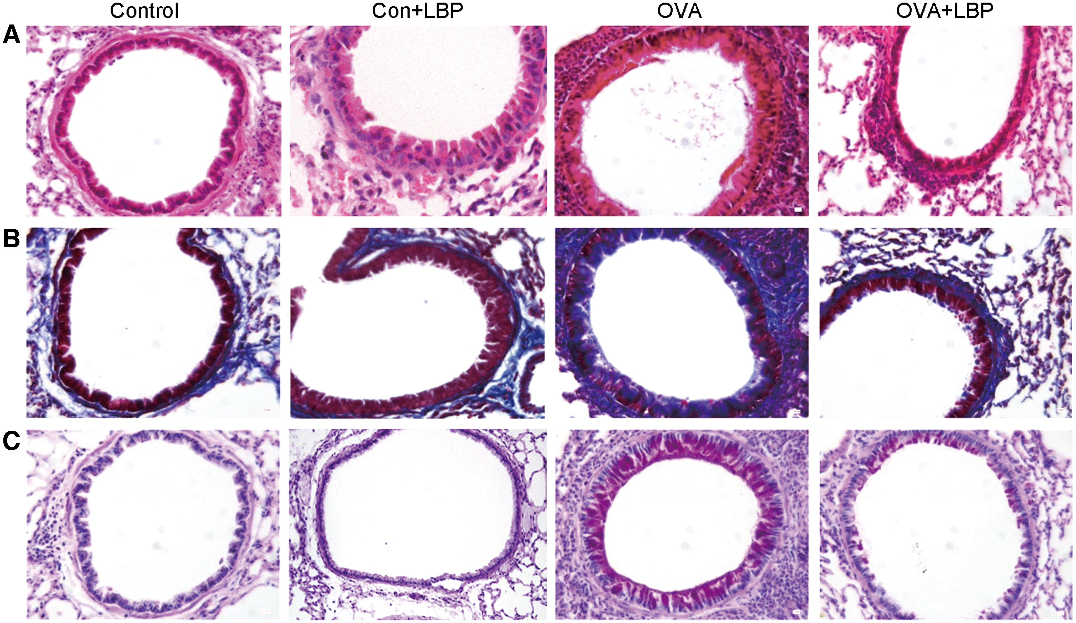
The effect of LBP intervention on pathological changes in the lung tissue. Histological assessment of airway inflammation and mucus secretion in the lung tissues was made in each group. A representative figure (original magnification 400 × ) of the lung tissue stained with H&E solution
In addition, we also tested the levels of inflammation-related factors in LBP-treated mice. The levels of TNF (P = .0002), MCP-1 (P < .0001), IL-6 (P = .0018), IL-4 (P < .0001), and IL-17A (P = .0101) in the plasma were increased in the OVA group relative to the control group. Th1-related cytokine, IFN-γ (P < .0001), was significantly decreased. The OVA+LBP group decreased the levels of TNF (P = .0068), MCP-1 (P = .0013), IL-6 (P = .0263), IL-4 (P = .0012), and IL-17A (P = .0003) in the plasma compared with the OVA group, except for IFN-γ (P = .0054). There was no statistical significance of inflammatory cytokines in the plasma in the Con+LBP group compared with normal mice (Fig. 2). Similar results were obtained in the BALF of mice (Fig. 3). These results indicate that LBP can alter the levels of inflammatory cytokines, such as Th1, Th2, and Th17. To some extent, LBP may inhibit the secretion of inflammatory factors and correct the Th1/Th2 imbalance in allergic asthma.
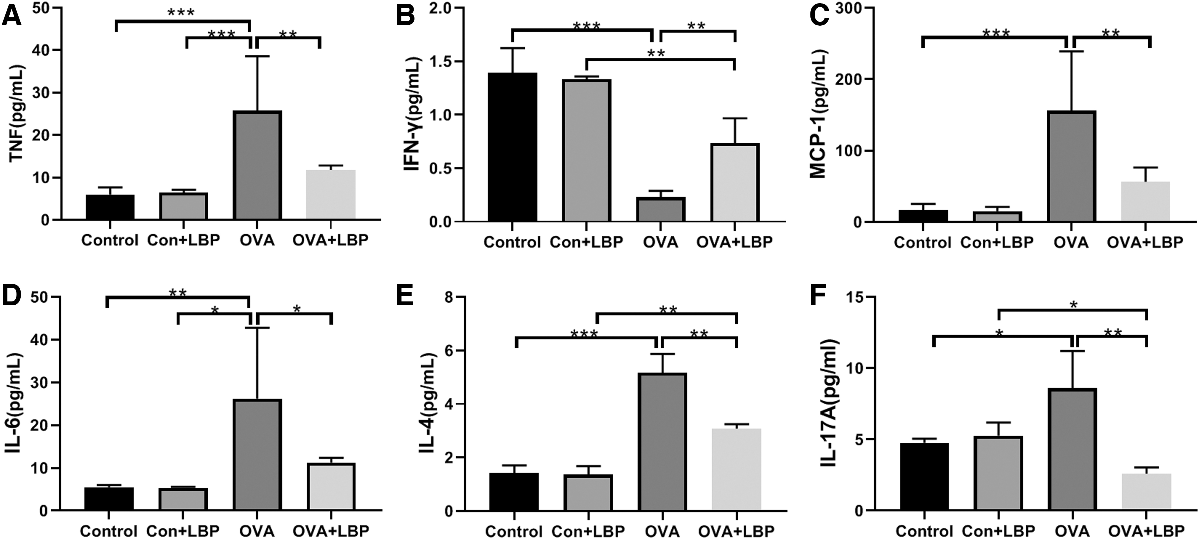
The effect of LBP on inflammatory cytokines of diverse groups. Plasma samples were collected for detection of TNF

Effects of LBP on inflammatory cytokines in different groups. BALF samples were collected for detection of TNF
In a word, LBP alleviated the occurrence of inflammation in airways in mice with asthma caused by OVA.
Administration of LBP inhibited airway remodeling in mice with allergic asthma
Airway remodeling is one of the important pathological characteristics of asthma. To investigate whether LBP improved airway remodeling in asthma mice, we performed Masson's trichrome staining of the lung tissues of the mice. We observed a large amount of blue collagen deposition around the bronchioles in the OVA group. However, the deposition of blue collagen decreased significantly in the OVA+LBP group. Moreover, these effects were comparable between the Con+LBP and control groups (Fig. 1B).
To assess the impact of LBP on goblet cell hyperplasia, the lung tissues were stained using PAS. The results showed that in the OVA model group, there were more mucinous goblet cells and a lot of mucinous substances secreted. In the OVA+LBP group, the number of myocytes and mucus decreased significantly. Intriguingly, there was no significant difference in LBP intervention in normal mice (Fig. 1C).
In summary, LBP significantly inhibited airway remodeling in mice with allergic asthma.
Administration of LBP modulated gut microbiota in mice with allergic asthma
Current evidence indicates that gut microbiota modulate the initiation and prognosis of asthma. 7 –10 We collected mice feces for 16S rDNA sequencing to investigate the impact of LBP on gut microbiota. Abundance of bacterial communities was estimated using Alpha diversity, which was evaluated by rarefaction curve. OTU number analysis revealed that the diversity and abundance of gut microbiota in the OVA group were the lowest, however, in the OVA+LBP group were the highest (Fig. 4A). In addition, the rarefaction curve presented similar results (Fig. 4B).

Alpha diversity analysis showing differences in terms of the abundance and diversity of the gut microbiota in the four groups.
Principal coordinate analysis (PCoA) was used to determine the composition of the bacterial population to characterize microbiota diversity. The PCoA indicated that there were significant changes in the diversity of gut microbiota of the OVA group relative to the control group (Fig. 5A). Bacterial flora were markedly different between the control group and the Con+LBP group (Fig. 5B). In addition, LBP regulated gut microbiota composition in mice with allergic asthma (Fig. 5C).

PCoA showed the difference in terms of species in fecal samples. PCoA of all samples by unweighted UniFrac distance.
In the subsequent analysis at the phylum level, Actinobacteria, Proteobacteria, Bacteroidetes, and Firmicutes comprised four dominant phyla in diverse group (Fig. 6A). The abundance of Bacteroidetes and Proteobacteria was comparable among the groups. In contrast, the proportion of Firmicutes was decreased in the control (P = .0008) and OVA+LBP groups (P = .0168) compared with the OVA group (Fig. 6C). The relative abundance of Actinobacteria in the OVA group was also higher than that in the control (P = .0009) and OVA+LBP groups (P = .0039) (Fig. 6D). Taken together, these findings revealed that the administration of LBP had major effects on Firmicutes and Actinobacteria, but a mild impact on Bacteroidetes and Proteobacteria.

Relative abundances of microbial species at the phylum and genus levels in the feces of mice.
Analysis of the top 10 bacteria at the genus level showed that the number of bacteria among the four groups was inconsistent (Fig. 6B). The proportions of Alistipes were lower in the control group (P = .0117), Con+LBP group (P = .0103), or OVA+LBP group (P = .0009) relative to the OVA group (Fig. 6E). The number of Lactobacillus was comparable between the Con+LBP group (P = .6201, Fig. 6F) and the control group but was lower in the OVA group than in the OVA+LBP group (P = .0025, Fig. 6F). The proportions of Bifidobacterium decreased in the Con+LBP group (P = .0327) or the OVA group (P = .0005) in relation to the OVA+LBP group (Fig. 6G). Moreover, the relative abundance of Clostridiales was markedly elevated in the control (P = .0003) and OVA+LBP (P = .0009) groups than in the OVA group (Fig. 6H). These results indicated that LBP caused marked changes in the number of genus members, especially for Alistipes, Lactobacillus, Bifidobacterium, and Clostridiales.
Gut microbiota influence the levels of inflammatory mediators
Further analysis was performed to characterize the relationships between abundance of the differential bacteria at the genus level and the level of inflammation-associated factors (Fig. 7). In plasma, the relative abundance of Lactobacillus was negatively related to TNF levels (P = .0174, Fig. 7A), IL-6 (P = .0069, Fig. 7C), and MCP-1 (P = .0104, Fig. 7D), but it exhibited a positive relationship with that of IFN-γ (P = .0334, Fig. 7B). Alistipes showed a positive correlation with levels of inflammatory factors, including TNF (P = .0160, Fig. 7E), IL-6 (P = .0330, Fig. 7F), and MCP-1 (P = .0461, Fig. 7H), respectively. Alistipes showed a inverse relationship with IFN-γ (P = .0082, Fig. 7G). The number of Bifidobacterium exhibited a positive relationship with IFN-γ (P = .0166, Fig. 7I) but negatively correlated with inflammation-related factors (TNF, P = .0359; MCP-1, P = .0353; IL-6, P = .0378) (Fig. 7J–L). The numbers of differential bacterial members were inversely related to BALF inflammation markers (data not shown). These data sets indicate that inflammatory markers and gut bacteria modulate allergic asthma.

Correlation analysis between the relative abundance (%) of gut bacteria and inflammatory induction.

Discussion
We investigated the therapeutic impact of LBP intervention on allergic asthma in mice. The data revealed that LBP efficiently eliminated asthma, by suppressing inflammation and normalizing dysbiosis of gut microbiota, suggesting that the intervention has preventive and treatment potential.
Asthma is manifested by chronic inflammation of airways 22 and is caused by multiple mechanisms. The main characteristics of asthma are related to specific Th2 cell responses, airway hyperresponsiveness, airway eosinophilia, and mucus secretion. 23 Despite progress in treatment, including inhaled β-adrenergic bronchodilators, corticosteroids, and leukotriene modulators, it is ineffective in some patients. 6 It is undeniable that antibiotics regulate the intestinal microflora, but the question is whether some antibiotics can change the composition of the microflora of asthmatic patients, or correct their disorders, so as to prevent or improve asthma.
Prebiotics have been shown to ameliorate asthma, 24,25 but the exact mechanisms still are currently unclear. Herein, supplementary LBP was demonstrated to dramatically alleviate allergic asthma throughout a series of routine parameters, including pathological examination, inflammatory indicators, and gut microbiota, which was consistent with previous study. 26 We found that the administration of LBP improved the general condition of mice with allergic asthma. Furthermore, H&E staining of the lung tissue revealed that LBP significantly improved airway inflammation, which is consistent with prior studies showing the anti-inflammatory effect of LBP. 27 –29
In addition, airway remodeling is commonly observed in allergic asthma. In the pathological process of airway remodeling, levels of extracellular matrix (ECM) protein and proliferation of goblet cells are the key essential factors. 30 The results of Masson and PAS showed that LBP reduced mucus secretion, ECM protein deposition, and goblet cell proliferation in the lung tissue, concurring with a prior report. 31
We subsequently assessed the protective mechanism of LBP on asthma by measuring inflammation biomarkers in plasma and BALF. The common features of asthma include tissue remodeling, airway hyperresponsiveness, and Th2-type inflammation. 32 –34 Upregulated levels of inflammatory factors IL-4 and IL-6 have been demonstrated to induce allergic inflammatory diseases. 4,5 The immunopathology of asthma confirms that Th1 cells prevent inflammation in response to allergens and suppress allergic asthma. 35 In the present study, we reveal that LBP treatment altered Th1/Th2 factors, for example, IL-4, IL-6, and IFN-γ levels. LBP reduced secretion of IL-4- and IL-6-associated with Th-2, and elevated levels of IFN-γ-associated with Th-1 in asthmatic mice. The levels of TNF are increased in BALF and sputum in some patients with asthma. 36 MCP-1, a pivotal inflammatory cytokine, was markedly different between the OVA+LBP group and the OVA group, revealing that LBP suppressed asthma inflammation partly by modulating MCP-1 expression. It has been shown that Th17 regulates asthma by modulating the development of airway hyperreactivity, airway remodeling, and allergic inflammation at various stages. 37 In this study, we found that IL-17A in the OVA+LBP group were significantly reduced compared with the OVA group. Collectively, our data reveal that LBP suppressed allergic asthma via decreasing a series of related inflammatory cytokines in asthma.
Dysbiosis of gut microorganisms has been implicated in the initiation of asthma. 9,10 Our microbial sequencing results demonstrated that LBP may resolve dysbiosis of gut microbiota in asthma by elevating the number of crucial bacteria (Lactobacilli and Bifidobacteria) and downregulating Alistipes at genus level, thereby reducing intestinal mucosal damage. Growing evidences have revealed that LBP may increase proportions of Lactobacillus and Bifidobacterium. 15 Bifidobacterium inhibit the production of cytokines that promote inflammation. 38 –40 Further studies showed that LBP increased the population of Lactobacilli, suggesting that LBP ameliorates allergic asthma by preventing the colonization and adhesion of pathogenic bacteria, regulating intestinal immune response and maintaining the role of intestinal barrier. 41,42 There are several species within Alistipes genus. Sometimes, it is one of the commensal bacteria in healthy subjects, but our results indicated that Alistipes participates in pathogenesis of asthma, which was consistent with a previous study. 43
In addition, further analyses of inflammatory indicators and gut microbiota revealed that Lactobacillus was negatively correlated with TNF and IL-6 as in previous research. 44 Furthermore, these results confirmed that LBP alleviated allergic asthma via elevated beneficial (Lactobacillus and Bifidobacterium) and decreased Alistipes. However, detailed multilevel interactions among immune cells (γδT cells, regulatory T lymphocytes, and NK cells), bacterial families, and their metabolites remain elusive. How these bacterial candidates and their metabolites (e.g., butyrate, succinate) affect inflammation through the immune system is unknown and needs further research.
In summary, our study shows that LBP can ameliorate allergic asthma through actions such as alleviating lung pathological changes, improving airway inflammation, and regulating intestinal flora. It was found that LBP can regulate Th17, Th1, and Th2 cells balance in asthmatic mice model, specifically the secretion of cytokines (TNF, IFN-γ, MCP-1, IL-4, IL-17A, and IL-6). It has been proved that LBP can elevate Bifidobacterium and Lactobacillus in the intestine of asthmatic mice. According to our current research results, there is a correlation between different bacteria and inflammatory cytokines. Taken together, these results suggest that LBP may improve asthma by alleviating airway inflammation and regulating intestinal microflora, potentially providing a theoretical basis for LBP as a cheap intervention to control allergic asthma. Further investigations are needed to assess the impact of different intestinal flora on asthma and to further explore the mechanism of LBP intervention on asthma.
Footnotes
Author Disclosure Statement
No competing financial interests exist.
Funding Information
We thank the National Natural Science Foundation of China (31560258), Ningxia Hui Autonomous Region Science and Technology Support Program (2015BY033), Ningxia Natural Science Fund Project (NZ17159, 2018A0402), Ningxia High School first-class Disciplines (West China first-class Disciplines Basic Medical Sciences at Ningxia Medical University; NXYLXK2017B07), and Ningxia Medical University Special talent project (XT201401).