




Abstract
Studies have confirmed that Angelica sinensis, which is a famous medicinal food in China, can effectively alleviate the symptoms of ulcerative colitis (UC) in rats. However, as the major water-soluble ingredient, the specific effects of A. sinensis polysaccharide (ASP) on UC and potential mechanisms were uncertain. In this study, we aimed to elucidate the protective effects of ASP on dextran sulfate sodium (DSS)-induced UC and to further explore the mechanisms. ASP could significantly ameliorate the symptoms of weight loss, disease activity index score, and colon shortening caused by DSS. ASP treatment also significantly suppressed the myeloperoxidase activity in colon tissues. Furthermore, after ASP administration, the expression of the proinflammatory cytokines (interleukin [IL]-6, IL-1β, and tumor necrosis factor alpha) induced by DSS was remarkably suppressed, and there was a definite improvement in the expressions of tight junction proteins, such as zona occludens 1, occludin, and claudin-1. In addition, the results of apoptosis experiments showed that the apoptotic events were noticeably reduced after ASP treatment. Taken together, these results suggested that ASP may be a potential natural agent against UC.
Introduction
Ulcerative colitis (UC) is one of the major clinical phenotypes of inflammatory bowel disease, a complicated disease caused by the interaction between genetic and environmental factors that affects mucosal homeostasis and triggers inappropriate immune responses. 1 Currently, various modern medicines have been used for clinical treatment of UC, such as glucocorticoids, 5-aminosalicylic acid, and immunosuppressive agents are effective, but their side effects and high recurrence rate are also inevitable. 2,3
Natural products have been a source of alternative or complimentary medicine for UC treatment in recent years, 4 with less side effects and low recurrence rate. Previous studies have shown that adjuvant medicinal plant extracts and dietary ingredients are beneficial for relieving UC symptoms. 5,6 Furthermore, increasing evidence has shown that polysaccharides purified from Chinese herbal medicine have become an important natural product for alleviating the symptoms associated with UC. Different polysaccharide, from varied sources like Hericium erinaceus, 7 Ganoderma lucidum, 8 baker's yeast, 9 and bacteria 10 have been reported to play positive roles in UC treatment.
The clinical treatment of UC with integrated Chinese and Western medicine methods is widespread in China. 11 Angelica sinensis (Oliv.) Diels (Chinese Danggui) commonly known as dong quai, has been traditionally used in Chinese medicinal formulation for a long time, and is also commonly used as a dietary supplement in Europe and America. 12 Studies have confirmed that Danggui Decoction can effectively alleviate the symptoms of UC, 13 suggesting that the water-soluble active ingredients in A. sinensis can contribute to the treatment of UC. A. sinensis polysaccharide (ASP), the major water-soluble ingredient, has gained remarkable attention over the past decades due to its exquisite pharmacological effects and low toxicity. Previous report has shown that ASP can reduce the symptoms of UC, 14 but the mechanisms behind this protective effect were uncertain. Therefore, the specific effects of ASP on UC and the potential mechanisms need to be further studied.
During the process of UC, activated and infiltrated leukocytes produce proinflammatory cytokines to trigger an inappropriate inflammatory response, 15 which plays a key role in the pathogenesis of UC. Tumor necrosis factor alpha (TNF-α) is earliest endogenous mediator of UC, and plays play an important role in intestinal mucosal inflammation. 16 Subsequent to TNF-α, there are incremental increases in the levels of interleukins (ILs) such as IL-1β and IL-6, which were also detected in UC patients, and their concentrations were similar to TNF-α in relationship to the degree of inflammation. 17 However, whether ASP can regulate proinflammatory cytokine expression in UC or not remains unclear.
Although the pathogenesis of UC remains unknown, there is no doubt that intestinal mucosal barrier dysregulation and increased paracellular permeability are important factors in the etiology of UC. 18 Tight junction (TJ) barriers are important components of the intestinal barrier and are typically constructed from transmembrane proteins such as zonula occludens-1 (ZO-1), occludin, claudins, and connective adhesion molecules. 19 Therefore, TJ dysfunction may lead to intestinal mucosal barrier dysregulation, which results in the development of UC. In addition, intestinal epithelial cells (IECs) act a vital role in maintaining mucosal homeostasis and structural integrity of intestinal barrier. They can promote the expansion of mucosal immune responses and the persistence of chronic intestinal inflammation. 20 Therefore, whether ASP could improve intestinal epithelial barrier function and regulate IEC apoptosis through the above-mentioned mechanisms require for further investigation.
In this study, we tried to elucidate the protective effects of ASP on UC. To further explore the mechanism underlying the protective effect, we studied from three aspects, including inflammatory response, intestinal barrier function, and IEC apoptosis through the dextran sulfate sodium (DSS)-induced mouse colitis model.
Materials and Methods
Materials
ASP was obtained and characterized according to our previous studies. 21 Antibodies against Bax, Bcl-2, ZO-1, occlaudin, claudin-1, and β-actin were purchased from Cell Signaling Technology (Danvers, MA, USA). DSS was purchased from MP Biomedical (Santa Ana, CA, USA). All other chemical reagents were analytical reagent grade.
Animals
Male 6–8-week-old BABL/C mice were used in the study. All procedures involving animals were conducted in strict accordance with the Guidelines of the Institutional Animal Care and Use Committee of Tongji Medical College and the National Institutes of Health Guide for the Care and Use of Laboratory Animals (permit number: SYXK [HuBei] 2016-0018), Tongji Medical College, Huazhong University of Science and Technology.
Induction of colitis by DSS
All mice were acclimatized for 7 days and fed standard chow and water ad libitum. Then, the mice were randomly divided into three groups: control group (n = 8), model group (DSS-induced colitis, n = 8), and ASP-treated group (200 mg/kg, n = 8). The experimental period was from day 0 to 21. The control group was given distilled water, while the other groups were given 2.5% (wt/vol) DSS from day 15 to 21. In addition, the ASP (200 mg/[kg·day])-treated group was administered by gavage with ASP from day 1 to 14, respectively. Body weight, stool consistency, and hematochezia were observed every day. On day 22, all mice were sacrificed and colon lengths were measured. Colon tissues were collected for histology and detection of the expression levels of RNA and protein. The colon tissues were stored at −80°C.
RNA extraction and quantitative real-time polymerase chain reaction analysis
All the experimental procedures were performed as reported in the previous report. 22 In brief, the total RNA of colon tissues was isolated using Trizol RNA extraction kit (Thermo Fisher Scientific, USA) following the manufacturer's protocol. Then, cDNA was synthesized using the PrimeScript™ RT Master Mix (Takara, Otsu, Japan). Real time polymerase chain reaction was performed using qPCR SYBR Green Master Mix in a standard operation. The relative expression levels were normalized to the housekeeping gene β-actin.
Determination of Caco-2 paracellular permeability
Caco-2 cells were seeded into 12-well polycarbonate transwell plates at a density of 1 × 10⁵ cells and grown for 21 days to form a confluent monolayer (pore size: 0.4 μm and surface area: 1.12 cm2). The integrality of the Caco-2 cell monolayer was verified by measuring the transepithelial electrical resistance (TEER) value using a Millicell-ERS volt-ohmmeter (Millipore Company, USA). 23 When the monolayer cell models were completed, the cells were divided into three groups: control group (treated with phosphate-buffered saline), model group (treated with 2 μg/mL lipopolysaccharide (LPS)), and ASP group (2 μg/mL LPS and 100 μg/mL ASP). The TEER was measured at 0, 2, 4, 6, and 8 h after treatment. The data were set to 100% with 0 h as the reference value.
Western blot analysis
Caco-2 cells were planted into six-well plates for 24 h and treated with 2 μg/mL LPS together with ASP (100 μg/mL) for 2 days. Total proteins from Caco-2 cells and colon tissues from mice were extracted for Western blot analysis.
Immunohistochemistry analysis
The resected colon was immediately fixed in 4% paraformaldehyde overnight and embedded in paraffin for histopathology and immunohistochemical staining. For the histopathological analysis, colon tissue sections were cut into 5 μm sections, and then stained with hematoxylin and eosin. The IECs were observed under a light microscope. The caspase-3 and TJ protein (ZO-1, occlaudin, and claudin-1) immunohistochemical assay were carried out under the manufacturer's protocol.
Myeloperoxidase
Colon tissues were excised, weighed, and homogenized, and then, myeloperoxidase (MPO) activity in the colon homogenates was determined using an MPO colorimetric activity assay kit (Sigma-Aldrich, Deisenhofen, Germany) according to the instructions.
Apoptosis determination
Apoptosis was assessed in colon tissue sections by a terminal deoxyribonucleotidyl transferase-mediated dUTP nick-end labeling (TUNEL) assay according to the manufacturer's instructions (Roche, Shanghai, China), as previously reported.
Statistical analysis
All data were presented as mean ± standard deviation for three replicates for each prepared sample. The statistical difference between the groups was analyzed using analysis of variance (ANOVA) and considered significant with P < .05. The corresponding markers in figures were defined as *P < .05 and **P < .01 versus control group, and # P < .05 and ## P < .01 versus model group, respectively.
Results
ASP ameliorated the symptoms of DSS-induced UC
We observed changes in body weight, disease activity index (DAI) scores, and colon length to assess the severity of UC symptoms (Fig. 1A–D). The mice did not show any symptom in the first 3 days, but those in the model group had diarrhea and loose feces by the fourth day. Bloody feces were found in three mice in the model group on day 19 and in all mice on day 21. Moreover, the body weight decreased significantly (20%) in the model group, but body weight loss relief (increased 3%) was detected in mice of the ASP-treated group (Fig. 1A and Supplementary Table S1). As a result, the DAI score of the ASP group was significantly lower compared with the model group (Fig. 1B). We also measured the colon length, which was often seen as an indirect indicator of colonic inflammation. The results showed that ASP markedly ameliorated DSS-induced colon shortening (Fig. 1C, D). According to Figure 2, the mice of model group displayed the most severe infiltration of inflammatory cells, disruption of surface epithelium, and loss of crypts. However, these changes were improved noticeably by ASP administration.
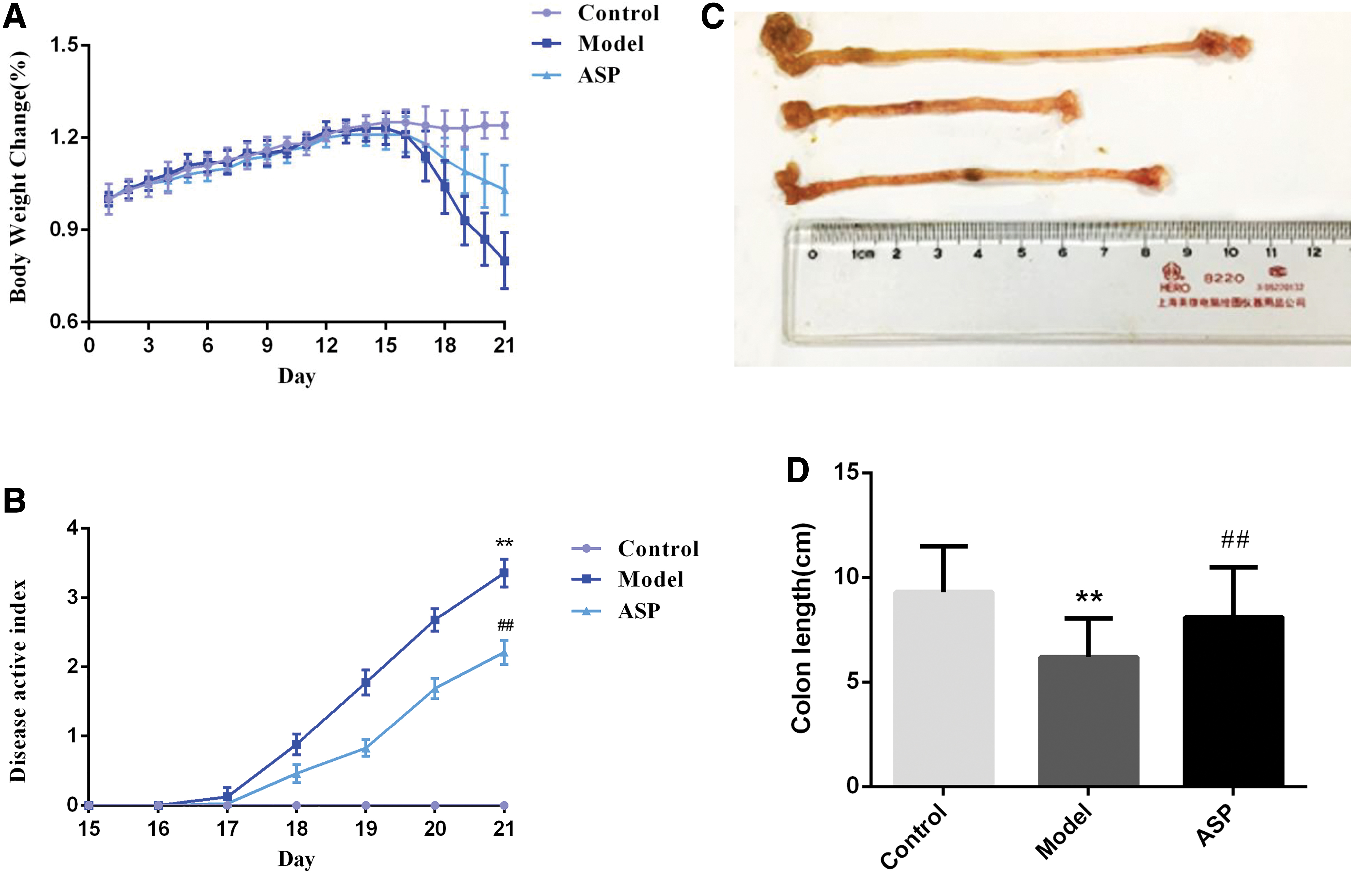
ASP ameliorated the symptoms of DSS-induced ulcerative colitis.

The effect of ASP on the morphology of colon tissues by H&E staining in DSS-induced colitis. DSS, dextran sulfate sodium; H&E, hematoxylin and eosin. Color images are available online.
ASP inhibited the proinflammatory cytokines in DSS-induced UC
To understand the mechanisms by which ASP alleviated inflammation in the UC model mice, we examined the levels of proinflammatory cytokines, such as IL-6, IL-1β, and TNF-α in colon tissues. As shown in Figure 3, in the model group, the mRNA expression of IL-6, IL-1β, and TNF-α in colon tissues was significantly increased (P < .01). However, the ASP treatment significantly suppressed the expression of the three proinflammatory cytokines induced by DSS.
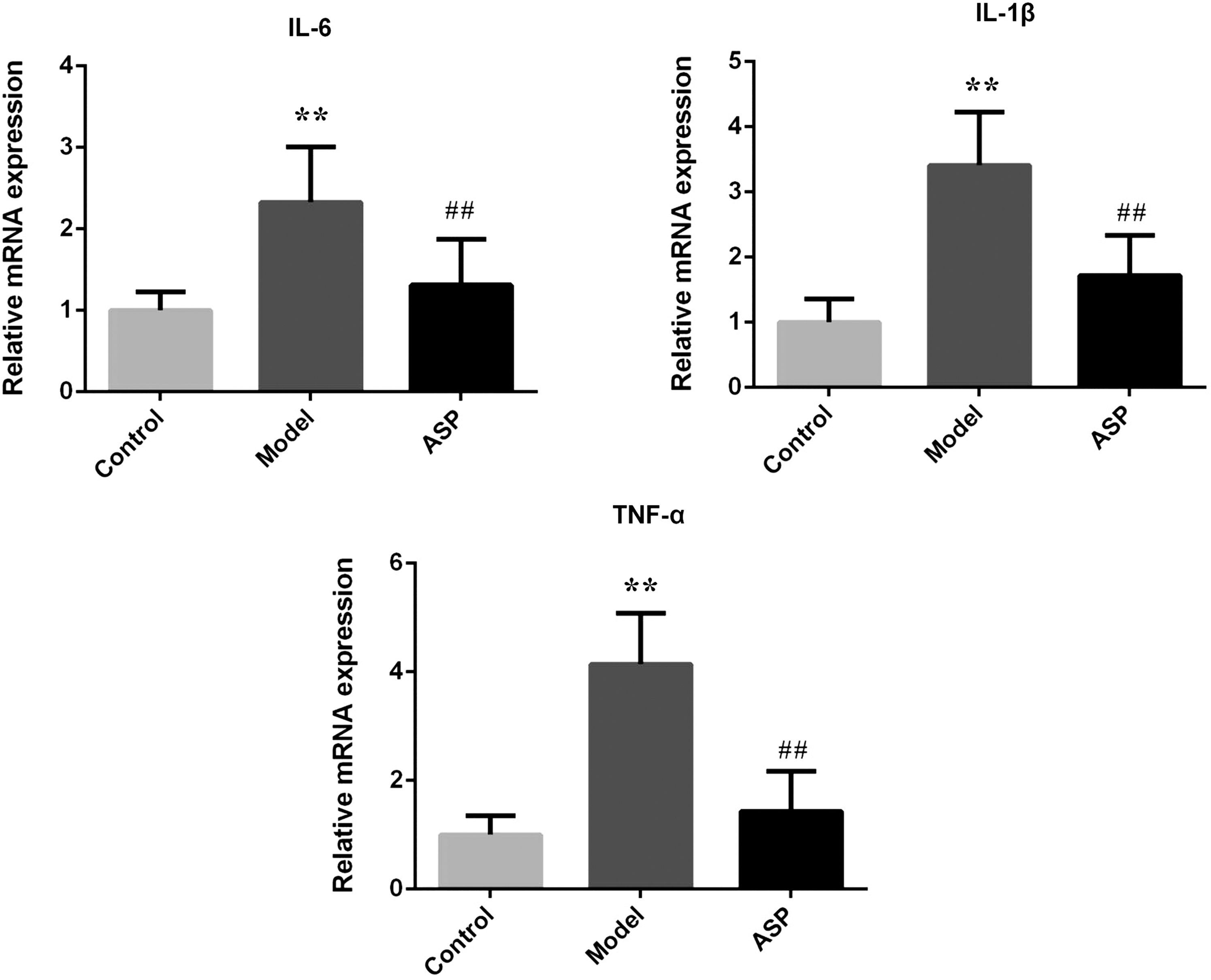
ASP inhibited the proinflammatory cytokine (IL-6, IL-1β, and TNF-α.) mRNA levels in DSS-induced ulcerative colitis. All of the data represent the mean ± SD (n = 8) and **P < .01 versus control group,
MPO activity
As shown in Figure 4, the result demonstrated that MPO activity in the model group was significantly elevated when compared with the control group. After treatment with ASP, the MPO activity significantly decreased.
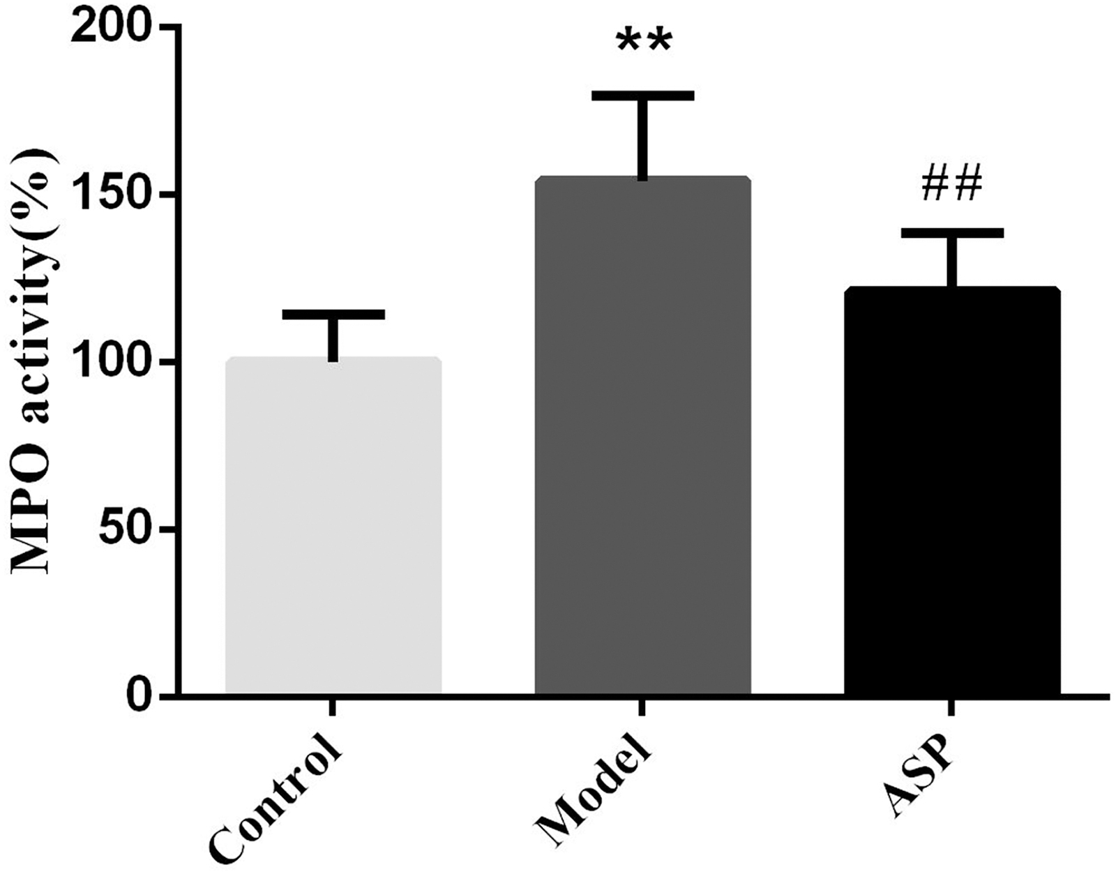
Effect of ASP on MPO activity of colon tissues. All of the data represent the mean ± SD (n = 8) and **P < .01 versus control group, ## P < .01 versus model group. MPO, myeloperoxidase.
ASP relieved epithelial TJ disruption in DSS-induced UC
To investigate the effect of ASP on epithelial TJ proteins, immunohistochemistry (IHC) staining and Western blot analysis of colons were performed. Figure 5A showed the IHC staining of TJ proteins, indicated by brown color in colon sections. The staining in the model group revealed less brown color; therefore, they exhibited less TJ protein expression in colons than control and ASP groups. As shown in Figure 5B, the expressions of TJ proteins (ZO-1, occludin, and claudin-1) significantly decreased in the model group. ASP-administered mice exhibited much higher expressions of the three TJ proteins. These findings revealed that ASP remarkably reduced epithelial TJ disruption in DSS-induced UC.
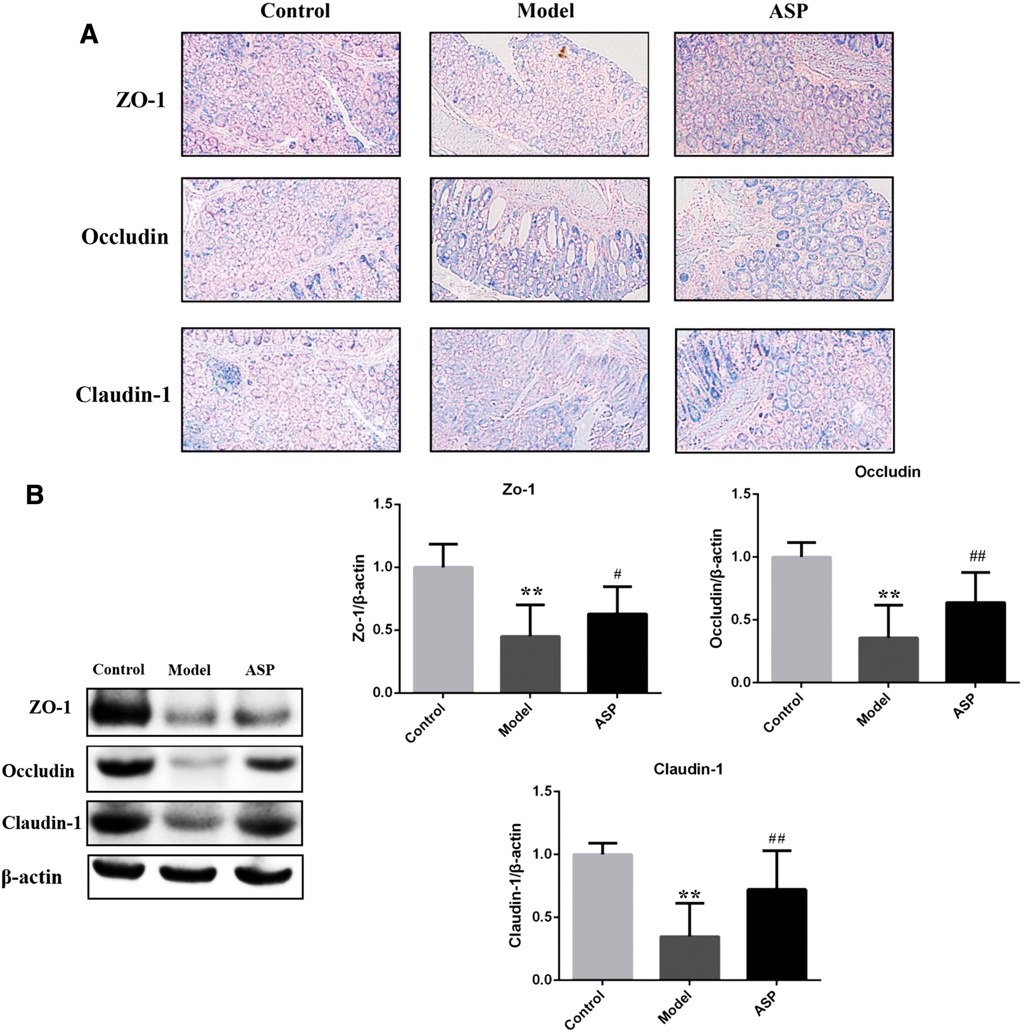
ASP relieved epithelial tight junction disruption in DSS-induced ulcerative colitis.
ASP reduced IEC apoptosis and improved proliferation in DSS-induced UC
We further employed the TUNEL method to detect apoptotic changes in colon tissues (Fig. 6A). In the TUNEL assay, apoptotic cells were indicated by brown color in colon sections. The model group demonstrated an increase in apoptotic events compared with the control group. However, the apoptotic events decreased after ASP treatment. At the same time, caspase-3 immunohistochemical analysis was used to detect proliferation and apoptosis in colon tissues (Fig. 6B). As expected, the results showed an enhanced caspase-3 immunostaining (caspase 3-positive cells showed brown color) in the model group. Then, the ratio of caspase-3 positive cells in the ASP-treated group was dramatically reduced. To better understand the antiapoptosis effect of ASP on UC, we measured the expression levels of Bcl-2 and Bax using the Western blot method (Fig. 6C), which are important apoptosis marker genes. The results showed that the expression of Bax/Bcl-2 was significantly decreased after ASP treatment compared with the model group.
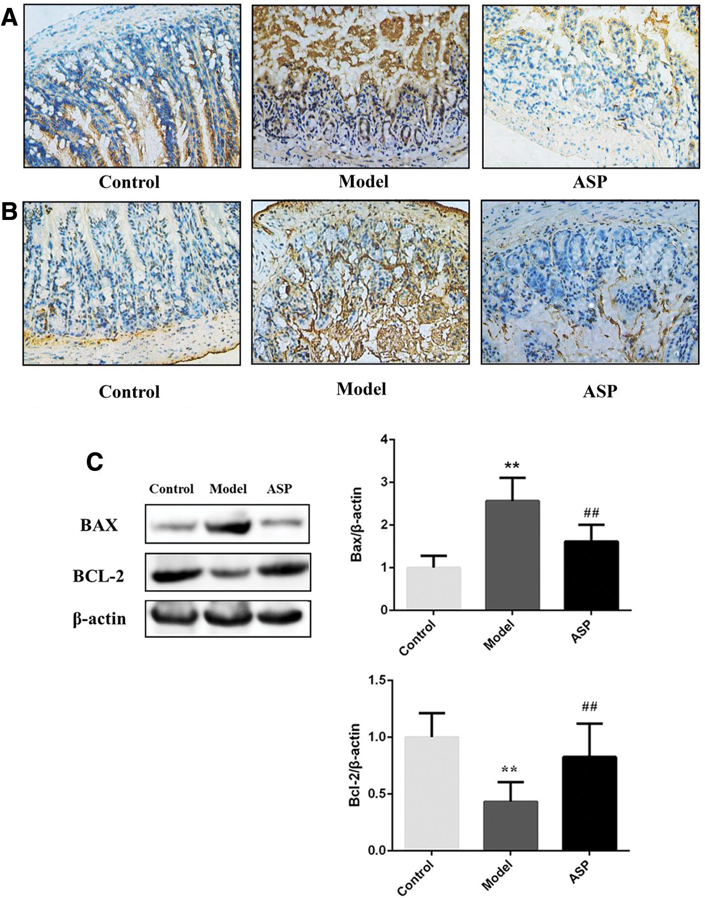
ASP reduced intestinal epithelium cell apoptosis and improved proliferation in DSS-induced
ASP ameliorated the function of intestinal mucosal barrier and reduced cell membrane permeability
To evaluate the effect of ASP on improving the permeability of the intestinal mucosal barrier, a single-layer Caco-2 cell epithelial inflammation model was induced by LPS. After giving ASP, the TEER values measured at different time points are shown in Figure 7A. The TEER value decreased by 56% at 8 h in the LPS group, whereas the ASP group was relatively the same as that of the control group. The result illustrated that ASP could ameliorate the intestinal mucosal barrier function and reduce cell membrane permeability. To further investigate the effect of ASP on intestinal barrier function of Caco-2 cells, Western blot analysis was performed. As shown in Figure 7B, the expressions of TJ proteins significantly decreased in the LPS group. After ASP administration, there was a definite improvement in the expressions of the three TJ proteins.
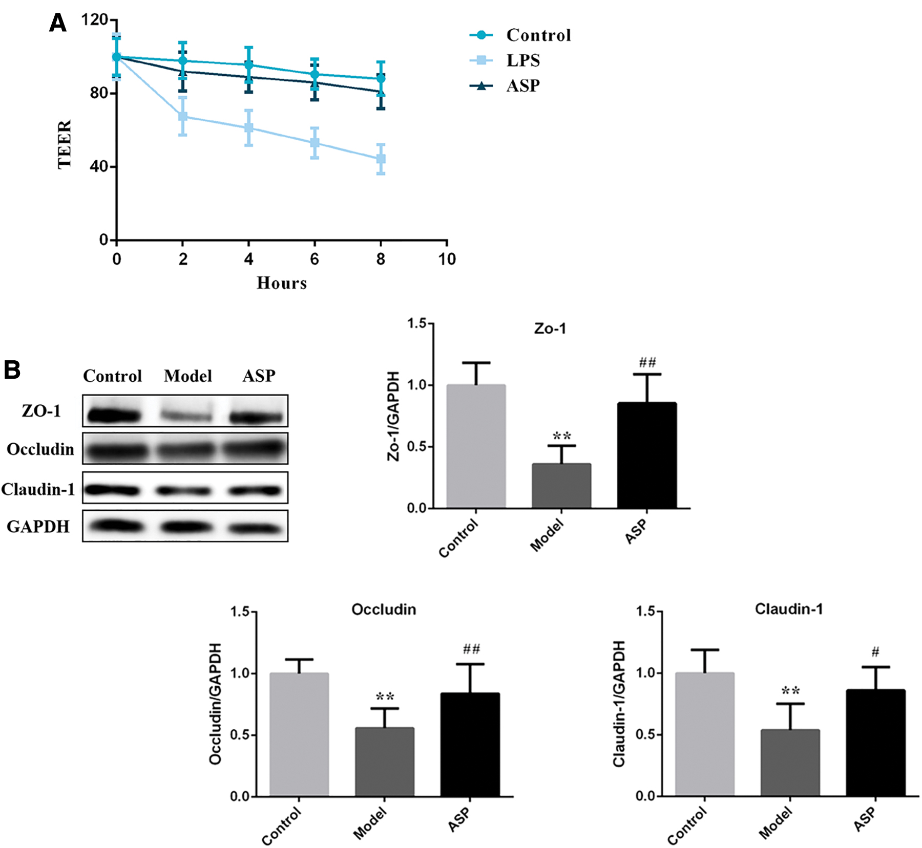
ASP ameliorated the function of intestinal mucosal barrier and reduced cell membrane permeability.
Discussion
Considering the dangers of UC to human health, it is essential to find alternative or supplementary therapeutic drugs for UC. In this study, we verified that ASP could alleviate the symptoms of UC in a mouse model of the disease. The protective effect of ASP is related to the inhibition of proinflammatory cytokines and apoptosis of intestinal cells. Furthermore, we identified that the intestinal barrier function was significantly ameliorated in mice after ASP treatment.
The DSS-induced model is often employed to mimic the pathological process of human UC, which is accompanied by a general inflammatory process directly associated with the symptoms of body weight loss, hemorrhagic, diarrhea, and intestinal histopathologic changes. 24 Unsurprisingly, ASP could significantly ameliorate the symptoms of weight loss, DAI score, colon shortening, and epithelial injury caused by DSS. We also observed that ASP treatment significantly repressed the MPO activity in colon tissues, which is closely related to neutrophil infiltration. These results suggested that ASP might provide a potential alternative to prevent or delay the progression of UC.
Much evidence suggests that the occurrence and development of UC are closely associated with the proinflammatory cytokines. 17 Intense investigations have revealed that TNF-α abundantly expressed in the intestine and participates in the antiapoptotic biological process of UC patients. 25 It has been reported that excessive IL-1β leads to an increase in intestinal permeability, promoting the activation of dendritic cells and macrophages. 26,27 Several studies have illustrated that blockade of IL-6 signal transduction in chronic intestinal inflammation led to remarkable inhibition of colitis activity. 28,29 Therefore, we investigated the mRNA expressions of IL-6, IL-1β, and TNF-α. These results indicated that ASP could ameliorate UC by inhibiting the expression of proinflammatory cytokines.
Emerging research has indicated that restoring and maintaining intestinal barrier function contribute to improving the defensive function of intestinal mucosa, promoting disease alleviation and reducing the recurrence rate of UC. 18 In recent years, many researchers have pointed out that the down-regulation of TJ proteins causes the colon wall to be attacked by intestinal pathogens and toxins in UC patients, which was considered to be a vital event in the pathogenesis of intestinal inflammation. 30 LPS could increase TNF-α production through TLR4/NF-κB. 31 It can lead to neutrophil infiltration, promote the production of mitochondrial oxidants, and stimulate the inflammatory response 32 ; therefore, it is usually a key component of the inflammation model. In our study, we verified the protective effect of ASP on the intestinal mucosal barrier through in vitro and in vivo studies. The results showed that LPS could damage the intestinal mucosal barrier and increase paracellular permeability, whereas ASP appeared to protect the integrity of TJ network by regulating the expression of TJ proteins such as ZO-1, occludin, and claudin-1, thereby balancing barrier integrity and protecting against cell membrane permeability.
Currently, conventional treatment strategies for UC are mainly aimed at controlling the inflammatory process. 33 The improvement of intestinal mucosal barrier function in the course of UC is seldom addressed, which leads to delays in improving many adverse effects and results in slow wound healing, prolonged usage of drug, and high recurrence rates. ASP addressed these issues and significantly promoted the expression of TJ protein, which in turn protected the intestinal barrier function and accelerated the healing of chronic ulcers, suggesting that the combination of ASP with current therapies may be a better choice for the treatment of UC.
IECs can form a selective physical barrier by connecting adjacent epithelial cells with TJ, thereby preventing harmful substances from entering the body and regulating the paracellular transport. 34 Previous reports have suggested that DSS is directly toxic to IECs in basal crypts, which increases IEC apoptosis. 35 There was also evidence illustrating that apoptosis signals can activate immune cells by releasing multiple chemokines to promote the progress of inflammation in vivo. 36 In this study, the data suggested that the protective effect of ASP on UC might be due to regulating IEC apoptosis and promoting mucosal healing.
In some sense, inflammatory cytokines, intestinal barrier, and IEC apoptosis form an interactive network, 37 which can be interrelated in the pathogenesis of UC. Our results also confirmed that ASP could protect against UC through the synergistic effect of inhibiting the expression of proinflammatory cytokines, decreasing the IEC apoptosis, and improving the intestinal mucosal barrier dysfunction.
In summary, our results demonstrated that ASP inhibited inflammatory cytokine expressions, decreased apoptosis of IECs, and promoted TJ protein expression in DSS-induced UC. Furthermore, these three aspects also exhibited cross-talk in the inflammatory response. More important, ASP could protect the intestinal barrier function and accelerate the healing of chronic ulcers, suggesting that combination of ASP with current interventions may be a better choice for the treatment of UC.
Footnotes
Acknowledgments
We appreciate the staff from the Analysis and Testing Center of Huazhong University of Science and Technology for their technical assistance.
Author Disclosure Statement
No competing financial interests exist.
Funding Information
This research was supported by National Key R&D Program of China (2017YFC0909900) and the National Natural Science Foundation of China (Grant No. 81403205).
Supplementary Material
Supplementary Table S1
References
Supplementary Material
Please find the following supplemental material available below.
For Open Access articles published under a Creative Commons License, all supplemental material carries the same license as the article it is associated with.
For non-Open Access articles published, all supplemental material carries a non-exclusive license, and permission requests for re-use of supplemental material or any part of supplemental material shall be sent directly to the copyright owner as specified in the copyright notice associated with the article.
