
Abstract
In this study, we explored whether the use of Streptococcus thermophilus LM1012 (TL-LM1012) as a safe probiotic exerts hepatoprotective effects by suppressing oxidative stress and inflammation in vitro and alleviating aspartate aminotransferase (AST), alanine aminotransferase (ALT), and lactate dehydrogenase (LDH) production in vivo. In a series of safety tests, TL-LM1012 was found to have a negative response to hemolysis and biogenic amines, as well as susceptibility to antibiotics. TL-LM1012 protected cell viability and suppressed cytotoxicity by inhibiting oxidative stress and induced heme oxygenase-1 and superoxide dismutase activity in a dose-dependent manner in diesel exhaust particulate matter (DEPM)-treated HepG2 cells. Moreover, proinflammatory cytokines, including tumor necrosis factor-α, interleukin (IL)-6, and IL-1β, were suppressed in DEPM-treated splenocytes. In DEPM-treated mice, oral administration of TL-LM1012 regulated AST, ALT, and LDH production in the serum after 14 days of treatment. These findings indicate that TL-LM1012, a safe probiotic, provides a potent preventive or therapeutic effect against liver disease caused by air pollution.
Introduction
Air pollution, caused by particulate matter (PM), poses a critical problem to human health worldwide. According to the World Health Organization (WHO), there are ∼3 million deaths each year due to air pollution. 1,2 PM exposure can worsen various diseases, including pulmonary mortality, cardiovascular disease, and even liver disease. One of the major components of PM is diesel exhaust particulate matter (DEPM), which was designated as a group 1 human carcinogen in June 2012. Although DEPM has extensive sizes of distributions, its main component includes nanosized particles (diameter below 50 nm) consisting of polycyclic aromatic hydrocarbons (PAHs) and nitro-PAHs and small amounts of other elements. 3 –5
Recent studies have shown that nanosized particle-rich DEPM causes hepatotoxicity, increasing oxidative stress and inflammation following intravenous or intratracheal administration, as well as acute and chronic airway disease, including lung cancer, allergic rhinitis, and bronchial asthma-like diseases. 6 In the human body, the liver plays a critical role in general homeostasis and has the largest mass, weighing 968–1860 and 603–1767 g in healthy men and women, respectively. It has various functions, including regulating carbohydrates and fat metabolism, bile acid synthesis, protein production, and glucose storage. However, continuous exposure to drugs, chemical agents, xenobiotics, and environmental pollutants results in liver injury by reactive oxygen species (ROS) and inflammatory cytokines, such as tumor necrosis factor-α (TNF-α), interleukin (IL)-6, and IL-1β. 7,8
Generally, probiotics consist of living microorganisms that have a beneficial effect on the health of the host when administered at adequate doses. They help to balance the gut microbiota, which affects the host health status. An imbalanced microbiota of the gastrointestinal tract can cause metabolic and/or inflammatory diseases, such as obesity, diarrhea, constipation, irritable bowel syndrome, inflammatory bowel diseases, autoimmune diseases, and certain cancers. 9 In contrast, recent results have suggested that the safety of probiotics is important to prevent or treat these diseases, or for use in foods and dietary supplements. Some of the factors that have been previously addressed include pathogenicity, infectivity, toxicity, and virulence factors. 10,11
The gut-liver axis is regarded as the connecting link between the gastrointestinal tract and the liver, both anatomically and functionally. Alterations in this axis can cause nonalcoholic fatty liver disease (NAFLD), including steatosis and nonalcoholic steatohepatitis (NASH), by compromising the integrity of the intestinal mucosal barrier (so-called leaky gut). The barrier plays a critical role in protecting against the infiltration of pathogenic bacteria, bacterial products, harmful toxins, and pathogen-associated molecular patterns, as well as damage-associated molecular patterns. Many studies have suggested that probiotics improve and modulate the intestinal barrier function. 12 For example, it has been previously reported that Streptococcus thermophilus and Lactobacillus acidophilus activate tight junction proteins to promote the integrity of the intestinal barrier. Similarly, Lactobacillus rhamnosus GG was found to regulate inflammation and apoptosis in intestinal epithelial cells. 13
In this context, some studies have shown preventive or therapeutic effects of probiotics against NAFLD. For example, VSL#3, a mixture of probiotic bacteria, was found to significantly suppress the hepatic inflammatory response and serum alanine aminotransferase (ALT) levels in vivo. 14 –16 In a clinical study, Lepicol, a mixture of probiotic bacteria, induced a reduction in serum aspartate aminotransferase (AST) and altered the intrahepatic triglyceride content. 17 In a single dose using lactic acid bacteria, Lactobacillus fermentum was found to reduce liver fat accumulation and decrease the levels of total cholesterol, triglycerides, uric acid, and insulin resistance. 18
In this study, we evaluated whether S. thermophilus LM1012 (TL-LM1012) can prevent liver damage caused by DEPM-induced inflammation and oxidative stress in vitro and in vivo.
Materials and Methods
Materials
Dulbecco's modified Eagle medium (DMEM), Rosewell Park Memorial Institute (RPMI) medium, fetal bovine serum (FBS), penicillin, and streptomycin were purchased from Life Technologies (Carlsbad, CA, USA). The expression of TNF-α, IL-1β, and IL-6 was determined by enzyme-linked immunosorbent assay (ELISA) kits (R&D Systems, Minneapolis, MN, USA). 3-(4,5-dimethylthiazol-2-yl)-2,5-diphenyltetrazolium bromide (MTT) and diagnostic kits to determine the activity of serum ALT and AST were purchased from Sigma-Aldrich (St Louis, MO, USA). The lactate dehydrogenase (LDH) release and LumiMaxTM superoxide dismutase detection kits were obtained from Roche Applied Science (Indianapolis, IN, USA) and Stratagene (La Jolla, CA, USA), respectively.
Preparation of TL-LM1012
TL-LM1012 used in this study was isolated from dairy products (KFCC 11771P). The strain was stored at −80°C in 15% skim milk and 10% glycerol mixture as the cryoprotectant before use. Cell propagation was performed in a 5 L fermenter containing 3 L growth medium, consisting of 20 g/L glucose, 10 g/L soy peptone, 20 g/L yeast extract, 1 g/L sodium acetate, 1 g/L K2HPO4, 0.05 g/L MnSO4, 0.1 g/L MgSO4, and 1 g/L
Preparation of DEPM suspension
National Institute of Standard and Technology (NIST) standard reference material (SRM 2975) was obtained from Sigma-Aldrich. 19 SRM2975 are DEP collected from a diesel-powered industrial forklift. A detailed description of the particles can be confirmed at NIST. DEPM stock (1 mg/mL) was suspended in culture medium and sonicated for 2 min to minimize particulate aggregation. Before HepG2cells were seeded, sonicated DEPM (1 mg/mL) was diluted into final concentration of 100 μg/mL with serum-free DMEM medium.
Safety test of TL-LM1012
For the hemolysis test, TL-LM1012 was cultured at 37°C for 18 h in MRS broth before streaking on 5% sheep blood agar. Based on the methods of Bover-Cid and Holzapfel, a biogenic amine test was performed to detect the levels of cadaverine, tyramine, histamine, and putrescine. 20 For the antibiotic resistance test, TL-LM14012 was grown at 37°C for 48 h in a broth containing agar Iso-Sensitest (90%) and MRS (10%), according to the Clinical and Laboratory Standards Institute (CLSI). The determination of concentration-dependent susceptibility was performed according to the European Food Safety Authority (EFSA). 21
Cell culture
Human hepatoma HepG2 cells were purchased from the American Type Culture Collection (Manassas, VA, USA). The cells were cultured in DMEM containing 10% FBS, streptomycin (100 μg/mL), and penicillin (100 U/mL) at 37°C with a humidified atmosphere of 5% CO2. Splenocytes were collected from sacrificed normal rats following an acclimation period of 1 week. The spleens were removed and single cells were prepared in RPMI-1640 medium (Life Technologies) containing 10% FBS and 100 U/mL penicillin/streptomycin with slight modifications. 22 The cells were treated with various concentrations (1 × 10⁵, 5 × 10⁵, and 10 × 105 CFU/mL) of TL-LM1012 or DEPM for various time periods.
Animal treatments and hepatotoxicity assessments
Male ICR mice (5 weeks of age) were purchased from Samtako (Osan, Korea) and adapted to the controlled conditions for temperature (22 ± 2°C) and humidity (50% ± 5%) on a 12:12 h light/dark cycle. The research protocol conformed with the Guidelines for the Care and Use of Laboratory Animals published by the US National Institutes of Health (NIH Publication No. 85-23, 1996). All experimental procedures were approved by the Committee on Ethics of Animal Experiments of International University of Korea (Ethics No., IUK-M-1710/02). Eighteen male mice were randomly designated to three experimental groups of six mice each. To determine the ability of TL-LM1012 to protect against DEPM-induced hepatotoxicity, the mice were administered intragastrically (i.g.) with TL-LM1012 or DEPM in saline at 1 × 10⁶ CFU/g or 100 mg/kg once daily for 14 consecutive days. Twenty-four hours after DEPM administration, the mice were anesthetized with CO2 and their blood was removed by cardiac puncture to determine the serum ALT, AST, and LDH levels. The animals were then euthanized by cervical dislocation. The hepatotoxicities were assessed by quantifying the serum ALT, AST, and LDH levels. The serum ALT, AST, and LDH levels were measured using spectrophotometric diagnostic kits (Sigma Co., St. Louis, MO, USA). For the measurement of body and organ weights, the mice were weighed on days 0 and 14. At the end of the experiment, the organs, including the liver, spleen, and kidney, were immediately removed and weighed.
Determination of cytotoxicity
Cell viability was measured using either the MTT or LDH cytotoxicity assay. HepG2 cells (1 × 10⁶ cells) were seeded in 96-well plates containing 100 μL DMEM medium and 10% FBS, and then incubated overnight. The cells were pretreated with various concentrations of TL-LM1012 (1 × 10⁵, 5 × 10⁵, or 10 × 105 CFU/mL) for 1 h followed by stimulated with DEPM (100 μg/mL) or without DEPM for 24 h. The supernatants were used in the LDH assay and the absorbance was measured at 490 nm using a microplate reader (Varioskan; Thermo Electron, Waltham, MA, USA). MTT solution (5 mg/mL in PBS) was added for 4 h at 37°C and 5% CO2. The medium was then discarded and the resulting formazan crystals were dissolved in 200 μL DMSO. The absorbance of each well was determined at 550 nm using a microplate reader (Varioskan; Thermo Fisher Scientific, Waltham, MA, USA).
Measurement of ROS production
The ROS production in HepG2 cells was determined using the redox sensitive fluorescent dye H2DCFDA. After treatment with 100 μg/mL DEPM or vehicle for 6 h, the cells were treated with 25 μM H2DCFDA for 20 min. The cells were washed twice with phenol-red-free DMEM containing 1% FBS. The fluorescence was then identified on a fluorescence reader (Varioskan; Thermo Electron Co.) by measuring the emission at 530 nm after excitation at 485 nm.
Measurement of TNF-α, IL-1β, and IL-6 levels
Rat primary splenocytes were pretreated with various concentrations of TL-LM1012 (1 × 10⁵, 5 × 10⁵, or 10 × 105 CFU/mL) for 1 h, then stimulated with DEPM (100 μg/mL) for 3 h (TNF-α) or 24 h (IL-1β and IL-6). The levels of TNF-α, IL-1β, and IL-6 in the culture supernatants were measured using the appropriate ELISA kits (R&D Systems).
Determination of antioxidant enzymes
The activity of heme oxygenase (HO)-1 and superoxide dismutase (SOD) was measured according to the manufacturer's instructions, as described previously. 22,23 In brief, HO-1 assay was performed at the end of each treatment. 24 The cell lysates were collected in 0.5 mL solution, including ice-cold 0.25 M sucrose and 50 mM potassium phosphate buffer (pH 7.4), and then were centrifuged at 200 g for 10 min. The supernatants were centrifuged at 9000 g for 20 min, and then further centrifuged at 30,000 g for 60 min. After being suspended in 50 mM potassium phosphate buffer (pH 7.4), the amount of the total protein in the cell pellets was quantified by bicinchoninic acid kit. Of cell lysate (200 μL), 500 μg/mL was incubated with 0.2 mM of the substrate hemin, 0.5 mg/mL rat liver cytosol as a source of biliverdin reductase, 0.2 mM MgCl2, 2 mM glucose-6-phosphate, 1 U/mL glucose-6-phosphate dehydrogenase, 1 mM NADPH, and 50 mM potassium phosphate buffer (pH 7.4) at 37°C for 2 h. After the reaction was stopped by applying chloroform (600 μL), the absorbance of the chloroform layer was determined by spectrophotometry. Bilirubin formation was calculated from the difference in absorption at 464 and 530 nm. To determine the SOD activity, xanthine and xanthine oxidase were used to generate superoxide radicals that reacted with 2-(4-iodophenyl)-3-(4-nitrophenol)-5-phenyl tetrazolium chloride to form a red formazan dye. The absorbance was then determined at 505 nm.
Statistical analysis
All data are expressed as the means ± standard deviations (SD) of triplicate experiments. Results from the animal study are expressed as the means ± SD (n = 6). The mean differences were made using analysis of variance followed by Dunnett's post hoc test. P-values <.05 were considered statistically significant.
Results
TL-LM1012 is a safe strain of probiotics
To evaluate whether TL-LM1012 is safe for probiotic use, we performed a hemolysis and biogenic amine production test, using Escherichia coli ATCC 25922 as a positive control. TL-LM1012 was found to have a negative response gamma hemolysis activity and biogenic amine production (Table 1), while E. coli ATCC 25922 showed beta hemolysis activity and a positive response for biogenic amine production.
Hemolysis and Biogenic Amine Production Activity of TL-LM1012 and Escherichia coli ATCC 25922
+, Positive; −, Negative.
To evaluate the susceptibility of TL-LM1012 to the selected antibiotics, we performed an assay for determining the minimal inhibitory concentration. As shown in Table 2, TL-LM1012 was found to be sensitive to the selected antibiotics compared to breakpoints of EFSA. Thus, our results collectively demonstrate that TL-LM1012 is completely safe for use as a probiotic for commercial applications.
Microbiological Cutoff Values (mg/L) of TL-LM1012
Including Lactobacillus delbrueckii, Lactobacillus helveticus.
Including Lactobacillus fermentum.
For Lactobacillus buchneri the cutoff for tetracycline is 128.
Including the homofermentative species Lactobacillus salivarius.
n.r., not required.
TL-LM1012 suppresses cell cytotoxicity and ROS production in vitro
To determine the effects of TL-LM1012 on cytotoxicity and oxidative stress induced by DEPM, we first performed a cell viability and LDH leakage assay for TL-LM1012 in HepG2 cells. As shown in Figure 1A and B, TL-LM1012 did not affect the cell viability or toxic events at a concentration of 1 × 10⁵, 5 × 10⁵, or 10 × 105 CFU/mL. Next, we evaluated whether TL-LM1012 maintains the cell viability by protecting cells against the cytotoxicity and oxidative stress induced by DEPM in HepG2 cells. TL-LM1012 was found to help in the recovery of cell viability (Fig. 2A), as well as decreasing the cytotoxicity (Fig. 2B) and production of ROS (Fig. 2C) in a concentration-dependent manner. These results indicate that TL-LM1012 has a protective effect against cytotoxicity and oxidative stress induced by DEPM in HepG2 cells.

Effects of TL-LM1012 cell viability and cell cytotoxicity. HepG2 cells (1 × 10⁶ cells) were seeded in 96-well plates containing 100 μL DMEM medium and 10% FBS, then incubated overnight. Cells were pretreated with various concentrations of TL-LM1012 (1 × 10⁵, 5 × 10⁵, or 10 × 105 CFU/mL) for 48 h. The cell viability was determined by MTT assays

Effects of TL-LM1012 on DEPM-induced cell viability. HepG2 cells were pretreated with various concentrations of TL-LM1012 (1 × 10⁵, 5 × 10⁵, or 10 × 105 CFU/mL) for 1 h, then stimulated with DEPM (100 μg/mL) for 24 h.
TL-LM1012 has anti-inflammatory effects in rat splenocytes
To determine the anti-inflammatory properties of TL-LM1012, we performed ELISA assay on splenocytes isolated from rat. Consequently, we found that DEPM increases the expression of inflammatory cytokines, including TNF-α, IL-6, and IL-1β. However, when pretreated with TL-LM1012, the expression of anti-inflammatory cytokines decreased in a concentration-dependent manner in rat splenocytes (Fig. 3A–C). These results demonstrate that TL-LM1012 alleviates inflammation induced by DEPM, which is one of the modulators of the immune response.

Effects of TL-LM1012 on inflammatory cytokine levels in DEPM-treated rat splenocytes. Rat primary splenocytes were pretreated with various concentrations of TL-LM1012 (1 × 10⁵, 5 × 10⁵, or 10 × 105 CFU/mL) for 1 h, and then stimulated with DEPM (100 μg/mL) for 3 h (TNF-α) or 24 h (IL-1α and IL-6). The expression levels of TNF-α
TL-LM1012 promotes heme oxygenase-1 and superoxide dismutase activity in vitro
HO-1, which is known to have cytoprotective, antioxidant, and anti-inflammatory properties, is a key enzyme. To evaluate HO-1 activity, we pretreated TL-LM1012 with DEPM in HepG2 cells, and confirmed that TL-LM1012 increases HO-1 activity in DEPM-treated HepG2 cells (Fig. 4A). Hence, SOD plays a critical role in regulating the ROS concentration in different cellular responses. To our surprise, TL-LM1012 increased the levels of SOD activity in a dose-dependent manner (Fig. 4B). Thus, TL-LM1012 alleviates inflammation and oxidative stress, promoting HO-1 and SOD activity in HepG2 cells.
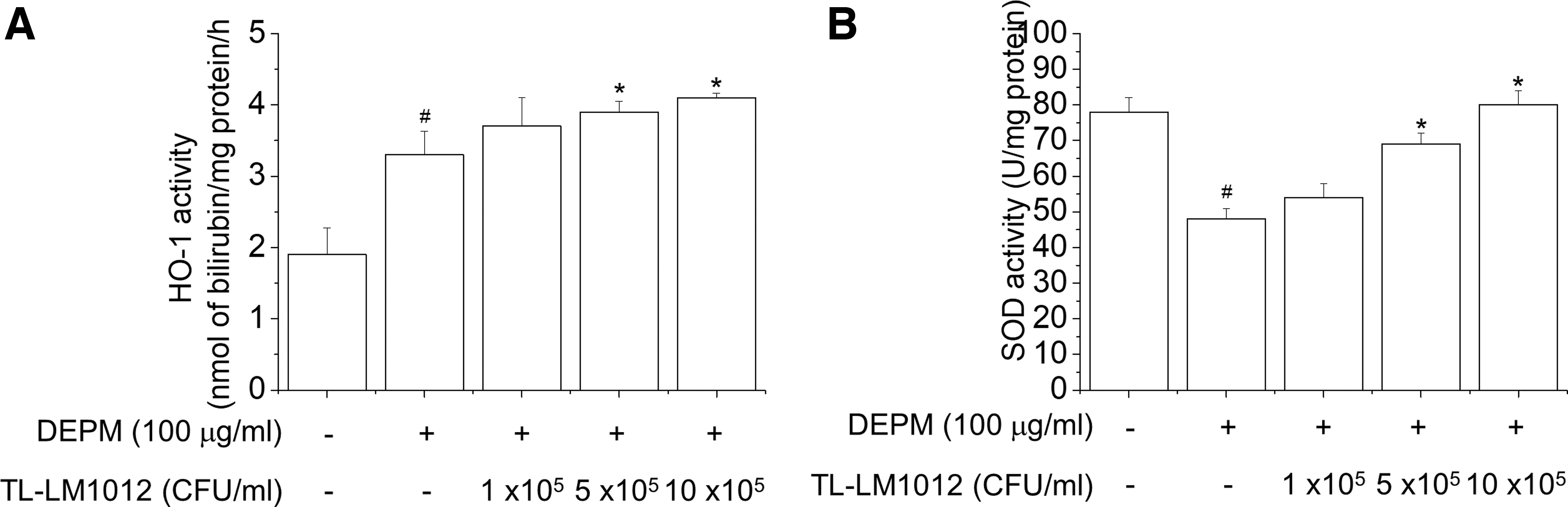
Effects of TL-LM1012 on HO-1 and SOD enzyme activity in DEPM-treated HepG2 cells. HepG2 cells were pretreated with various concentrations of TL-LM1012 (1 × 10⁵, 5 × 10⁵, or 10 × 105 CFU/mL) for 1 h, and then stimulated with DEPM (100 μg/mL) for 24 h. HO activity
TL-LM1012 protects against DEPM-induced liver injury in mice
To determine the hepatoprotective effects of TL-LM1012 under DEPM treatment, we performed an in vivo study measuring the biomarkers for liver damage in mice. We found that none of the groups underwent any changes in the body, liver, spleen, or kidney weights after 14 days (data not shown). As shown in Figure 5A–C, we found that DEPM increases the levels of AST, ALT, and LDH in the blood; however, TL-LM1012 decreases the factors of liver injury induced by DEPM in mice. Collectively, when DEPM-treated mice were orally administered TL-LM1012 on a daily basis, TL-LM1012 had a protective effect against liver injury.
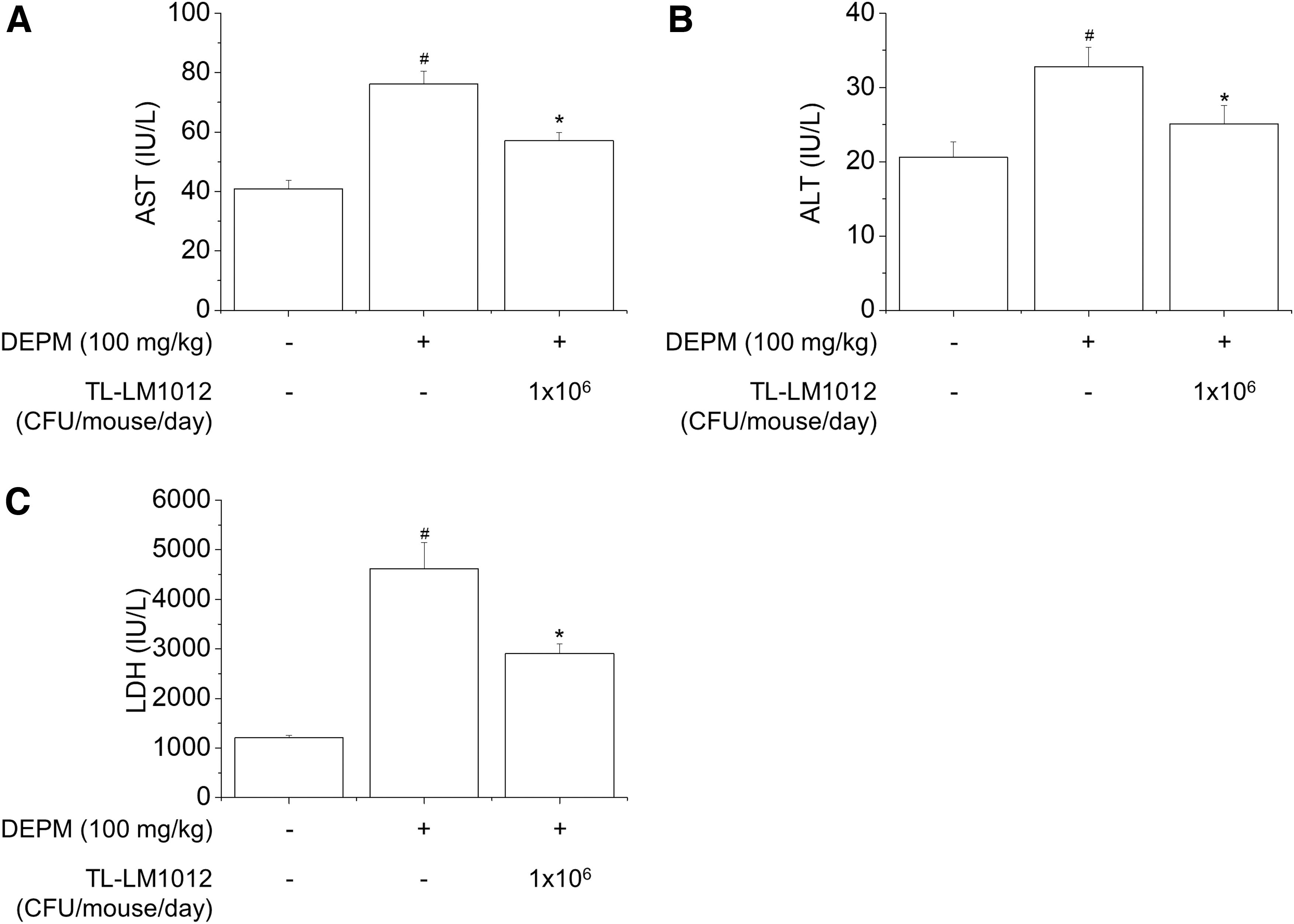
Effects of TL-LM1012 on DEPM-induced hepatotoxicity. Hepatotoxicity was determined by quantifying the serum levels of AST
Discussion
In the present study, we demonstrated that TL-LM1012 is safe for use as a probiotic for commercial applications. Furthermore, TL-LM1012 was found to have a preventive effect against DEPM-induced cytotoxicity, inhibiting the production of ROS by promoting HO-1 and SOD activity and inflammatory cytokine production (TNF-α, IL-6, and IL-1β) in vitro. In contrast, the increases in the levels of AST, ALT, and LDH in blood by DEPM, which result in liver damage, were found to decrease in vivo in the TL-LM1012-treated mice. Therefore, our results suggest that TL-LM1012 is a safe supplement for the prevention of liver damage caused by PM, such as DEPM.
Hemolysin is a toxin which destroys cells by means of cell membrane collapse, leading to cell death. When a red blood cell is exposed to hemolysin, its soluble components are eluted, followed by the formation of a red transparent liquid around red blood cells. This process is known as hemolysis. While α-hemolysis is considered to be a partial decomposition of the hemoglobin, β-hemolysis is considered to represent the complete breakdown of the hemoglobin. Alternatively, γ-hemolysis represents the lack of hemolysis. 25 As shown in Table 1, TL-LM1012 did not exert a hemolysis effect on cells, compared to the positive control of E. coli ATCC 25922. Biogenic amines have a low molecular weight and are basic nitrogenous and toxic compounds, removing the α-carboxyl group from the amino acids. It has been reported that some strains of lactic acid bacteria can produce biogenic amines, including histamine, cadaverine, tyramine, and putrescine. Therefore, the determination of biogenic amine-producing lactic acid bacteria is very important for use in dietary supplements, food products, and medicine. 26 –28 As shown in Table 1, TL-LM1012 showed a negative response to histamine, cadaverine, tyramine, and putrescine compared to E. coli ATCC 25922 (positive control).
The misuse and abuse of antibiotics for the treatment of bacterial infections represent the greatest threat. The most notable threat lies in the potential transfer of genes for acquired resistance to pathogenic bacteria or other lactic acid bacteria, rather than the genes for intrinsic resistance. Interestingly, many studies have shown that several species of lactic acid bacteria, such as Enterococcus, Lactobacillus, Lactococcus, Leuconostoc, Pediococcus, and Streptococcus, contain resistance genes, or show resistance against antibiotics. S. thermophilus has been reported to exert resistant activity on gentamycin, kanamycin, chloramphenicol, tetracycline, and streptomycin. 29 Although the EFSA cutoffs calculated here indicated that TL-LM1012 does not show resistance against antibiotics, further studies are needed to determine whether or not resistance genes can confer potentially acquired resistance at a genetic level, using polymerase chain reaction or whole-genome sequencing.
Although DEPM toxicity mainly affects the lungs due to its inhalation, several studies have demonstrated that it can also induce ROS by oxidative stress and regulate inflammatory cytokines through an acute phase response in the liver. 4,30 –32 Following oxidative stress, to maintain a cellular redox homeostasis, it is necessary to upregulate the expression levels of antioxidant enzymes, such as HO-1 and SOD. 33 Consistently, TL-LM1012 showed increased levels of HO-1 and SOD activity, reflecting its hepatoprotective effect on the recovery of cell viability, and the inhibition of LDH leakage. It is likely to have a counteractive effect. In contrast, the spleen, which acts as a blood filter, plays an important role in modulating the immune system against chemical reagents and microbial and viral infections. As shown in Figure 3, we found that TL-LM1012 suppresses the expression of proinflammatory cytokines (TNF-α, IL-6, and IL-1β) in DEPM-treated splenocytes isolated from rats. However, a mechanical analysis of the effects of transcriptional factors, including NF-κB and MAPKs, on oxidative stress and inflammation, such as the signaling molecules and events, need to be investigated further. 34
To the best of our knowledge, gut microbiota makes form an axis with liver as well as kidneys, brains, and other extraintestinal organs through portal circulation in homeostasis maintenance. 35 This symbiotic relationship allows the liver to be regulated and stabilized by metabolic, immune, and neuroendocrine crosstalk.
Under oxidative stress and/or inflammation in the gastrointestinal tract, unfavorable molecules, such as antigens derived from either pathogens or foods, lipopolysaccharide, peptidoglycan, and flagellin, are transported toward the liver through blood vessels, resulting in the development of various chronic liver diseases, including chronic hepatitis B and hepatitis C, alcoholic liver disease, NAFLD, NASH, cirrhosis, and hepatocellular carcinoma. 36 Some studies have suggested that lactic acid bacteria play a preventive and therapeutic role in liver disease induced by pathogens, chemical reagents, and toxins. 37 In accordance with these results, we discovered that TL-LM1012 plays a protective role against liver damage by restoring the levels of AST, ALT, and LDH in DEPM-treated mice. The weights of liver, spleen, and kidneys were measured, but no significant change was found (data not shown). Although further studies are required to confirm whether the hepatoprotective effects of TL-LM1012 are involved in regulating the microbiome in the gastrointestinal tract, or hepatocytes in liver, we suggest that it has indirect effects on liver damage, stabilizing gut microbiota in the intestinal tract.
In summary, as a dietary supplement, TL-LM1012 exerts hepatoprotective effects against oxidative stress and the inflammatory response in DEPM-treated animal cells, as well as reducing the levels of AST, ALT, and LDH in DEPM-treated mice. Our findings have critical implications on the development of healthy functional foods and medicine for the prevention of liver disease induced by air pollution.
Footnotes
Acknowledgments
We thank Yosep Ji and Whilhelm H. Holzapfel (Holzapfel Effective Microbes, HEM) for providing safety data of TL-LM1012.
Author Disclosure Statement
The Authors declare that there are no competing interests associated with the manuscript.
Funding Information
This research was supported by the Support Program for Creative Industry Institutes (Commercial Biotechnology Sophistication Platform Construction Program, R0003950), funded by the Ministry of Trade, Industry & Energy (MOTIE, Korea).