
Abstract
Alcohol induces liver injury related to oxidative stress and inflammatory responses. The purpose of this study was to investigate the hepatoprotective effect of Humulus japonicus extract (HJE) against alcohol–induced liver injury. Furthermore, we investigated the mechanisms of the protective effect of HJE on alcohol–induced liver injury. The pretreatment of HJE decreased the levels of aspartate aminotransferase, alanine aminotransferase, triglyceride, and total cholesterol in the plasma, suppressed the malondialdehyde, myeloperoxidase, and enhanced the activities of superoxide dismutase, glutathione, and catalase. The inhibitory effect of HJE against oxidative stress may be associated with the upregulation of nuclear factor erythroid 2-related factor 2 and its target gene heme oxygenase-1. Moreover, HJE inhibited the pro-inflammatory cytokines (tumor necrosis factor alpha, interleukin-1 beta) by downregulating toll-like receptor 4, myeloid differentiation primary response 88, and nuclear factor kappa B p65. These findings provide evidence for the elucidation of the hepatoprotective mechanisms for HJE.
Introduction
Long-term alcohol intake is one of the important factors in liver disease. 1,2 Liver inflammation, steatosis, and cirrhosis are often caused by chronic alcohol intake. 3,4 Moreover, cirrhosis may progress to hepatocellular carcinoma when it is accompanied by heavy alcohol intake. 5
Alcohol not only changed the gut permeability but also the translocation of bacteria-derived lipopolysaccharide (LPS) from the gut to the liver and increased the levels of circulating endotoxin in the portal blood and liver. 6 In a previous clinical study, the development of liver disease was associated with the increased circulating endotoxin level caused by alcohol. 7
Toll-like receptors (TLRs) are a class of transmembrane proteins, which play important roles as pattern recognition receptors. 8 TLRs activate antigen presenting cells to initiate innate immunity and to invade pathogens by sensing endogenous danger signals. 8 There are two different pathways in TLR signaling. The production of inflammatory cytokines is initiated by the myeloid differentiation primary response 88 (MyD88)-dependent pathway. The stimulation of interferon-β and the maturation of dendritic cells are associated with MyD88-independent pathway. 9 Several transcription factors such as nuclear factor kappa B (NF-κB) are also involved in TLR signaling. 8
Many studies have shown that TLR4 signaling is associated with alcohol–induced liver disease. 10 –12 Because of the change in gut permeability and the increase of LPS in the liver, the pro-inflammatory cytokines such as tumor necrosis factor alpha (TNF-α) and interleukin-1 beta (IL-1β) are increased through TLR4 signaling. 13 –15 Alcohol administration leads to the increase of TLR4 protein in hepatocytes, indicating that the TLR4 signaling pathway is strongly associated with alcohol–induced liver disease. 16
Many studies have shown that mitochondrial dysfunction, inflammatory factors, steatosis, and oxidative stress are involved in alcohol–induced liver injury. 17,18 Inflammation and oxidative stress play important roles in the pathogenesis of alcohol–induced liver damage. 17,19 It has been well known that ROS, H2O2 (hydrogen peroxide), O2 − (superoxide anion), and lipid peroxides caused by excessive alcohol ingestion are the main causes of liver injury. 20 –22 It has been known that there are two types of antioxidant factors to counteract against intracellular oxidative stress: enzymes, including glutathione peroxidase (GPx), superoxide dismutase (SOD), catalase (CAT), and nonenzymatic substances, including glutathione (GSH). 23,24 Increasing antioxidant capacity can protect hepatocytes against alcohol–induced liver injury effectively. 25 Recently, many studies reported that nuclear factor erythroid 2-related factor 2 (Nrf-2) has protective effects against oxidative stress by upregulating antioxidant enzymes such as heme oxygenase (HO-1). 26
Humulus japonicus is widely used as traditional medicine to treat not only dysentery, tuberculosis, pulmonary, diarrhea, and pneumonia but also to promote blood circulation. 27,28 H. japonicus has been reported to contain flavonoids, phenolics, lupulones, terpenes, flavone glycosides, phenolic compounds, alkaloids, and isoquinoline that were well known and have antioxidative and anti-inflammatory effects. 29 –34 Moreover, recent studies have revealed that the Humulus japonicus extracts (HJEs) have antioxidant properties. 29,31 Furthermore, a recent study reported that the aqueous extract of H. japonicus contains the flavone and luteolin. 35
Our previous study has demonstrated that HJE has protective effects in the LPS and d-galactosamine induced acute liver injury rat model. 36 However, there is no prior research about the protective effects of HJE against alcohol–induced liver injury. In this study, we used HJE to investigate how H. japonicus affects the progression of alcohol-induced liver injury, including anti-inflammation, antioxidant capacity, and lipid accumulation in the chronic alcohol ingestion rat model. Furthermore, we investigated the involvement of TLR4/MyD88/NF-κB signaling in modulating the protective effects of H. japonicus against alcohol-induced liver injury.
Materials and Methods
Reagents
Alcohol, silymarin, ethyleneglycol-bis-(β-aminoethylether)-N,N,N′,N′-tetraacetic acid (EGTA), ethylenediamine tetra acetic acid (EDTA), leupeptin, aprotinin, β-mercaptoethanol, phenylmethylsulfonylfluoride (PMSF), Triton X-100, and sodium dodecyl sulfate (SDS) were purchased from Sigma Aldrich Co., LLC (St. Louis, MO, USA). Ammonium persulfate, N,N,N′,N′-tetramethylethylenediamine (TEMED), nitrocellulose membrane, tris/glycine/SDS buffer, tris/glycine buffer, and RestoreTM western blot stripping buffer were purchased from Bio-Rad (Richmond, CA, USA). Tris-buffered saline and phosphate-buffered saline were purchased from Welgene, Inc. (Daegu, Republic of Korea). HJE was kindly provided by Prof. Ji Hoon Jeong of College of Medicine in Chung-Ang University (Seoul, Republic of Korea).
Preparation of HJE
Dried H. japonicus (10 kg) was extracted with 99% water (10 × ) at room temperature (RT) for 8 h. The extracted materials were precipitated at RT for 8 h. The crude water extracts were filtered through a 25 μm diameter filter paper. The filtered H. japonicus compounds were concentrated using a reduced pressure concentrator, finally 26 BRIX. The condensed H. japonicus compounds were spray dried with a final yield of 17%. The final H. japonicus extract had a medium level viscosity and was water soluble. The H. japonicus concentrated materials were stored at 4°C. All of these procedures were executed at Samjin Geinseng natural food company (Hong-chun, Republic of Korea).
Animals and experimental protocols
Sprague-Dawley male rats (7 weeks old, weighing 250–300 g) were randomly divided into six groups (n = 10 in each group) as follows: (1) control group, (2) alcohol–induced liver injury group, (3) positive control (silymarin) group, (4) HJE 8 mg/kg treated group, (5) HJE 16 mg/kg treated group, and (6) HJE 32 mg/kg treated group. Pathogen-free food and water were provided, and sterilized bedding was used for housing. The concentrations of HJE were selected based on a previous study. 36 The control group and alcohol–induced liver injury group were administered orally with 1 mL of normal saline, and the HJE group was orally gavaged with 1 mL of HJE for 42 days. Rats in positive control were treated with 10 mg/kg silymarin, which is a widely used liver protection agent for 42 days. 37 Silymarin was orally administered with normal saline as a solvent. Except the control group, rats in other groups were orally administrated with alcohol (7 g/kg) at 4 h before the administration of normal saline, HJE, or silymarin for 42 days. The concentration of alcohol was explored based on the previous study. 38,39 All experimental procedures were in accordance with the guidelines established by the Institutional Animal Care and Use Committee of Chung-Ang University (No. 2018-00006).
Liver function assay
The rats were anesthetized and sacrificed using CO2 gas after the last administration. Blood samples were obtained from the inferior vena cava for biochemical assay. The blood samples from the rats were contained in Vacutainer (BD, Plymouth, United Kingdom) and centrifuged at 1300 g for 10 min at RT to obtain plasma. The plasma levels of aspartate aminotransferase (AST), alanine aminotransferase (ALT), triglyceride (TG), and total cholesterol were investigated by the Raonbio, Inc. (Yong-in, Gyeonggi-do, Republic of Korea).
Protein isolation and western blot analysis
Obtained liver tissues were homogenized in ice in lysis buffer (3.15 × 10⁻³ g/mL tris-HCl [pH 7.4], EDTA, EGTA, 0.01 g/mL Triton X-100, 10−4 g/mL SDS, 10−5 g/mL aprotinin, 10 μg/mL leupeptin, 1 mM PMSF, and 0.7 μg/mL β-mercaptoethanol). The lysates were centrifuged at 16,200 g for 15 min at 4°C, and supernatants containing cytosolic proteins were collected for western blot analysis. Nrf-2, HO-1, TLR4, NF-κB, and β-actin and secondary antibodies (Santa Cruz Biotechnology, Inc., CA, USA) were used to detect protein expression levels. The results were analyzed by Quantity One analysis software (Bio-Rad Chemical Division, Richmond, CA, USA).
Determination of plasma pro-inflammatory cytokines
To measure TNF-α and IL-1β levels in plasma, we used Enzyme-Linked Immunosorbent Assay (ELISA) Kits from BioVision, Inc. (California, USA). The ELISA was performed according to manufacturer's protocol.
Measurement of activities of neutrophil infiltration, lipid peroxidation, and antioxidant enzymes
Liver tissues were obtained to make homogenites with cold saline. After centrifugation at 3000 g for 10 min at 4°C, the supernatant was collected to measure myeloperoxidase (MPO), malondialdehyde (MDA), GPx, GSH, CAT, and SOD. The activity of MPO was analyzed by assay kit (BioVision, Inc.). Lipid peroxidation was determined by measuring MDA production using the Lipid Peroxidation Assay Kit (BioVision, Inc.). The total GSH was determined using a Glutathione Detection Kit (BioVision, Inc.). GPx activity was determined using GPx Activity Colorimetric Assay Kit (BioVision, Inc.). SOD was determined using SOD Activity Assay Kit (BioVision, Inc.). CAT activity was determined using CAT Activity Assay Kit (BioVision, Inc.).
Histological evaluation
All groups were sacrificed, and liver tissues were collected for histological evaluation at the end of the experiment. Histological evaluation was performed in samples subjected to hematoxylin and eosin (H&E) staining. Pieces of liver tissue were fixed in 10% buffered neutral formalin for the H&E staining by dehydration with different concentrations of ethanol and xylene, replacing xylene with hot paraffin. The liver tissue was embedded in paraffin solution. The paraffin sections were cut into 5 μm thick sections using a sliding microtome. The sections were passed through water, alcohol, and xylene and then stained with H&E dye.
Stained tissues were observed with a Leica DMR 6000 microscope, and images were captured with a Leica DM 480 camera (Wetzlar, Germany). Images presented were photographed at 400 × .
Statistical analysis
Each treatment was analyzed from 10 rats (n = 10) in each group. Data are expressed as mean ± standard error of the mean. Statistical analysis was done using one-way ANOVA. Data were considered significant at *P < .05, **P < .01 versus control group, # P < .05, ## P < .01 versus alcohol group.
Results
Abnormality of liver function induced by alcohol was ameliorated by HJE
To evaluate the abnormality of liver function, we measured AST and ALT, which are well known indicators of liver function. The elevation of AST and ALT level is a crucial clinical sign of hepatocellular injury. In the liver injury group, AST and ALT levels were increased compared with control group after feeding alcohol for 6 weeks (Fig. 1A, B). HJE treatment reduced AST and ALT levels compared to the liver injury group. Total cholesterol and TG were analyzed to evaluate the effect of HJE on lipid contents (Fig. 1C, D). In the liver injury group, the plasma total cholesterol and TG concentration were significantly increased compared with the control group after 6 weeks of alcohol treatment, but plasma total cholesterol and TG concentration were lower in the HJE treated group compared to the liver injury group.

Effect of HJE on AST, ALT, TG, and total cholesterol. Hepatic function was assessed by plasma levels of
HJE prevented alcohol–induced neutrophil infiltration and lipid peroxidation
The MPO level is an index of neutrophil infiltration. Livers from the alcohol–induced liver injury group had significantly increased MPO activity than the livers from the control group in Figure 2A. This increase was alleviated in the groups that fed alcohol with HJE (8, 16, 32 mg/kg). MDA level is an index of lipid peroxidation. Levels of MDA were increased significantly in the alcohol–induced liver injury group compared to the levels from the control group in Figure 2B. This increment was attenuated in the livers from rats fed all doses of HJE (8, 16, 32 mg/kg).
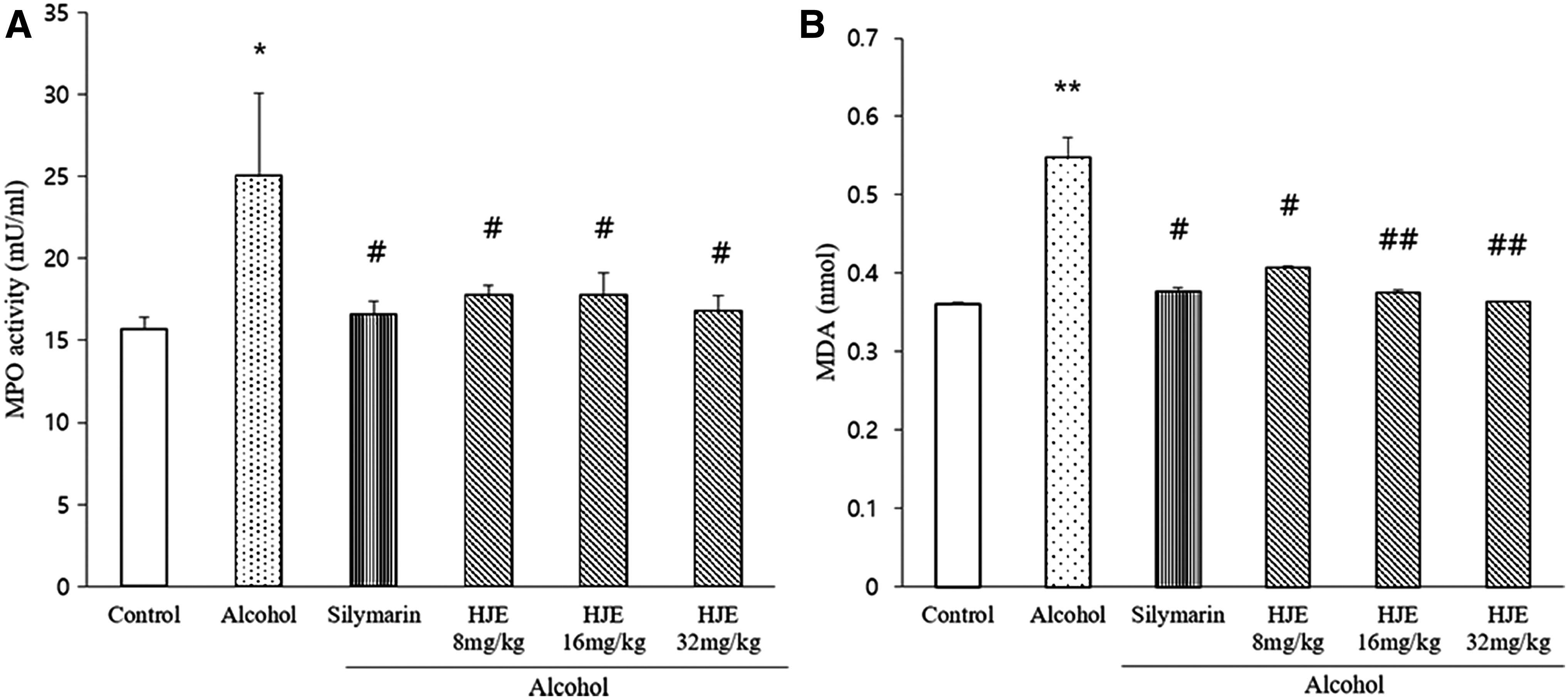
Effect of HJE on MPO and MDA level. Effect of HJE on
HJE restored the reduced GSH levels induced by alcohol
GPx activity and GSH concentrations in liver tissues were investigated in this study. There was no difference between the GPx activity of the control group and the alcohol–induced liver injury group as shown in Figure 3A. GSH is a primary index of oxidation in cells. Figure 3B shows that the GSH concentration was significantly decreased in the alcohol–induced liver injury group compared to the control group. The pretreatment of HJE with alcohol increases the GSH level compared to the alcohol–induced liver injury group.
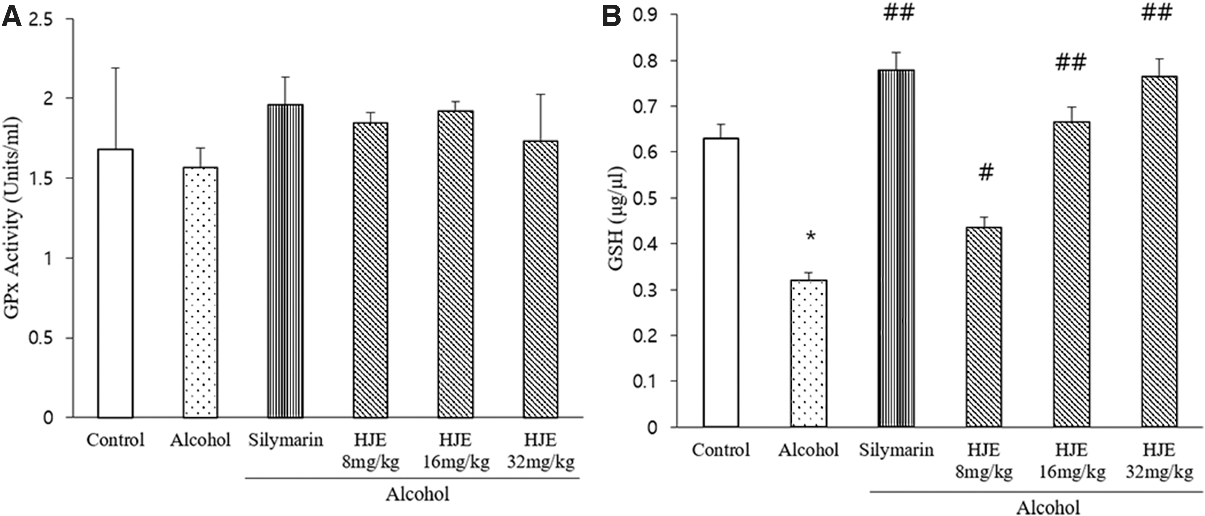
Effect of HJE on GPx and GSH levels. Effect of HJE on
HJE ameliorated alcohol–induced oxidative stress
Oxidative stress induced by excessive alcohol ingestion plays a critical role in liver damage. The activities of the antioxidant enzymes, CAT and SOD, were measured to assess antioxidant status. The activities of CAT were decreased in the alcohol–induced liver injury rat model compared with the control group (Fig. 4A). HJE and silymarin significantly attenuated these decreased enzyme activities in the liver. Furthermore, HJE treatment restored the decreased SOD activities as shown in Figure 4B.

Effect of HJE on CAT and SOD level. Effects of HJE on
HJE inhibited alcohol–induced inflammatory response
As shown in Figure 5A and B, TNF-α and IL-1β levels in the alcohol–induced liver injury rat model were remarkably increased. Treatment with silymarin significantly attenuated the concentration of TNF-α and IL-1β. Treatment with HJE at 16 and 32 mg/kg significantly suppressed the alcohol–induced increased TNF-α levels. Treatment with 32 mg/kg of HJE significantly attenuated the increased levels of IL-1β induced by the alcohol treatment.
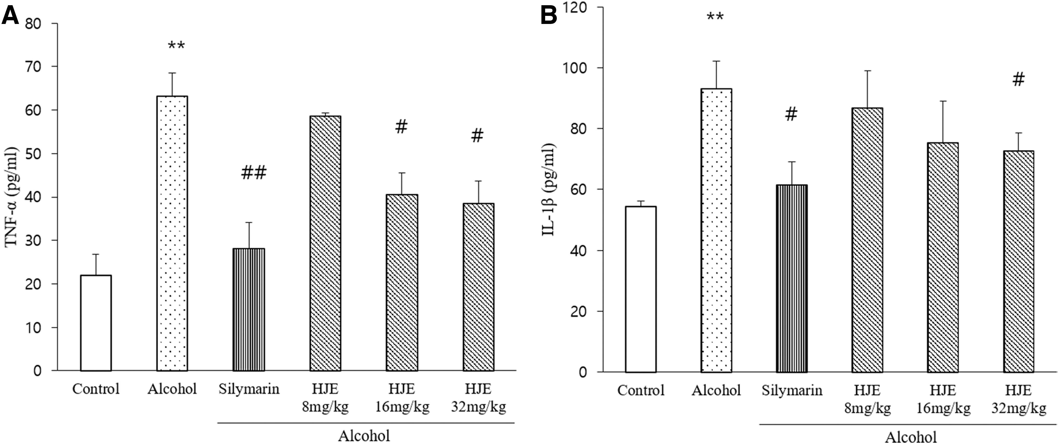
Effects of HJE on pro-inflammatory cytokines. Effect of HJE on
HJE improved alcohol–induced liver pathological changes
As shown in Figure 6, these results showed that changes in histology induced by the alcohol treatment led to increased inflammation, necrosis, and architecture collapse. Liver damage was suppressed by the pretreatment with silymarin compared with the control group. When pretreated with HJE, the liver damage was reduced as shown in Figure 6.

Effect of HJE on histopathology. Figure showed photomicrographs (200 × ) of liver tissue of control and experimental animals from each group. Sections were stained with hematoxylin–eosin. Histological changes of liver tissues were observed under microscope 200 × .
HJE modulated the expressions of Nrf-2 and HO-1 in liver
To determine the mechanism of the hepatoprotective effect of HJE on alcohol–induced liver injury, the protein expression of Nrf-2 and HO-1 was measured. The expressions of Nrf-2 and HO-1 in the liver injury group were reduced in comparison to the control group (Fig. 7). This reduction was alleviated by the pretreatment of HJE and silymarin. These data show that HJE attenuates the oxidative stress by upregulating Nrf-2 and HO-1.
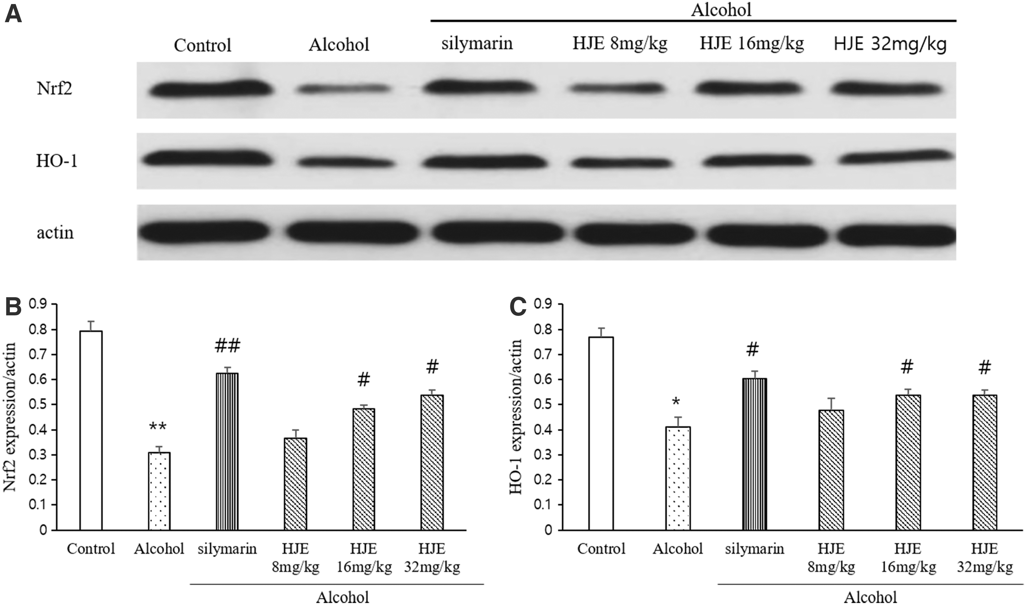
Effect of HJE on alcohol–induced oxidative stress. Effect of HJE on alcohol–induced oxidative stress.
HJE modulated the expressions of TLR4 and NF-κB in the liver
To investigate the role of the TLR4/NF-κB inflammatory signaling pathway on the anti-inflammatory effect of HJE, the expression of NF-κB was analyzed through western blotting (Fig. 8). While the protein expression levels of TLR4 and MyD88 and its downstream NF-κB were enhanced in the alcohol–induced liver injury group, silymarin and HJE treatments decreased the expressions of those proteins. These results suggest that the anti-inflammatory effect of HJE on alcohol–induced liver injury is mediated by the suppression of the TLR4/MyD88/NF-κB pathway.
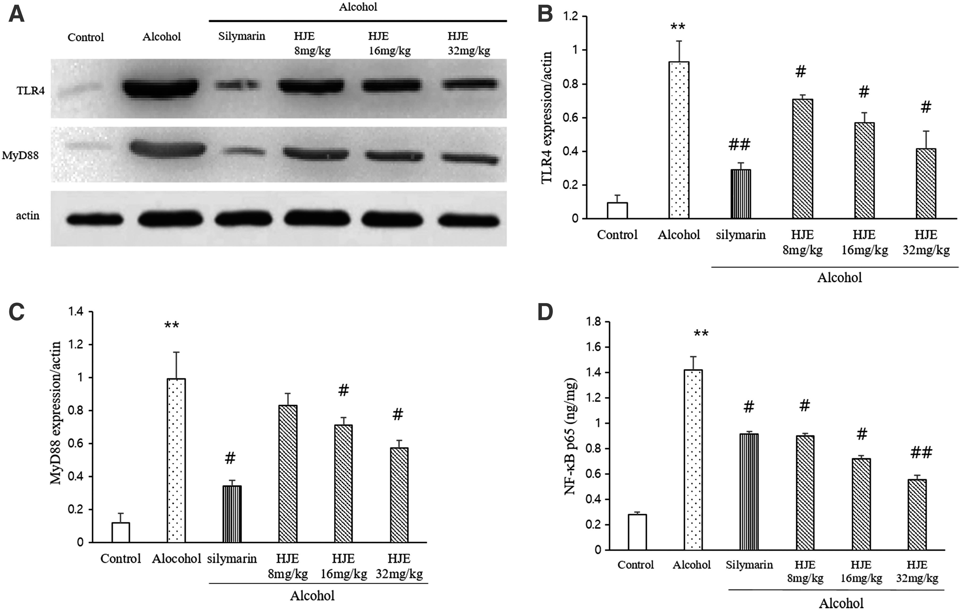
Effects of HJE on TLR4 signaling pathway and NF-κB.
Discussion
The specific mechanism of alcohol–induced liver disease still remains unclear. Many studies have demonstrated that the inhibition of inflammation, suppression of oxidative stress, and the amelioration of fatty degeneration have been considered to be the promising therapeutic strategies for alcohol liver disease. Therefore, the aim of this study was to seek out potential therapeutic agents against the alcohol–induced liver injury based on these strategies.
The levels of plasma AST and ALT are among the most sensitive biochemical markers when evaluating liver function. It has been known that abnormal increase of AST and ALT can lead to necrosis of hepatocytes. 40,41 The pretreatment of HJE alleviated the increase of these indicators, which means that HJE can improve the hepatic function damaged by alcohol intake.
Through histological analysis, the effect of HJE on improvement of liver injury induced by alcohol was evaluated. Alcohol induced abnormal hepatocyte structure such as inflammation and necrosis. This abnormal structure of hepatocytes was ameliorated by the pretreatment of HJE. Taken together, the hepatoprotective properties of HJE were evident from the inhibition of inflammatory reactions in the liver section of rats treated with various dosages.
Excessive alcohol consumption can damage the hepatocyte mitochondrial function, interfere in the lipid oxidation reaction, and further inhibit the peroxidase activity, leading to the lipid accumulation. 42,43 Moreover, alcohol can increase the mobilization of fatty acids in the liver by stimulating peripheral steatolysis and promote TG synthesis. 44 In line with these findings, this study suggested that the lipid metabolic disorder occurred after alcohol administration, accompanying with the increase of TG content and total cholesterol. According to this study, the HJE effectively improved the hepatic injury by decreasing TG and total cholesterol contents.
Neutrophils are well known to release the pro-inflammatory cytokines and chemokines and play a crucial role in liver damage. 45 –47 Through MPO activity assay, we showed that the HJE treatment attenuated neutrophil infiltration into the liver tissues induced by alcohol.
It has been well known that the oxidative stress is involved in the molecular mechanism underlying the pathogenesis of alcohol–induced hepatic injury. 48 During the ingestion of alcohol, the metabolism of alcohol accelerates and forms acetaldehyde to promote the ROS generation. 17,49 MDA, as the final product of lipid peroxidation, is a marker of the oxidative stress. 50 This study showed that the HJE lowered the overproduction of MDA. SOD and CAT are crucial enzymatic antioxidants to regulate the cellular redox balance. 48 This study showed that the pretreatment with HJE could reverse the reduction of the activities of SOD and CAT, revealing that HJE could protect liver against alcohol as counteracting oxidative stress through the inhibition of MDA and increasing SOD and CAT.
GSH is synthesized by glutamate-cysteine ligase and GSH synthase. 51 GSH depletion by itself induces oxidative stress in cells and induces apoptosis in cells. 52 This study showed that alcohol administration induced GSH depletion, and the pretreatment with HJE restored GSH content. Although alcohol administration did not change the GPx activity in this model, alcohol administration can induce oxidative stress through GSH depletion. Administration of HJE inhibited the decrease in GSH levels, and this result revealed that HJE had a protective effect against oxidative stress caused by alcohol–induced GSH depletion.
Moreover, Nrf-2 and HO-1 protein expressions were measured in this study to examine the mechanisms of antioxidant effects of HJE in alcohol–induced liver injury model. Nrf-2, as a transcription factor, which induced antioxidant and detoxification enzymes has been known as a therapeutic target for alcohol–induced liver injury. 17,53 HO-1, the downstream protein of Nrf-2, plays a crucial role in strengthening the resistance capacity when the liver is damaged. This study showed that the HJE pretreatment increased the expression of HO-1, as well as Nrf-2. This indicates that the antioxidant effect of HJE on oxidative stress induced by alcohol in liver was associated with the HO-1/Nrf-2 pathway.
It is known that excessive alcohol ingestion activates the TLR4/MyD88/NF-κB signaling pathway due to the overproduction of ROS. 54,55 Other studies also suggested that alcohol can increase circulating endotoxin level, which may induce TLR-mediated inflammation. 56,57 TLR4 can identify the molecular patterns which are associated with liver injury. 58,59 Activation of the TLR4 leads to the translocation of NF-κB from the cytoplasm to the nucleus. When stimulated, NF-κB triggers the production of inflammatory mediators such as TNF-α, IL-1β, and IL-6. The release of TNF-α, IL-1β, and IL-6 is thought to be the key factor in the pathogenesis of the alcohol–induced liver injury. 60 –62 In our previous study, we investigated the hepatoprotective effect of HJE on LPS–induced liver injury model, and we expected that HJE can ameliorate the TLR4 signaling stimulated by alcohol administration. 36 In this study, we found that alcohol ingestion significantly elevates TLR4, MyD88, and NF-κB p65, as well as TNF-α and IL-1β. As expected, the HJE pretreatment attenuated the TLR4, MyD88, and NF-κB p65 expression, thus suppressing the formation of inflammatory cytokines. This suggested that the HJE provides anti-inflammatory effects against the alcohol–induced liver injury by suppressing the TLR4/MyD88/NF-κB pathway.
In conclusion, our study clearly indicated that the HJE suppressed oxidative stress and inflammatory response. The protective effect of HJE under the conditions of our experiments might involve the prevention of oxidative response through HO-1/Nrf-2 pathway and the prevention of inflammatory response through TLR4/MyD88/NF-κB pathway, resulting in lower stimulation of hepatic oxidative stress and inflammation.
Footnotes
Author Disclosure Statement
No competing financial interests exist.
Funding Information
This study was supported by the Basic Science Research Program through the National Research Foundation of Korea (NRF), funded by the Ministry of Education, Science and Technology (Grant NRF-2019R1F1A1062070).