
Abstract
Intestinal mucosal barrier dysfunction is involved in the pathogenesis of inflammatory bowel disease, including ulcerative colitis (UC). Xinhuang tablets (XHTs) have been prescribed for several kinds of inflammatory diseases, including UC, whereas its possible underlying molecular mechanisms had never been explored. Mouse model of UC was constructed by DSS treatment and followed by XHT treatment. Disease activity index, histopathological of colonic tissue, tumor necrosis factor-alpha (TNF-α), and serum amyloid A (SAA) levels in serum were further assessed. The underlying mechanism was further explored by determination of the expression of epithelial tight junction-related protein. XHT administration ameliorated dextran sulfate sodium (DSS)-induced clinical symptoms, colonic histological injury, and decreased the circulating levels of TNF-α and SAA. Moreover, XHT treatment significantly increased the protein levels of zona occludens (ZO)-1, whereas decreased the levels of phosphorylation of Elk-1. In conclusion, this study confirmed the therapeutic effects of XHT treatment on UC in a DSS-induced mouse model, and indicated that by increasing expression of epithelial tight junctions and decreasing phosphorylation of Elk-1 might be one of the underlying mechanisms of XHT treatment on UC.
Introduction
Ulcerative colitis (UC) is a form of inflammatory bowel disease (IBD) characterized by chronic inflammation involving the colon. UC affects millions of individuals worldwide. 1,2 Patients with UC might experience significant symptoms such as severe abdominal pain, diarrhea, and rectal bleeding, and develop complications leading to the need for surgical intervention. 3 Chronic colonic mucosal inflammation, as seen in inadequately controlled UC, is associated with an increased risk of developing colorectal cancer. 4 –8 Available therapies for UC are not completely effective in inducing and maintaining remission in all patients. Moreover, many of these current therapies have the potential for adverse side effects. 9 –11 Therefore, there remains a need for the development of additional safe and effective therapies for patients with UC.
Previous studies have demonstrated that the colonic mucosal barrier plays a fundamental role in the pathogenesis of UC. 12,13 Colonic mucosal barrier consists of epithelial cells and the intercellular tight junctions (TJs) that are formed by claudins, occludins, junctional adhesion molecules, and zona occludens (ZO). 14 –16 TJ disruption gives rise to paracellular barrier disturbance and increased mucosal permeability, which is regarded as a promoter of mucosal inflammation in UC. 17 –20 The TJs are dynamic highly regulated structures. One such intracellular regulator is the transcription factor Elk-1. Activation of Elk-1 through phosphorylation leads to an increase in the permeability of the TJ barrier. 21 –23 Thus, this protein might serve as a therapeutic target for UC. 19 –24
Traditional Chinese medicines (TCMs) have drawn attention as potential therapies for a variety of inflammatory disorders based upon their widespread use in China and the possibility they have fewer adverse effects as compared with modern medicines. 25 Xinhuang tablet (XHT) is a commercially produced TCM formulation consisting of several Chinese herbs (e.g., Herba Sarcandrae, Radix Notoginseng), Calculus Bovis, and so on. According to TCM practices, XHT has several properties: heat and dampness eliminating, blood activating, stasis dissolving, and swelling and pain relieving. Therefore, XHT formulation had been widely used as a remedy for arthritis and various inflammatory states. 26,27 The goal of this study was to confirm the therapeutic effects of XHT on UC using a dextran sulfate sodium (DSS)-induced murine model.
Materials and Methods
Materials and reagents
DSS (molecular weight, 36,000–50,000 Da) was from MP Biochemicals (Solon, OH; cat no.: 0126011090-500 g) and stored at room temperature. The T-PER™ Tissue Protein Extraction Reagent, bicinchonic acid (BCA) assay kit, and the mouse serum amyloid A (SAA) ELISA kit were obtained from Thermo Fisher Scientific (Grand Island, NY). myeloperoxidase (MPO) and p-Elk-1 antibodies were purchased from Abcam (Cambridge, MA). ZO1, Elk-1 and Occludin antibodies were obtained from GeneTex (Irvine, CA). β-actin antibody and horseradish peroxidase (HRP) conjugated secondary antibodies were purchased from Cell Signaling Technology (Beverly, MA). The mouse tumor necrosis factor-alpha (TNF-α) ELISA kit was obtained from BioLegend (San Diego, CA). All the other chemicals were obtained from Sigma-Aldrich (St. Louis, MO) unless otherwise noted.
Preparation of XHT
XHTs were provided by and authenticated by the sole manufacturer Xiamen Traditional Chinese Medicine Pharmaceutical Co. Ltd. (Xiamen, China; Chinese FDA, approval no. Z35020063). XHT stock solution was prepared by dissolving ground XHT in normal saline (NS) to a concentration of 75 mg/mL before use.
Mouse model of UC and XHT treatment
Male BALB/cJ mice weighing 25–28 g were obtained from the Jackson Laboratory (Bar Harbor, ME). Mice were housed under pathogen-free conditions with an automated 12 h/12 h light/dark cycle and allowed access to standard diet and water ad libitum. Experiments were performed after a 2- to 5-day acclimatization period. All animal procedures and protocols were approved by the Institutional Animal Care and Use Committee at Case Western Reserve University (CWRU IACUC, protocol no. 2015-0158). All animal experimental procedures were performed in accordance with the standard operating procedure of the Case Western Reserve University Institutional Animal Care and Use Committee. Fifteen mice were randomized to three groups (n = 5): control, DSS, and DSS + XHT groups. To induce colonic inflammation, DSS (3% [w/v] dissolved in distilled water) was administered through the drinking water from day 1 to day 7 to the appropriate groups of mice and intragastric administration of 200 μL of saline daily from day 1 to day 11. The control group received standard drinking water and intragastric administration of 200 μL of saline daily for 11 days. The DSS group received DSS water and intragastric saline. The DSS + XHT group received DSS and intragastric administration of 200 μL XHT solution for 11 days. We have used this protocol in similar experiments. 25
Disease activity index
Disease activity index (DAI) is calculated using change in body weight, presence of fecal blood, and consistency of the feces (Table 1), 25,28 which is used to measure the degree of colonic inflammation. The DAI was determined every other day by an investigator blinded to the treatment groups.
Disease Activity Index Score
The disease activity index was calculated as the sum of the scores for weight loss, stool consistency, and rectal bleeding.
Serum and tissue collection
At the end of the experiments, mice were anesthetized and blood was collected through cardiac puncture using heparinized syringes. Serum was separated by centrifugation at 5000 rpm for 5 min at 4°C and stored at −80°C until use. Mice were euthanized after blood collection. Colons were removed, and lengths measured. The colons were cut into two pieces and processed for histological examination and quantification of protein expression.
Histopathological evaluation
Colonic samples were fixed in 10% buffered formalin overnight, paraffin embedded, sectioned, and stained with hematoxylin and eosin (H&E). The colonic sections were scored for the severity of inflammation by two pathologist blinded to the experimental groups (Table 2).
Histological Scores
Measurement of TNF-α and SAA
The serum levels of TNF-α and SAA were measured using commercially available ELISA kits following the manufacturer protocol. A standard curve was created for each assay using known concentrations of TNF-α and SAA. Plates were read at 450 nm using a Fluostar Omega Spectrofluorimeter (Thermo Fisher Scientific). All samples were measured in duplicate. Results are presented as mean and standard deviation (SD).
Western blot analysis
Total protein was extracted from the colonic tissue by homogenization using T-PER lysis buffer solution with 1% phosphatase inhibitor and 1% protease inhibitor cocktail and subsequent centrifugation at 14,000 rpm in 4°C for 15 min. Protein concentrations were assessed using a BCA protein assay kit. Equal amounts of protein were separated on 12% Tris-glycine gels and transferred onto NC membranes. Membranes were blocked with SuperBlock buffer (Thermo Fisher Scientific) for 1 h, and incubated with a primary antibody against occludin (rabbit, polyclonal, 1:1000, Proteintech, 13409-1-AP), ZO-1 (rabbit, polyclonal, 1:1000, GeneTex, GTX108613), Elk-1 (rabbit, polyclonal, 1:1000, GeneTex, GTX101263), p-Elk-1 (rabbit, polyclonal, 1:1000, Abcam, ab218133), MPO (rabbit, monoclonal, 1:1000, Abcam, ab208670) or β-actin (rabbit, polyclonal, 1:2000, CST, 4967) overnight at 4°C. After washing in TBST buffer, the membrane was incubated with the appropriate anti-rabbit IgG, HRP-linked antibody (1:5000, CST, 7074) and visualized through enhanced chemiluminescence detection with ChemiDoc™ MP Imaging System (Bio-Rad, Hercules, CA).
Statistical analysis
All the data were analyzed with the SPSS software (22.0) for Windows. One-way analysis of variance (ANOVA) were performed to determine the significance of the differences among the groups, followed by LSD's test or Dunnett's test. P values <.05 were considered statistically significant.
Results
XHT alleviates DSS-induced UC in mice
We investigated whether XHT improves clinical manifestations of DSS-induced UC in mice. Validating the DSS colitis model, by the end of the experimental period, the group of mice receiving DSS had significantly lower weights (Fig. 1A; 77.99 ± 6.25 vs. 99.45 ± 4.45, P < .05), higher DAI (Fig. 1B; 9.20 ± 1.79 vs. 0.40 ± 0.55, P < .05), and shorter colons (Fig. 2; 5.54 ± 0.02 cm vs. 8 ± 0.01 cm, P < .05) than the control group. Evaluation of the effect XHT demonstrated, on day 11 of the experiment, that the treated mice with DSS-induced colitis weighted more than those in the DSS group (Fig. 1A; 86.58 ± 6.42 vs. 77.99 ± 6.25, P < .05.) and, as shown in Figure 1B, an overall lower disease activity, as reflected by the lower DAI score (4.20 ± 1.79 vs. 9.20 ± 1.79, P < .05). Similarly, the colons in the XHT-treated mice were longer (Fig. 2; 7.14 ± 0. 02 cm vs. 5.54 ± 0.02 cm, P < .05). Taken together, these data demonstrate that XHT significantly alleviates the clinical manifestations of DSS-induced UC in mice.
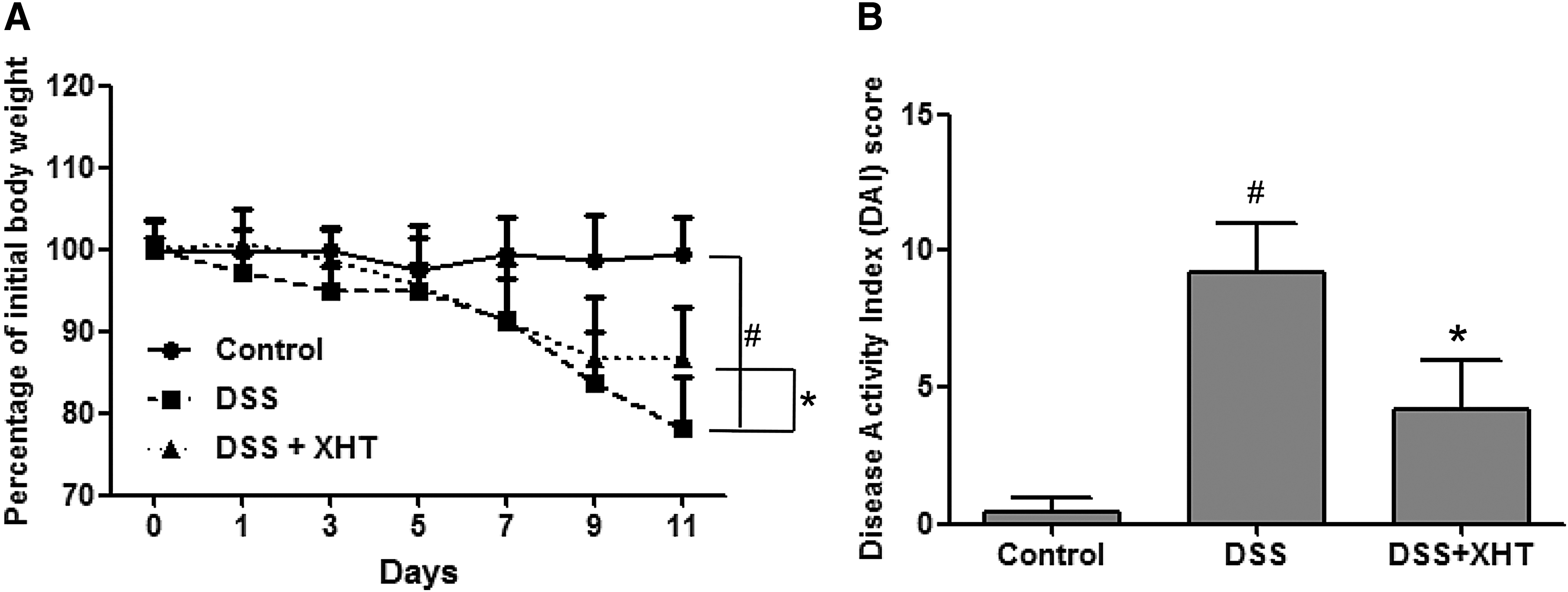
Effect of XHT on clinical manifestations in the DSS-induced UC mouse model.

Effect of XHT on colon length in the DSS-induced UC mouse model.
XHT ameliorates colonic inflammation in DSS-induced UC
To further evaluate the therapeutic effect of XHT on DSS-induced UC in mice, we assessed the histology of the colonic mucosa. As shown in Figure 3, DSS administration resulted in colonic mucosal ulceration, crypt distortion, and inflammatory cell infiltration that were significantly ameliorated by XHT therapy. The quantitative histological score for inflammation was lower in the mice treated with XHT (4.4 ± 1.34 vs. 8.6 ± 1.52, P < .05). Furthermore, XHT-treated mice had reduced levels of MPO (neutrophil marker) 29 within the colonic mucosa as compared with untreated mice with DSS-induced UC (Fig. 5). These data demonstrate that XHT treatment specifically ameliorated the colonic inflammation induced by DSS.
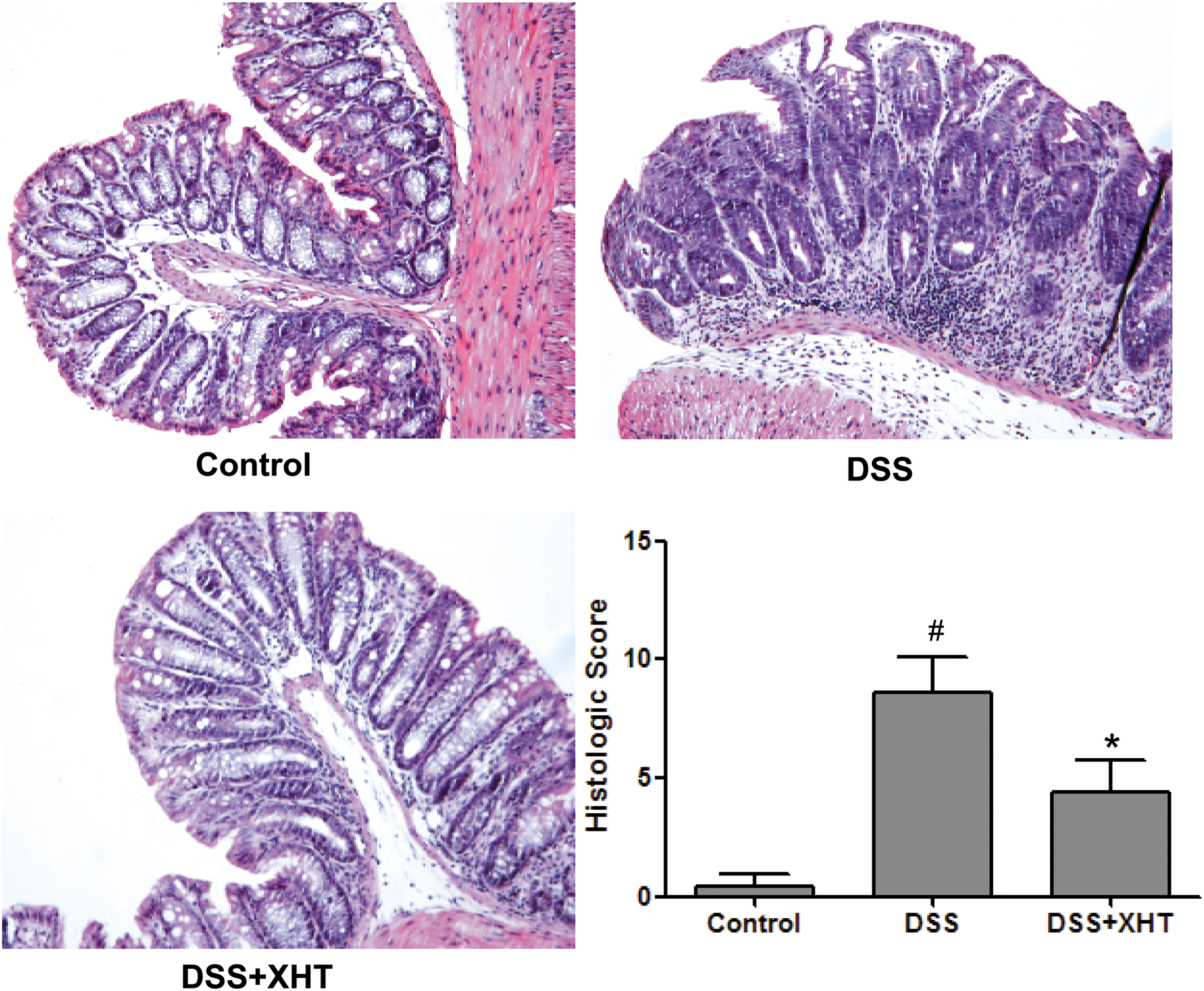
Effect of XHT on colonic histological changes in the DSS-induced UC mouse model. Representative microscopic images of colonic tissue sections (magnification, × 100) and histopathological score for each experimental group. Data shown are the means ± SD (error bars) from five individual mice per group. # P < .05, versus controls; *P < .05, versus mice stimulated with DSS but not treated with XHT. Color images are available online.
XHT reduces the levels of circulating mediators and markers of inflammation in DSS-induced UC
Circulating levels of TNF-α (a critical inflammatory cytokine 30 ) and SAA (an inflammatory marker) are elevated in patients with UC and mice with DSS-induced UC. XHT treatment obviously reduced the levels of serum TNF-α and SAA (Fig. 4; P < .05, versus DSS group), which were increased in the mice with DSS-induced UC (Fig. 4; P < .05, versus control group). Taken together, these data provide further evidence that XHT may ameliorate the inflammation of DSS-induced UC in mice.

Effect of XHT on the serum levels of TNF-α and SAA in the DSS-induced UC mouse model.
XHT regulates the expression TJ proteins and phosphorylation of Elk-1 in DSS-induced UC
To explore the underlying mechanism of the observed therapeutic effect of XHT on UC, this study evaluated the expression of occludin and ZO-1 in colonic tissue of mice from each experimental group. As shown in Figure 5, the decreases in the expression levels of occludin and ZO-1 mediated by DSS were reversed with XHT administration. Also, XHT reversed the DSS-induced increase in phosphorylation of Elk-1.

Effect of XHT on expression levels of MPO, p-Elk1, Elk1, ZO-1 and Occludin in colonic tissues of the DSS-induced UC mouse model.
Discussion and Conclusion
Recently, there have been significant advances in our understanding of the pathogenesis of IBD and, in turn, the development of novel therapeutics for these diseases. 31 However, many patients with IBD do not completely respond to therapy and develop adverse effects from these medications. Therefore, there is a need for alternative interventions. The anti-inflammatory effects of many natural products have drawn increasing attention due to the relatively low toxicity. 25 XHT, a well-known traditional Chinese formula, is used in clinical settings for the treatment of several inflammatory disorders, including arthritis, hepatitis, and cholecystitis. 26,27 However, whether XHT has a therapeutic effect in IBD has yet to be investigated.
We used the DSS mouse model of intestinal inflammation in this study. Thus, the model is widely accepted and is used extensively in research into potential therapeutic agents for UC and other IBDs. The DSS-induced UC mouse model is characterized by colonic inflammation resulting in weight loss, bloody diarrhea, shortening of the colon, intestinal mucosal ulceration, and an inflammatory systemic response. 28 In this study, we found that XHT ameliorated these changes. XHT was effective in preventing DSS-induced colonic inflammation as demonstrated by significant differences in weight loss, disease activity (e.g., DAI), colonic length, and mucosal histological scores. Moreover, the colonic levels of MPO and circulating levels of TNF-α and the inflammatory marker SAA were reduced in the XHT-treated mice, thus demonstrating the anti-inflammatory properties of XHT.
We hypothesized that the effect of XHT, in part, would be through maintenance of the intestinal mucosa tight junctions as the colonic mucosal barrier plays a pivotal role in intestinal homeostasis and its disruption is part of the inflammatory cascade in UC. Therefore, we evaluated the expression of proteins comprising the TJs: occludin and ZO-1. As previously described, the level of intestinal epithelial TJ proteins was reduced in the DSS-induced UC model. However, the level of occludin and ZO-1 was maintained to near normal levels in those mice treated with XHT. To further explore the potential mechanism by which XHT affects intestinal permeability we evaluated its effect on the activation of Elk-1. Elk-1 is a transcription factor that is activated (through phosphorylation) by TNF-α and plays a role in TNF-α-induced increases in intestinal mucosal permeability. We found that in the XHT-treated mice, the phosphorylated form of Elk-1 was reduced. We do not know if this is a direct effect of XHT on Elk-1 or the end result of reduced inflammation and circulating levels of TNF-α.
In conclusion, this study demonstrates that XHT ameliorates the clinical symptoms and colonic histopathological changes, and improves the intestinal barrier function by regulating the tight junction proteins in the DSS-induced UC mouse model, suggesting that XHT has the potential to serve as a therapeutic agent for the treatment of UC.
Footnotes
Author Disclosure Statement
No competing financial interests exist.
Funding Information
This study was supported by 100 Talents Program of Fujian Province (2018).