
Abstract
Eye diseases have a negative impact on the eyesight quality of the world population. The age-related macular degeneration (AMD) draws special attention since it is a chronic disorder characterized by oxidative and inflammatory damage to the retinal epithelial pigment, which triggers progressive vision loss. In the Brazilian Amazon, Astrocaryum aculeatum is an Amazonian fruit (Tucumã) used by riverside communities in traditional medicine to treat a number of ailments
Introduction
Age-related macular degeneration (AMD) is a serious chronic disease that leads to severe visual loss in the elderly in developed and developing countries. 1 The main mechanism for the development of AMD is oxidative stress; and although this disease still has no effective treatment, 1,2 bioactive compounds could improve eyesight condition. 3 Therefore, the search for natural products that can prevent or mitigate eye diseases may be a strategy to develop new supplements or therapeutic drugs.
Tucumã is a widely distributed fruit in the Amazon biodiversity and is traditionally consumed by the riverside population in culinary and as medicine. 4 However, little is known about the benefits of Tucumã for human health. Considering that its seeds are big and stuck to the fruit pulp, this study aimed to produce a seed extract from the pulp and seed.
Astrocaryum aculeatum, popularly known in Brazil as Tucumã, native of the palm tree that belongs to the Arecaceae family, is an attractive fruit with a yellow-orange coloration (Fig. 1). It is distributed in the northern regions of South America and north of Brazil. This fruit is popularly consumed in natura, often used in the preparation of ice cream, sweets, and alcoholic liqueurs by riverside communities in the Brazilian Amazon. 4

Astrocaryum aculeatum popularly known as Tucumã is an Amazonian fruit. Produced by palm tree that belongs to the Arecaceae family, is an attractive fruit yellow-orange coloration.
The Tucumã fruit is rich in vitamin A, phenolic compounds, fibers, and edible oils rich in saturated fatty acids. 2 According to Cabral et al, 5 catechin belongs to the most abundant subgroup of phenolic compounds and is responsible for the antioxidant potential of these fruits.
Tucumã is used by riverside communities in their traditional medicine to treat a number of ailments. However, there are few studies that investigate the pharmacological properties of Tucumã and they are generally limited to its anti-inflammatory, 6 antimicrobial, 7 anticarcinogenic, 8 and antioxidant 5 activities. For this reason, the effect of Tucumã in chronic diseases, such as AMD, using retinal pigment epithelium (RPE) cells should be considered relevant.
AMD is a progressive, degenerative disease of the retinal epithelium that is a common cause of blindness and visual disability in people over the age of 50 in developed countries. 1,2 Worldwide, the number of affected people will be 196 million in 2020, and this figure is expected to increase to 288 million in 2040. 9
This morbidity is associated with cumulative oxidative stress that results in RPE cell damage; with advanced age, this condition may worsen due to the high energy demand of RPE and the inefficiency of antioxidant enzymes. 10 Over the years, these diseases have a negative impact on the quality of life of the elderly, favoring the development of depression, social isolation, falls, and hospitalizations.
Since eye diseases such as AMD are the leading causes of visual disability in the elderly population of developed countries, 1,2 the present study assessed the potential effect of Tucumã on processes associated with these diseases. For this investigation, in vitro protocols were conducted to analyze (1) the effect of chemically characterized ethanolic Tucumã pulp/seed extract on RPE cells, (2) the cytopreventive effect of Tucumã on RPE cells exposed to high concentrations of superoxide anion, (3) the antioxidant effect and preventive DNA damage on RPE cells exposed to high concentrations of superoxide anion.
Materials and Methods
Tucumã fruit samples and extraction
This study has been previously authorized by the Brazilian Ministry of the Environment (SisGen, No. A08E59B). Fruits of A. aculeatum were acquired from the Public Market of Manaus-AM/Brazil. Ethanolic extracts were prepared as described by Souza-Filho et al. 8 with modifications. Peel and pulp were manually removed from fresh fruits, titrated, and placed in amber bottles containing an absolute ethanol solution, at a ratio of 1:5. Extraction was performed over 4 days with shaking once a day; homogenates were subsequently filtered through Whatman No. 1 filter paper. The ethanol was removed using a rotary evaporator (IKA RV 10 Digital V-C Distilling Rotary Evaporator, 30–40°C at 115 rpm). Extracts were then lyophilized and stored at −20°C until further analysis.
Chemical characterization
The phenolic extract profile was determined and quantified using ultra-high-performance liquid chromatography (HPLC) coupled with diode-array detection and mass spectrophotometry (MS) (HPLC-DAD-MS), with an SIL-20 AHT autosampler, an LC-20 ADT pump, a DGU-20A3R degasser, a CBM-20A integrator, and a UV-Vis SPD-M20A DAD detector, a CTO-20A oven, and a C-18 column (4.6 mm × 250 mm) packed with 5-μm diameter particles. Analytical parameters were set using the negative ion mode, with spectra acquired over a mass range from m/z 50 to 1200. The optimum values of the electrospray ionization-MS parameters were as follows: capillary voltage, 4500 V; drying gas temperature, 215°C; drying gas flow, 10.0 L/min; nebulizing gas pressure, 5.0 Bar; collision radio frequency, 150 Vpp; transfer time, 70 ls, and prepulse storage, 5 ls. Automatic MS/MS experiments were performed by adjusting the collision energy values as follows: m/z 100, 20 eV; m/z 500, 30 eV; and m/z 1000, 35 eV, using nitrogen as the collision gas. The MS data were processed using the Data Analysis 4.0 software (Bruker Daltonics, Germany). The mobile phases were A: acetic acid 2% (pH 2.0), and B: methanol: acetic acid: distilled water at 18:1:1, following an elution gradient of 0 min: 20% of B, 0–25 min: 50% of B, 25 min: 20% of B, 30 min: 20% of B (end of run), at a slow rate of 0.8 mL/min. Peaks were identified by comparison with the retention time and mass spectrum from the software library, and obtained by internal standards. Samples were tested in triplicate.
Cell culture conditions
Experiments were performed using the commercially available adult human RPE cell line ARPE-19 (CRL-2302™; ATCC). Cells were cultured in Dulbecco's modified Eagle's medium (Gibco™; Thermo Fisher Scientific; Grand Island, NY, USA) with fetal bovine serum (VITROCELL) to a concentration of 10%, and supplemented with 1% penicillin/streptomycin (VITROCELL; 10 mg/mL) and amphotericin B (VITROCELL; 0.25 μg/mL) in a humidified incubator at 37°C with 5% carbon dioxide (Thermo Scientific™; Forma™ Series 3 Water Jacketed). After treatment, cells were detached using Trypsin/EDTA (0.25%; Gibco; Thermo Fisher Scientific). Experiments were carried out in 96- or 6-well plates according to the protocol.
In vitro protocol
Cells were treated with different concentrations of Tucumã extract, to assess its cytotoxicity (5, 10, 50, 100, and 500 μg/mL) for 24 h, and then exposed to the oxidizing agent Paraquat dichloride hydrate (Sigma-Aldrich) at a concentration of 30 μM for 6 h—the experimental model of AMD was developed according to the protocol previously carried out by our group. 3
The concentration of 50 μg/mL Tucumã extract was used to perform the oxidative and molecular tests.
Cell viability assays
Cell viability was determined by the MTT assay (3-[4,5-dimethylthiazol-2yl]-2,5-diphenyltetrazolium bromide). Cells were seeded in 96-well plates (1 × 10⁵ cells/well), after treatment. They were washed once with phosphate-buffered saline (PBS; 0.01 M, pH 7.4). The MTT reagent (Sigma-Aldrich) dissolved in PBS was added, and plates were incubated for 1 h at 37°C, at a final concentration of 2 mg. Formazan crystals generated by the reduction of MTT salt by metabolically active cells were released from the cells through the addition of dimethyl sulfoxide, and absorbance was measured at 560 nm using a 96-well microplate reader (SpectraMax® i3 Multimode Plate Reader; Molecular Devices). The protocol was carried out according to Fukui et al. 11 with adaptations by our laboratory.
Flow cytometry for apoptosis/necrosis analyses
Apoptosis and necrosis were assayed using the fluorescein isothiocyanate (FITC) Annexin V Apoptosis Detection Kit I (BD Pharmingen™-556463; San Jose, CA, USA) following the manufacturer's instructions. Cells were analyzed using a BD Accuri™ C6 Plus (BD Pharmingen). The protocol was carried out following the manufacturer's instructions. Briefly, cells were seeded in six-well plates at 1 × 10⁶ cells/well. After treatment, they were detached using Trypsin/EDTA (0.25%), washed twice with PBS, and resuspended in 1 × binding buffer at a concentration of 1 × 10⁶ cells/mL. Then, 100 μL of the solution containing 1 × 10⁵ cells were transferred to a new culture tube, and cells were stained with 5 μL of FITC Annexin V and 5 μL of propidium iodide. Cells underwent a smooth vortexing and were incubated in the dark at room temperature (25°C) for 15 min. Following incubation, 400 μL of 1 × binding buffer was added to each tube, and cell fluorescence was measured by flow cytometry in a BD Accuri C6 BD Biosciences flow cytometer.
Morphology
Cell morphology was examined under phase-contrast microscopy (Nikon Eclipse Ts2) 4 × /0.13, microscopy lens model 4 .0/0.13, WD16.5.
Oxidative markers
To assess the antioxidant activity of Tucumã extract, oxidative markers were measured. (1) For thiobarbituric-acid reactive species levels, cells were mixed with malondialdehyde and thiobarbituric acid and then heated in a boiling water bath for 60 min for lipoperoxidation assessment. Results were expressed as nmol. 12 (2) For nitrite levels, total cellular levels of nitrite were quantified using a colorimetric assay. Cells were incubated with Griess reagent for 10 min at room temperature. 13 Nitrite levels were expressed as micromoles of nitrite. (3) For the determination of superoxide anion, the levels of superoxide radical (O2 •) were measured using a colorimetric protocol. 14 The assay was based on the formation of formazan salt through reaction between nitroblue tetrazolium chloride and superoxide radical anion (O2 −). (4) For reactive oxygen species (ROS) measurements, biochemical methodology and flow cytometry were used. In the first, oxidation of dichloro-dihydro-fluorescein diacetate (DCFH-DA) to fluorescent 2′,7′-dichlorofluorescein (DCF) was measured to detect intracellular ROS. Results were expressed as micromoles of DCF. 15 For flow cytometry, which offers the advantage of being able to measure the intracellular fluorescence of cells in the culture, DCFH-DA was used. Cells were washed with PBS transfer, and immediately exposed to the prewarmed loading buffer and fluorescence was examined. The redox state of the sample was monitored by detecting the increase in fluorescence. Accumulation of DCF in cells was measured by an increase in fluorescence at 530 nm when the sample was excited at 485 nm. 16 Results were expressed by mean fluorescence intensity (MFI-DCFDA).
DNA damage quantification
The quantification of DNA damage was performed using the reagent PicoGreen (Quant-iT™ PicoGreen® dsDNA Reagent [Invitrogen®]). 17 After 10 min of incubation with PicoGreen at room temperature, the fluorescent signal was measured. A DNA quantification curve was generated using calf DNA purchased from Sigma-Aldrich. Results were expressed as a percentage of untreated cells.
Statistical analyses
Statistical analyses were performed with the GraphPad Prism software, version 6.0 (GraphPad Prism Software Company, 2014). One-way and two-way analysis of variance were used followed by Tukey's posthoc test. The variables were expressed as mean ± standard error of the mean and results were considered significant at P < .05. All experiments were independently performed in triplicate, and data analysis was performed according to good in vitro practices. Before statistical analyses, most data were normalized and transformed as percentage of control.
Results
Chemical characterization
The Tucumã ethanolic extract showed a chemical composition rich in caffeic acid, gallic acid, catechin, luteolin, quercetin, and rutin. The retention times were as follows: gallic acid; 3.7 min; catechin, 5.6 min; caffeic acid, 8.2 min; rutin, 13.9 min; quercetin, 20.1 min; and luteolin, 22.0 min. These compounds were identified by chromatographic analysis, and the most abundant was quercetin, followed by catechin, both of which are important antioxidant compounds (Table 1).
Phenolic Compounds Composition of Tucumã (Astrocaryum aculeatum) Extract
Results are expressed as mean ± standard derivations of triplicate.
Tucumã effect on RPE cells
In the first protocols, ARPE-19 cells were treated only with Tucumã extract for 24 h. To assess the cytotoxic effect of the extract, different concentrations were used (5, 10, 50, 100, and 500 μg/mL). The Tucumã extract showed no cytotoxicity effect (Fig. 2A). In addition, when the lowest concentrations (5, 10, and 50 μg/mL) were used, the extract did not cause apoptosis and necrosis in ARPE-19 cells (Fig. 2B, C).
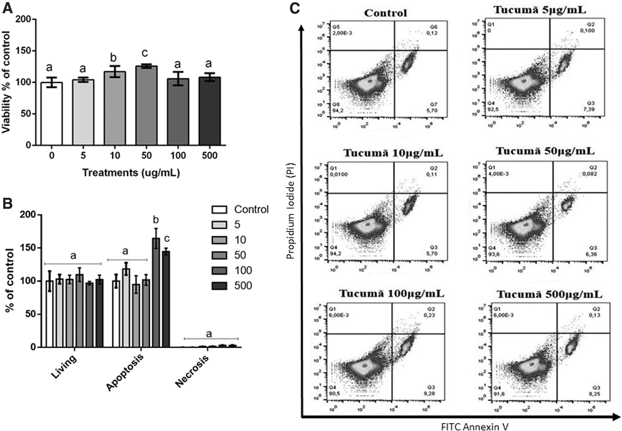
Effect of Tucumã extract on RPE cells. Different concentrations were tested (5, 10, 50, 100, and 500 μg/mL) for 24 h.
Cytopreventive effect of Tucumã extract on the AMD model
The cytopreventive effects of Tucumã on ARPE-19 cells exposed to Paraquat (30 μM) for 6 h were determined through the analysis of viability (Fig. 3A), necrosis, apoptosis (Fig. 3B), and cell morphology (Fig. 3C). As expected, Paraquat exposure triggered higher mortality levels in untreated control cells. Lower mortality effects were observed in ARPE-19 cultures supplemented with Tucumã concentrations. Cells exposed to 50 μg/mL showed significantly lower mortality than the negative control group.
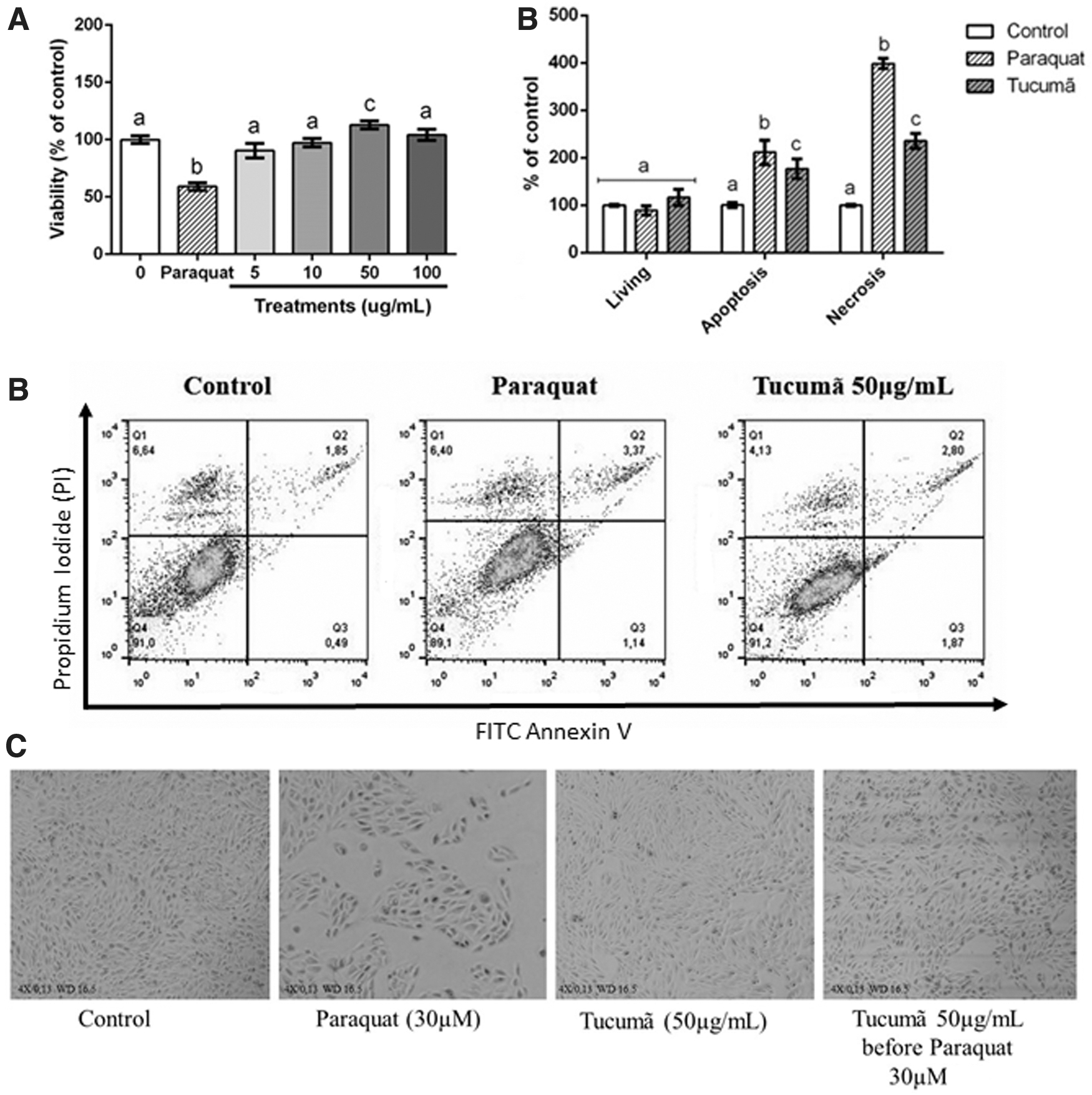
Effect of Tucumã extract in an AMD model. Different concentrations were tested (5, 10, 50, and 100 μg/mL) in AMD protocol (Tucumã extract treatment for 24 h before Paraquat 30 μM 6 h).
Paraquat was highly cytotoxic reducing viability, increasing necrosis and apoptosis, and causing important changes in cell morphology. These results indicated that the Tucumã extract had a cytopreventive action, decreasing cellular mortality.
Antioxidant effect of Tucumã extract on the AMD model
The antioxidant effects of Tucumã were determined by analyzing ROS, MFI-ROS, nitrite, lipoperoxidation, and superoxide anion. As expected, Paraquat increased all oxidative markers. A significant reduction in oxidative markers was observed in cells treated with 50 μg/mL of Tucumã extract (Fig. 4). These results suggest that the extract was able to prevent oxidative damage caused by Paraquat in ARPE-19 cells.
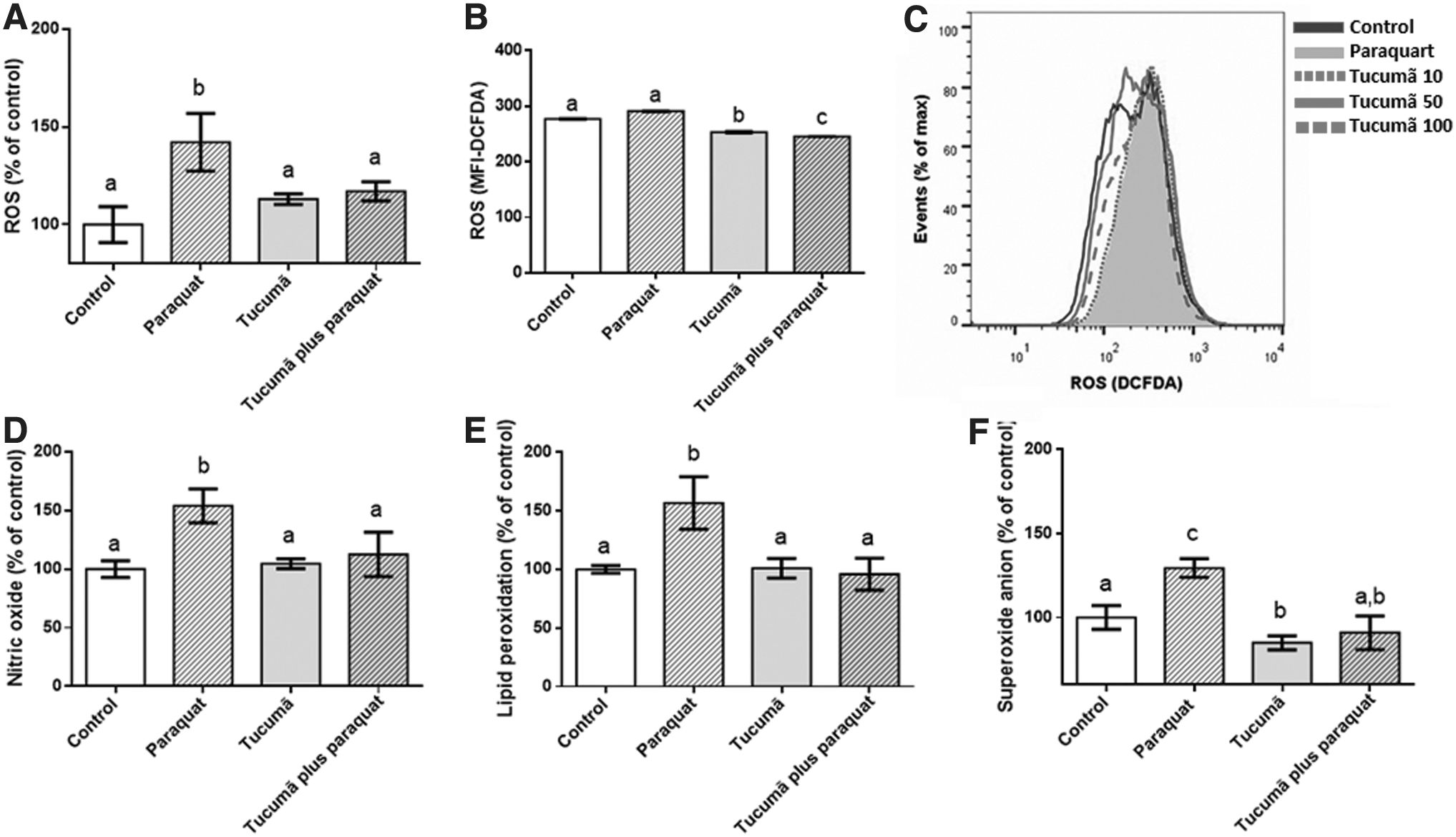
Antioxidant effect of Tucumã extract on apoptosis and necrosis in an AMD model.
Effects of the extract on the DNA
A complementary protocol was performed to confirm the potential biological effects of Tucumã on ARPE-19 cells exposed to high concentrations of ROS. DNA integrity is constantly challenged by endogenous and environmental genotoxic factors. In this investigation, the Tucumã extract significantly (P < .001) prevented DNA damage caused by Paraquat (Fig. 5).
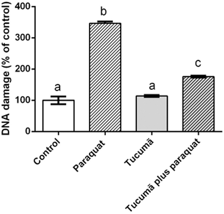
Effect of extract on DNA damage in a model of AMD. Tucumã extract treatment for 24 h before Paraquat 30 μM 6 h. Data are reported as mean + SEM. Different superscript letters (a, b, c) indicate statistically significant differences among treatments at P < .05.
Discussion
Visual dysfunctions are a global health problem often observed in the elderly population. As age progresses, there is a gradual reduction in retinal cell metabolisms and in antioxidant enzymes, 9 leading to the accumulation of oxidant and inflammatory substances that trigger cell damage and lead to visual dysfunctions. 10 Molecular damage is key to the development of AMD, a disease characterized by the progressive visual loss in the elderly population. 9 Several studies have proven that functional foods with a chemical matrix rich in phenolic compounds and carotenes can contribute to the prevention of cell damage in AMD models. 1,3 In this study, the potential effect of Tucumã extract to prevent molecular damage in retinal pigment epithelium (RPE) cells was presented.
Chemical analysis of the extract identified bioactive compounds that have biological effects on human health and could improve eyesight condition. 18 In this study, the high-resolution mass spectrometry of the bioactive compounds found in the Tucumã extract allowed the identification of six phenolic compounds: caffeic acid, gallic acid, catechin, luteolin, quercetin, and rutin. All these compounds have been previously described in these fruits. 5 –8
Furthermore, a study by Cabral et al. 5 shows that the ethanolic extract of Tucumã also presents compounds such as B-carotene, Kaempferol, and ellagic acid, which are respectively carotenoids, flavonoids, and polyphenols, in addition to the compounds described in our study. Also, Baldissera et al. 19 shows the presence of fatty acids in the chemical composition of Tucumã oil. In addition to the compounds found in our study, the other compounds described enrich the chemical matrix of the fruit, further strengthening its antioxidant activity.
The potential antioxidant properties of catechins have attracted a great deal of interest. Various catechins exert their antioxidant roles in retinal cells, and Karthikeyan et al. 18 suggest that catechin is involved in the transport of nutrients, ions, and metabolites, acting as a gatekeeper for the maintenance of retinal integrity. These compounds prevent various chronic diseases, metabolic disorders, such as cardiovascular diseases and cancer. 20,21
Regarding their chemical composition, phenolic compounds are the main set of secondary metabolites present in the Tucumã fruit. Catechin belongs to the most abundant subgroup of phenolic compounds and can be responsible for the antioxidant potential of these fruits. The effect of Tucumã on RPE cells was tested. The extract showed no cytotoxicity effect on cells treated only with extract for 24 h. The assays showed that the extract improved viability, reduced apoptosis and necrosis. Indeed, these results suggested that the extract is safe for in vitro investigations.
According to Kurutas, 22 the normal healthy cell metabolism can cause damage to nucleic acids, lipids, and proteins, which can affect cell health and viability or induce a variety of cellular responses through the generation of secondary reactive species, leading to cell death by necrosis or apoptosis. Nevertheless, Benhar 23 suggests that bioactive molecules can reduce oxidative damage as well as act directly to reduce cell death by necrosis. As previously shown, the extract used in this study has a chemical matrix rich in molecular antioxidants able to prevent oxidative and molecular damage, such as necrosis and apoptosis in healthy cells and in a model of AMD.
In the AMD protocol, when the RPE cells were exposed to high concentrations of pro-oxidant molecule, the Tucumã extract presented a significant cytopreventive effect, confirming the potential of this fruit to prevent diseases triggered by oxidation mechanisms, thus improving human health. With the AMD model, the in vitro protocols showed that the Tucumã extract improved viability and reduced necrosis and apoptosis in cells exposed to Paraquat. Also, the extract was efficient in reducing all the oxidizing markers tested, as well as protecting from DNA damage, showing that Tucumã has an important antioxidant effect and prevents damage to DNA.
In this investigation, the antioxidant potential may be related to the large amount of catechins found in the chemical characterization. Kim et al. carried out a review 24 suggesting that several foods have preventive effects and antioxidant potential, thus preventing AMD. Such fruits and vegetables containing antioxidant components, such as vitamin C, α-carotene, and β-carotene, may have a protective effect against AMD. Similar to these, Tucumã has bioactive molecules that could contribute to reduce the oxidative effects on RPE cells. Thus, we can infer that food has an influence on the eyesight quality in the elderly.
The significant reduction in lipoperoxidation drew our attention (P < .001) because lipoperoxidation is a mechanism associated with a higher risk for AMD development, playing a critical role in drusenogenesis. 25 The RPE accumulates cholesterol either from phagocytosis of the photoreceptor outer segments, or from the ingestion of lipoproteins. 26 It is important to emphasize that some lipid-related pathological characteristics are shared with other age-related diseases such as atherosclerosis and Alzheimer's disease. 27,28 However, the Tucumã extract could positively influence homeostasis and improve patients' visual health.
Another major observation stemming from this study was the effects on DNA damage, a condition in which damaged cells lose developmental capacity. Several studies suggest that repeated cycles of ROS overproduction result in increased DNA damage, which in turn leads to impaired cellular function. 23 Therefore, the maintenance of DNA integrity can be crucial to the proper functioning of EPR cells and play an important role in the pathogenesis of ROS-related diseases. Unrepaired or misrepaired damage to DNA may contribute to aging, so DNA damage response can also be important in age-related diseases. 27 Hyttinen et al. 28 showed that repairing DNA damage and reducing lipid oxidation could be the key to prevention and/or treatment of AMD.
There is evidence that diet is a strong predictor of AMD development. In vivo and in vitro studies developed by our group showed that guarana, highly prevalent in the Amazon diet, contributed to the low incidence of visual diseases in the riverside population in Maués, Amazon, Brazil. 3 Ribeiro et al. 29 have shown that people living in the Amazon inland have a lower profile of diseases such as obesity, hypertension, and type II diabetes. These findings have raised questions about the relation of their lifestyle and diet with their longevity and decreased morbidity. 5,29
Considering that Tucumã has been used in traditional culinary and traditional medicine by the Amazonian population, the results described in this study open to the perspective that Tucumã acts effectively against oxidant molecules and AMD. Complementary in vivo studies could elucidate the mechanisms associated with the antioxidant and cytopreventive activity of Tucumã.
Footnotes
Acknowledgments
The authors would like to express their gratitude to Filomena Marafon, “Conselho Nacional de Desenvolvimento Cientıfico e Tecnológico” (CNPq), and “Coordenação de Aperfeiçoamento de Pessoal de Nível Superior” (CAPES).
Author Disclosure Statement
No competing financial interests exist.
Funding Information
This work was supported with grants (Grant Nos. 402325/2013-3, 490760/2013-9 and 311446/2012-4) from the “Coordenação de Aperfeiçoamento de Pessoal de Nível Superior” (CAPES).