
Abstract
This study evaluates whether some common beverages treated before coffee could protect or increase tooth staining caused by coffee. Initial color of 50 incisor teeth were measured with a spectrophotometer and recorded according to CIELAB color system. Teeth were randomly divided into five groups, water (control), milk, green tea, orange juice, and cola (n = 10) and were kept in selected beverage for 10 min. Immediately afterward, they were immersed in coffee and allowed to stand for 24 h. The treatment was repeated for 5 days. At the end of the fifth day, L*a*b* color measurements of the teeth were repeated. Calcium, phosphorus, potassium, and magnesium changes on representative teeth surfaces were also investigated with X-ray fluorescence spectroscopy. Color differences were calculated with both CIEab and CIE00 formulas. Groups were compared with Kruskal–Wallis test complemented by the Bonferroni correction and Mann–Whitney U test for pairwise comparisons (P = .05). The teeth submitted to coffee challenges after distillated water or beverages showed a perceptible color change. Soaking in cola or orange juice before coffee immersion caused severe tooth discoloration. All the beverages tested here were not able to protect the tooth from coffee staining. People should be informed that some acidic beverages consumed before a coffee can worsen the coffee-based tooth discolorations.
Introduction
Nowadays, esthetic appearance has become a crucial aspect for many people. Especially with the influence of social media, the interest in tooth bleaching is increasing day by day. The natural color of the human tooth crown is typically white, whereas there are slightly yellow and small red shades that reflect the dentin color under the transparent enamel. 1,2 Numerous discoloring factors put the whiteness of teeth and thus the esthetic appearance at risk. 3 In our daily routine, the oral environment is exposed to many factors that cause discoloration. Many foods and drinks have some discoloration agents in their ingredients. 4
To overcome this problem, several esthetic materials, various whitening systems have been introduced to the dental market and different treatment protocols have been developed. Dental professionals commonly concentrate on how to bleach discolored teeth for a pleasing white, beautiful smile. In accordance, in dental academia, color studies generally focus over bleaching methods and materials. Intense dark drinks like coffee or wine, and so on, are accepted as a common risk factors for tooth discolorations. 5 There are many studies in the literature examining the discoloring effect of coffee on materials and dental tissues 3,5 –8 and its treatment with the bleaching procedures. 4,9,10
However, bleaching applications may have numerous side effects, for instance, damaging the dental enamel, gum irritation, tooth sensitivity, and more. 11 –14 Since Hippocrates, medicine's first aim is to avoid damage to the patient, respecting the rule of “primum non nocere” (first, not harm). With this vision, it would be better to understand the details of tooth discolorations and try to prevent this common public problem.
In this study, we would like to assess the staining potential of coffee challenges after some common drinks. Beverages such as cola, milk, orange juice, and green tea are frequently consumed at meals or breakfast like coffee. We would like to examine if these beverages could diminish or increase the discoloring effect of coffee in vitro. To the best of our knowledge, the possible staining effect of coffee after such drinks was not assessed before.
The hypotheses tested were as follows: There will be no difference in the color of the teeth that were submitted to coffee after a beverage. There is no difference between distillated water–coffee, milk–coffee, green tea–coffee, orange juice–coffee, and cola–coffee beverage groups in terms of staining.
Materials and Methods
Ethics approval and tooth collection
The Clinical Researches Ethics Committee of Akdeniz University Faculty of Medicine reviewed and approved this study (11/12/2019—1155). Recently extracted, sound 50 human incisors were used in this study. Immediately after extraction, teeth were cleaned with a brush under distillated water and stored in a 10% chloramine solution. “After an examination with a 2,5 X dental loupe, the teeth with perceptible extrinsic or intrinsic staining's were excluded.”
Test groups and pH measurement
The teeth were randomly divided into five groups and numbered from 1 to 50 to measure the same sample in recurrent measurements (n = 10). Distillated water was used as control. Coffee and green tea were prepared according to manufacturer's recommendations. pH values of each drink was measured with a benchtop pH meter (Hanna Edge, Hanna Instruments, USA). Table 1 shows the preparation methods of coffee and green tea and the beverages used in this study.
Beverages Used in the Study
Treatment regimens and color assessments
The baseline CIELAB color values of each teeth was measured using a digital spectrophotometer (VITA Easyshade, VITA Zahnfabrik, Germany) with D-65 light against a neutral gray background. The shade was determined according to CIELAB color space, which expresses the color as three values; L*, a*, and b*, where L* symbolizes the value of 0 (black) to 100 (white), a* is the amount of red and green, and b* is the measure of yellow and blue. The middle third of each tooth were measured three times. After this baseline measurement teeth in all groups were immersed in the designated beverage for 10 min.
Teeth in each group were kept in the selected beverage for 10 min. Immediately afterward, they were immersed in coffee and stored for 24 h. The treatment was repeated every 24 h for 5 days. At the end of the fifth day, color measurements were repeated. Figure 1 represents experimental design flowchart.

Experimental design flowchart.
Color calculations
ΔL*, Δa*, and Δb* values were calculated as follows: ΔL* = L
2
* − L
1
*, Δa* = a
2
* − a
1
*, Δb* = b
2
* − b
1
* and ΔEab
and ΔE
00 values for each group were calculated with the following formulas:
In the CIEDE2000 formula parameters of KL , KC , and KH were set as 1. The 50%:50% acceptability thresholds were accepted as 2.7 for ΔEab and 1.8 for ΔE 00, as declared by Paravina et al. 15
X-ray fluorescence spectroscopy
For X-ray fluorescence (XRF) measurements an XRF spectrometer was used (NEX-CG Applied Rigaku Technologies, Inc., Austin, TX, USA). The system is equipped with a 50 W power palladium X-ray tube and four secondary targets of RX9, Cu, Mo, and Al.
We focused on four key elements: calcium (Ca), phosphorus (P), potassium (K), and magnesium (Mg). Ca and P are two major elements forming the hydroxyapatite molecules of human teeth and Mg and K are the two main elemental contents in coffee. Detected elements were calculated as percentage of 100%. Elemental changes between baseline and after beverage–coffee immersions were calculated in the representative teeth samples.
Statistical analysis
The overall color differences between baseline and final color values were the main outcome measure. The level of significance was defined as 0.05. Kolmogorov–Smirnov and Shapiro–Wilk tests showed that ΔEab and ΔE 00 data did not adhere to the normal distribution. Both data were analyzed by the Kruskal–Wallis test complemented with Bonferroni correction and Mann–Whitney U test for pairwise comparisons. All statistical analyses were conducted with SPSS for Windows (SPSS for Windows, Version 22; SPSS, Inc., Chicago, IL, USA). The expected statistical power for the chosen number of samples (n = 10) was 99% (G*Power 3.1.7, University Kiel, Germany).
Results
pH measurements
The pH values of tested beverages varied between 6.61 (distillated water) and 2.44 (cola). Table 2 provides the pH measurement results.
pH Values of Tested Beverages
Color changes
Figure 2 represents the color differences calculated with CIEab and CIE00 formula. In both CIEab and CIE00 color difference calculations, all the teeth group-submitted coffee challenges (10 min beverage immersions and then 24 h coffee immersions, repeated for 5 days) were above 2.7 ΔEab and 1.8 ΔE 00 50%:50% acceptability thresholds. Details of L*a*b color coordinate changes after coffee challenges are given in Figure 3. The representative images of each group are given in Figure 4.
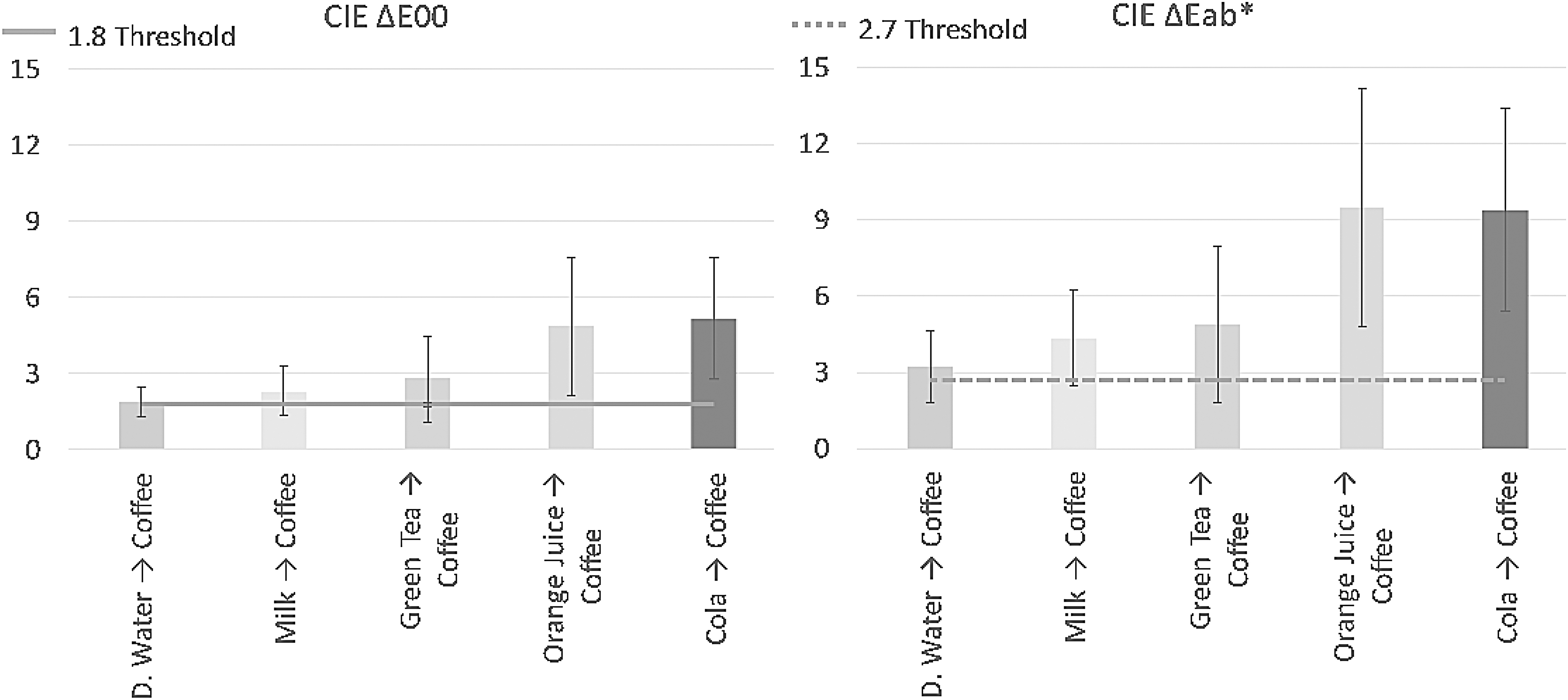
Mean ΔE 00 and mean ΔEab color change values after beverage–coffee immersions. Solid line represents 1.8 threshold for ΔE 00 and dashed line represents 2.7 threshold for ΔEab .

Mean changes in CIE L*, CIE a*, and CIE b* values after beverage–coffee immersions. CIE, The International Commission on Illumination.

The representative images of each group.
Statistical comparison of test groups
Figure 5 presents the pairwise comparisons graphically. In both color difference calculation methods, orange juice–coffee and cola–coffee groups showed statistically significant differences compared with the control group (P < .05) However milk–coffee and green tea–coffee group results were not significantly different with controls in both calculations (P > .05). ΔEab * and ΔE 00 color difference methods were diverse when comparing orange juice–coffee group with milk–coffee and green tea–coffee groups. ΔEab * calculations pointed a difference, whereas ΔE 00 formula was not.
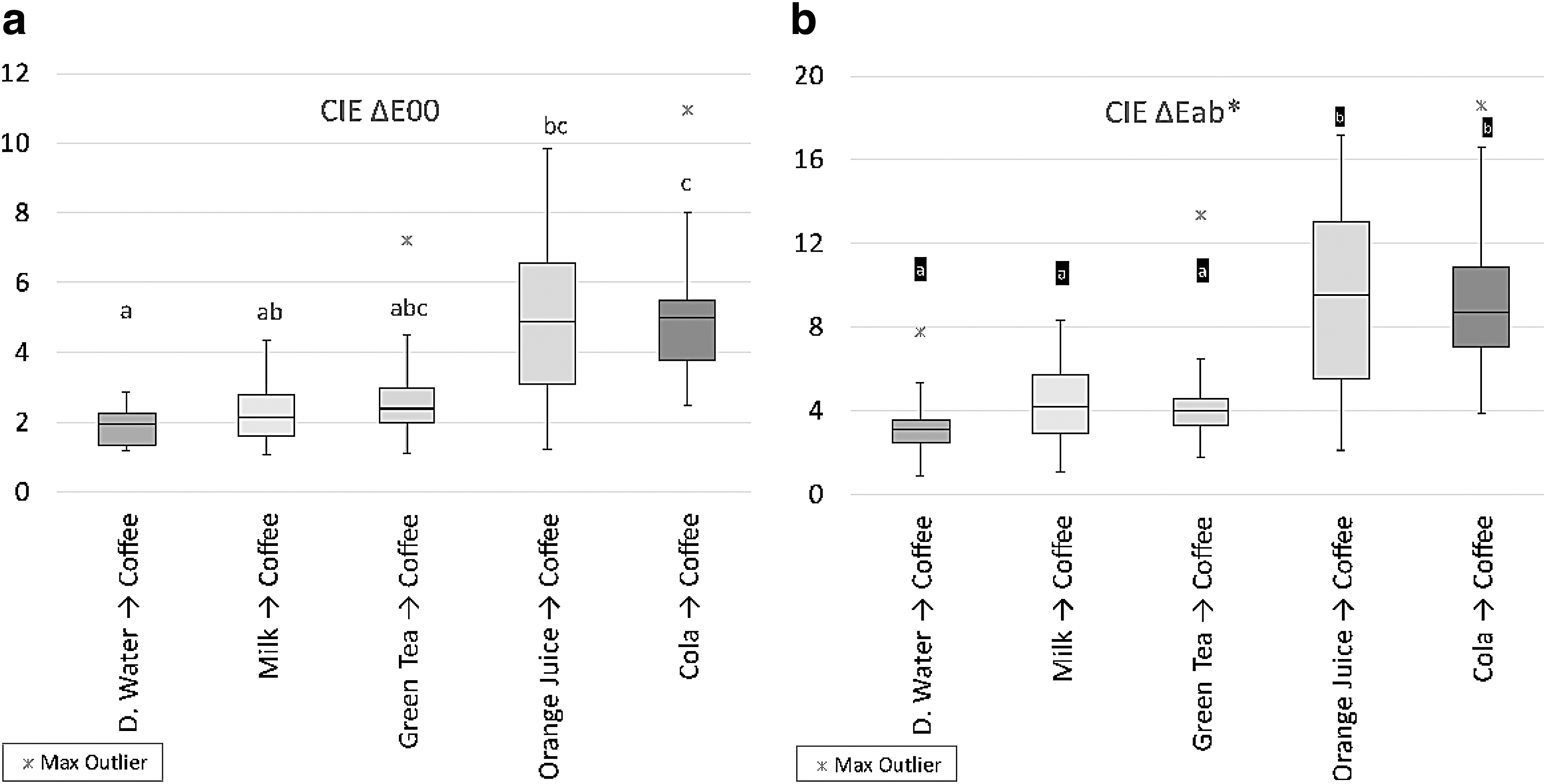
Box plot graph of median ΔE
00 and ΔEab
values.
Discussion
Tooth staining is a public dental problem and it is well known that coffee is one of the major causes of this coloration. The aim of this study was to investigate the effect of some common beverages on coffee-based staining. The null hypotheses were as follows: there will be no difference in the color of the teeth that were submitted to coffee after a beverage, and there is no difference between tested groups in terms of staining. These hypotheses were partly rejected because in both ΔEab * and ΔE 00* calculations, there were statistically significant differences in samples treated with orange juice–coffee and cola–coffee groups when compared with the control group (Fig. 5). In addition, the ΔEab * and ΔE 00* values of all tested groups were above 50%:50% acceptability thresholds (ΔE 00 = 1.8 and ΔEab = 2.7) (Fig. 2).
In our study, the least discoloration was detected in distilled water group; however, it was still above the 50%:50% acceptable limits with two calculation methods (ΔE 00 = 2.31, ΔEab = 1.90). The acceptable thresholds were arranged owing to the previous studies of Paravina et al. 15,16 The acceptable thresholds mean that half the observers consider a color change to be unacceptable at ΔE 00 = 2.31 and ΔEab = 1.90, whereas the other half will determine this color difference acceptable.
To detect color changes, the CIELAB system (ΔEab ) calculation formula is commonly used; however, it was just shown that the CIEDE2000 system is more compatible with visual color alteration perception and acceptance because the color difference equation (ΔE 00) is adjusted by light, hue, and chroma values. 17,18 In this study we made calculations with both methods and got similar results in terms of color differences. The difference was only with the results of orange juice group. ΔEab formula gave a statistically significant difference when orange juice group was compared with milk and green tea groups, whereas ΔE 00 formula did not (Fig. 5). This could be attributed to calculation differences of these formulas. ΔEab values were always higher than ΔE 00 records. As stated previously, the CIEDE2000 color difference formula is better correlated with visual perception than the CIELAB and it is recommended in dental in vitro research and in vivo dental color measurement. 15,19
XRF analysis is a direct and nondestructive elemental examination method. 20,21 In dental studies this technique is commonly used for chemical characterization of the dental hard tissues and material surfaces. 22 Here we used XRF technique to observe the mineral changes on tooth surfaces. Dental tissues contain many kinds of minerals; however, Ca and P are the main elements forming the hydroxyapatite structure. Decalcification of hydroxyapatite is the first step of dental erosion. 21,23
Coffee is a worldwide popular beverage and is consumed by millions of people every day. 24 The role of coffee consumption in preventing some prevalent diseases validates its classification to functional beverage. 25,26 However, coffee contains a lot of chromogens and deposition of these particles on teeth surface promotes tooth staining. 7,26,27 Tannins are one of these important constituents and are responsible for tooth discoloration. This very reactive polyphenol has a significant protein affinity that would cause tooth coloration by the effect of conjugated double bonds that interact through dental enamel by ion exchange. 28,29
Coffee immersion for 1 week was considered to simulate about 1 year of coffee consumption, with the theory that one cup of coffee is drank for 30 min every day. 30 In our study, we kept the extracted teeth in coffee for a shorter period (5 days) to reflect a similar long-term coffee consumption. We observed increased red and yellow saturation after coffee storage (Fig. 3). These results are in agreement with prior studies. 28,31 The coloring property of coffee also comes from the roasting procedure that thermally breaks monosaccharides into a brown-colored caramel substance that reacts with chlorogenic acids to produce brown-black pigments and the low pH value of coffee is linked to the formation of acetic acid during the roasting procedure. 10,32 After brewing, these pigments are dispersed in acidic medium (pH 4.5–6.5) as water-soluble complexes. 33 The acids in coffee influence its staining ability 6 and immersion in coffee may also increase surface roughness leading to further stain update. 27,34 Coffee can decrease Ca/P ratio and may induce decalcification. 21 In XRF analysis we observed slight elemental decrease in Ca and P levels.
In everyday life, tooth surfaces are constantly exposed to acid attacks. Saliva is an important protector that promotes enamel remineralization with its buffering systems. However, these demineralization and remineralization processes could result in dental pigmentation with the contribution of pigmented diet. Pigmented beverages, like coffee and tea, are known to color dental enamel as a result of long-lasting and frequent exposure. 28,31 In our study the most intense discolorations were measured in orange juice–coffee and cola–coffee groups. The acidic nature of these beverages could be the reason of that consequence. 28,35
Milk is a nutrient-rich beverage frequently consumed at breakfast. It is also a functional drink because of its anti-cariogenic 36 and remineralizing properties. There are several studies in the literature about the increase of remineralization by casein phosphospeptides and amorphous calcium phosphate complexes (CPP-ACP) in cow's milk. 37,38 We observed an increase in Ca and K levels in XRF analysis. CPP-ACP in the milk may be the cause. In this study immersion in milk could not block the staining effect of coffee. ΔE 00 and ΔEab * values were >50%:50% acceptable limits (ΔE 00 = 2.31, ΔEab = 4.88) (Fig. 2). The milk tested here is a full fat milk that includes 3.5–4% fat and the pH was 6.51, which is closer to neutral than coffee (pH = 5.11). Lee et al. investigated whether adding milk to the tea decreases the ability of tea to color teeth surfaces. Unlike our results they suggested that the addition of milk may be an effective way of reducing extrinsic tea coloring. 39 They stated that casein could be in charge of avoiding tea-induced staining. The differences in methodology of these two studies could be the reason for this diversity.
Like milk, green tea has not been found to have a protective effect on tooth staining (ΔE 00 = 2.31, ΔEab = 2.80) in this study. Green tea is brew of dried plant Camellia sinensis leaves. It is higher in protective polyphenols and possesses antioxidant, antimutagenic, antibacterial, and anti-inflammatory properties. Green tea is a spring of fluoride and some other trace elements. 40 In addition, the epigallocatechin gallate (EGCG), a polyphenol existing in the green tea, 41 has the ability to inhibit the expression and activity of metalloproteinases (MMPs) like MMP-2 and 9. 23,42 Lopes et al. evaluated the color change of novel composite restorations, submitted to challenge of thermocycling in coffee, after treatment with EGCG and concluded that the former EGCG treatment did not cause a significant color difference at the dentin–resin interface. 23 The pH of green tea is 6.60, which is again closer to neutral.
Observing the elemental changes on enamel surfaces with XRF spectroscopy may have some limitations because the staining of teeth surfaces is a long-term, multifactorial process. However, these elemental fluctuations can present prominent evidences. Acidic beverages have apparent erosive effects on dental structures and restorative materials. 43 –46 Hydroxyapatite crystals form the vast part of tooth enamel and Ca and P are the main components of this structure. 47 As given in Figure 6, highly acidic beverages, orange juice and cola, caused Ca and P reductions on tooth surfaces in this study. Significant color changes were also detected in these two groups (P < .05).
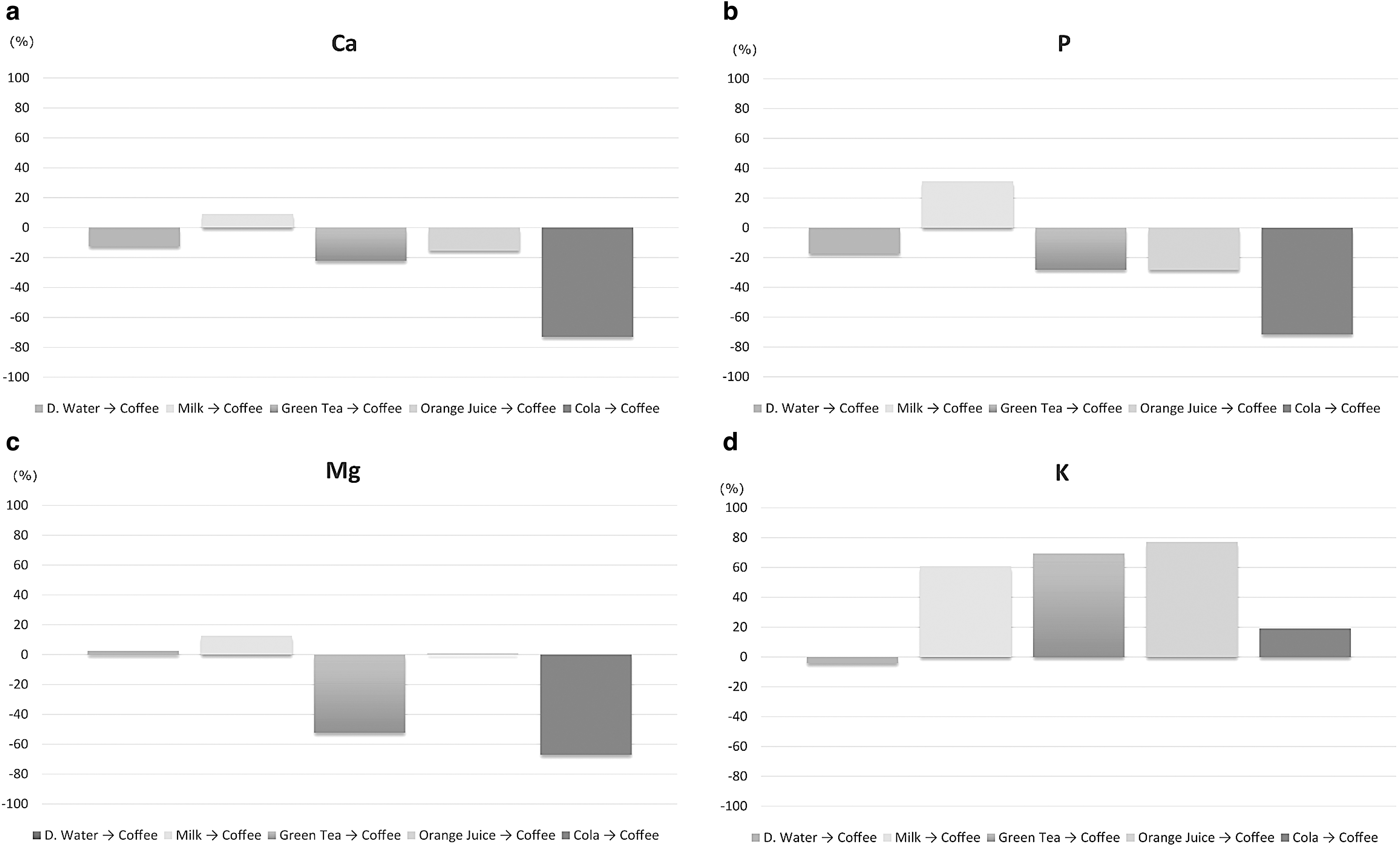
XRF results of elemental changes (%) on tooth surfaces before and after immersions.
In this study the pH of the cola and orange juice was 2.4 and 3.5, respectively. Cola includes food grade phosphoric acid and the main organic acid found in orange juice was citric acid. Erosive beverages can create more rough enamel surfaces and rough surfaces are associated with intense discolorations. 4 During demineralization, remineralization process minerals and pigments can structurally incorporate into enamel structure and weak, rough enamel surfaces are more prone to staining. In a previous study it was shown that the cola with a pH of 2.74 and orange juice with a 3.74 pH had significantly reduced the surface hardness of enamel. 43 In addition it was shown that exposure of enamel slabs to acidic beverage decreases the hardness of enamel. 48 Conversely it was also found that rinsing enamel slabs with milk have a rehardening effect on enamel surfaces. 49 In this study we observed the rise of Ca and P ions after milk–coffee immersions. The color changes in this group was also found to statistically lower than cola and orange juice with CIEab calculations (P < .05)
The color changes observed here could not be directly linked to the staining effect of only coffee. The beverages applied before coffee could also have a cumulative effect on staining. This study is an in vitro study and could not directly mimic the clinical conditions. The immersion periods are extended periods compared with the in vivo situation and factors such as saliva, microbial biofilm, tooth brushing, and so on could not be possible to consider. However, the findings of this study have some important conclusions about the staining effect of coffee. Shifting the focus to early intervention and prevention of tooth staining would be more effective than consuming time and finance to bleach heavily discolored teeth.
Within the limitations of this in vitro study it could be concluded that coffee could cause intense tooth staining especially when the tooth surface is affected after consuming other beverages. Acidic beverages like orange juice and cola could worsen coffee-based discolorations. In addition, highly acidic components in beverages, such as phosphoric acid in cola, can cause decalcifications on the tooth surfaces and have an erosive effect. None of the beverages tested here were able to protect the teeth from coffee staining.
Footnotes
Author Disclosure Statement
No competing financial interests exist.
Funding Information
This work was supported by The Scientific Research Projects Coordination Unit of Akdeniz University. Project Number: TDH-2019-5020.