
Abstract
The main aim of this study was to determine and compare the antimicrobial effect of hibiscus acid and a commercial 0.12% (w/v) chlorhexidine mouthrinse against Streptococcus mutans, Streptococcus sanguinis, Capnocytophaga gingivalis, and Staphylococcus aureus, and to determine the effect on bacterial cell membrane permeability and the toxicity of hibiscus acid in a mouse model. Hibiscus acid was obtained from acetone extract of Hibiscus sabdariffa calyces. Chlorhexidine (0.12% w/v) mouthrinse was purchased from a local pharmacy. The antimicrobial activity of hibiscus acid and mouthrinse were determined using the gel diffusion technique. The minimum inhibitory concentration (MIC) and minimum bactericidal concentration (MBC) of the solutions were determined using the broth dilution method. The effect on bacterial cell membrane permeability of hibiscus acid and mouthrinse was determined by crystal violet assay. The toxicity of hibiscus acid was investigated in a mouse model (registration number: UAEH2019-A1-S-8288). Hibiscus acid and mouthrinse showed antibacterial activity against all oral pathogenic bacteria. However, hibiscus acid showed a lower antibacterial effect compared with chlorhexidine mouthrinse. The MIC and MBC for hibiscus acid were 3 and 5 mg/mL, respectively, and was between 30 and 50 μg/mL for mouthrinse. The crystal violet test results indicate that hibiscus acid and mouthrinse alter the permeability of the bacterial membrane. Finally, hibiscus acid did not show toxicity in mouse studies.
Introduction
Dental biofilm is formed on a hard surface, such as dental enamel, orthodontic restoration appliances, or dental prosthetics. Altered biofilm homeostasis, overgrowth, or an increased number of acid-forming bacteria leads to the development of the most common diseases of the oral cavity: dental caries and periodontal disease. 1
Chlorhexidine gluconate is a topical antiseptic with wide efficacy against bacteria, yeasts, and viruses, which is increasingly recommended in infection prevention and treatment protocols. 2 This antiseptic is often used as a gel formulation at concentrations between 0.12% and 5% (w/v) with bactericidal and bacteriostatic properties, and is considered the “gold standard” of oral antiseptics. 3 Likewise, it reduces bacteria in oral epithelia and biofilms. 4,5
However, prolonged use at concentrations that are slightly elevated compared with the recommended levels, cause adverse effects; the most commonly reported are alterations of taste, effects on oral mucosa such as pain, irritation, slight flaking and ulceration or erosion of the mucosa, and tongue burning. 6 Therefore, the daily use of chlorhexidine is not recommended for the control of oral pathogens and dental biofilm formation.
There are indications that plant extracts and their components have significant antibacterial activity against oral bacteria. 7 Owing to the increase in infections caused by bacteria, research has focused on medicinal plants, as they are a rich source of antimicrobial agents. 8 A plant that has hardly been considered for application in the dental area, and for which there are reports of its antimicrobial potential, is Hibiscus sabdariffa, and little attention has been paid to organic acids such as hibiscus acid. Recently, we reported that one of the main compounds responsible for the antimicrobial effect of hibiscus calyces is hibiscus acid. 9
Hibiscus acid may have an antimicrobial effect against bacteria in the oral cavity. There have been no studies that address this bactericidal potential using hibiscus acid, and this compound may be an alternative to the use of chlorhexidine, which has adverse effects. Furthermore, the toxicity of hibiscus acid is not currently known. For this reason, the objective of this study is to determine and compare the antimicrobial effect of hibiscus acid and its effect on the permeability of the bacterial membrane on pathogenic oral bacteria as well as to determine the toxicity of hibiscus acid in a mouse model.
Methods, Results, and Discussion
Calyces of Hibiscus sabdariffa grown in Mexico in the state of Guerrero were used. We use the variety of H. sabdariffa named “Tecoanapa” with registration number JAM-004-260210 in the National Catalog of Varieties of Vegetables of Mexico (SNICS, 2021). 10
Hibiscus acid was obtained from an acetonic extract exactly as described by Portillo-Torres et al. 9 With this procedure, we obtained 13 g of hibiscus acid for every 1 kg of dried H. sabdariffa calyces.
The identification of the hibiscus acid and verification of its purity was carried out by proton nuclear magnetic resonance (1H NMR) spectroscopy according to the methodology that we previously reported. 9 The NMR spectroscopy analysis was performed using deuterated acetone (acetone-d6; Sigma-Aldrich, Toluca, Mexico) to solubilize the crystals, in a 400 MHz NMR spectrometer (Jeol, Tokyo, Japan). The acquired spectra were analyzed using the MestReNova 2009 Software (version 6.0.2-5475; Mestrelab Research S.L., Santiago de Compostela, Spain). The 1H NMR spectra of the hibiscus acid crystals are shown in Figure 1. Only signals that correspond to the hibiscus acid atoms and its structure, and the signal of the solvent used for the NMR analysis are observed.

NMR spectrum at 400 MHz in acetone-d 6 of purified crystals obtained from Hibiscus sabdariffa calyces. NMR, nuclear magnetic resonance.
Most of the proton signals (
1
H) appeared between δ
H 0 and 12. The signal observed at δ
H 2.05 corresponds to the acetone-d
6 used to dissolve the presumptive crystals of hibiscus acid. The other signals were δ
H: 5.34 (
1
H, singlet [s], C
To determine the toxicity of hibiscus acid, the LD50 in mice was determined according to the methodology described by Lorke 11 ; in brief, 8-week-old male CD1 mice were used, which were obtained from the Bioterium of the Autonomous University of the Hidalgo State (UAEH) and kept under standard light/dark conditions with water and food provided ad libitum, and three animals used per dose group. It is important to note that the work with mice was evaluated and approved by the Institutional Review Board (IRB) named as UAEH Ethics Committee for the Care and Use of Laboratory Animals with an IRB number: UAEH2019-A1-S-8288. The oral administration of 10, 100, and 1000 mg/kg of hibiscus acid was carried out and the mortality of the animals was observed during the following 3 days. No animal mortality was observed during the 3 days of observation.
As there was no mortality, the second phase of the study was continued, where doses of 1600, 2900, and 5000 mg/kg of weight were administered to the animals, following the scheme described earlier. There was no mortality in these groups of animals, so it can be established that the LD50 of hibiscus acid is >5000 mg/kg of weight and it can be considered as a minimally toxic compound. It is important to note that the experimental protocol involving mice was analyzed and approved by the UAEH ethics committee for the care and use of laboratory animals.
For determination of the minimum inhibitory concentration (MIC) and minimum bactericidal concentration (MBC), we used solutions in sterile distilled water of hibiscus acid (100 mg/mL), amoxicillin (50 μg/mL), and a commercial 0.12% (w/v) chlorhexidine mouthrinse (Bexident® For Gum Care, ISDIN, México). Tubes of tryptic soy broth (TSB, Bioxon, Becton Dickinson, Mexico) at 35 ± 2 °C for 24 h were prepared with different concentrations of hibiscus acid (1–50 mg/mL), commercial 0.12% (w/v) chlorhexidine mouth-rinse (1–100 μg/mL) and amoxicillin (0.01–1 μg/mL) by dilution from an antimicrobial stock solution. Sterile distilled water was used as a negative control.
The tubes were inoculated with a suspension of 1 × 10⁵ CFU/mL of fresh cultures (TSB, 24 h/35°C) of Capnocytophaga gingivalis (American Type Culture Collection [ATCC] 33624), Staphylococcus aureus (ATCC 25923), Streptococcus mutans (ATCC 25175) or Streptococcus sanguinis (ATCC 10556) strains. All inoculated tubes were incubated at 35 ± 2°C for 24 h. The MIC was the last dilution of the compound that inhibited bacterial growth without observing turbidity in the tubes.
To determine the MBC, TSB tubes containing the lowest concentration of hibiscus acid, chlorhexidine mouthrinse, or amoxicillin with no turbidity were inoculated into trypticase soy agar (TSA) using the pour plate technique and incubated at 35 ± 2°C for 24–48 h. The MBC was defined as the lowest concentration of hibiscus acid, chlorhexidine mouthrinse, or amoxicillin that showed no colony growth in TSA.
The MIC of hibiscus acid for the four oral strains was 3 mg/mL and the MBC was 5 mg/mL, whereas for 0.12% (w/v) chlorhexidine mouthrinse, the MIC for S. mutans, S. aureus, and S. sanguinis was 30 μg/mL and for C. gingivalis was 40 μg/mL, compared with the MBC, which was 30 μg/mL (S. mutans), 40 μg/mL (C. gingivalis), and 50 μg/mL (S. aureus and S. sanguinis; Table 1). In contrast, the MIC and MBC values of the positive control for the four bacterial strains were <0.1 μg/mL (Table 1).
Minimum Inhibitory Concentration, Minimum Bactericidal Concentration, and MBC/MIC Ratio of Hibiscus Acid, 0.12% (w/v) Chlorhexidine Mouth Rinse and Amoxicillin Against Four Oral Pathogenic Bacterial Strains
Average for three replicates are illustrated.
MIC and MBC units: mg/mL for hibiscus acid; μg/mL for 0.12% (w/v) chlorhexidine mouth rinse and amoxicillin.
MBC, minimum bactericidal concentration; MIC, minimum inhibitory concentration.
The negative control did not show any inhibitory effect. A study to analyze the susceptibility of 80 oral streptococcal strains revealed that the MIC and MBC values of chlorhexidine were in the range of 0.12–15.63 and 1.95–62.50 μg/mL, respectively. 12 The highest MIC value for chlorhexidine for Streptococcus mitis was 15.63 μg/mL, whereas the lowest value for S. mutans was 0.12μg/mL. Regarding MBC, the highest chlorhexidine value for Streptococcus gordonii and S. mitis was 62.50 μg/mL. 12
Although the antimicrobial effect exhibited by hibiscus acid was less than that exhibited by chlorhexidine and much less than that of the antibiotic control, the antimicrobial effect of hibiscus acid is generally within the range of effect exhibited by plant extracts or isolated compounds of plants, as reported by different researchers. 13
Although the objective of a study with plant antimicrobials is to identify compounds with a high antimicrobial effect for use in medicine or food, the reality is that compounds with a high antimicrobial effect are not always obtained. However, compounds with a medium or low antimicrobial activity obtained from plants are of interest as they have the potential to be used in products that do not require a high antimicrobial effect, such as mouthrinses.
The antimicrobial effect of hibiscus acid and chlorhexidine was determined to be bactericidal or bacteriostatic. A compound is considered bactericidal when the MBC/MIC ratio is ≤4 and bacteriostatic when it is >4. 14 Hibiscus acid and chlorhexidine exhibited bactericidal activity against the four oral pathogenic strains (Table 1).
Finally, a test of the alteration of the permeability of the bacterial membrane was performed according to that described by Devi, 15 with some modifications. TSB tubes (5 mL) were inoculated with 100 μL of S. mutans, S. aureus, S. sanguinis, and C. gingivalis and incubated at 35 ± 2°C for 6 h; these bacterial suspensions were subsequently centrifuged at 4500 g for 20 min. The washed bacterial cell suspensions were incubated with different concentrations of hibiscus acid and 0.12% (w/v) chlorhexidine mouthrinse, and the treated bacterial cells were finally harvested by centrifugation at 4500 g for 5 min before being suspended in phosphate-buffered saline containing crystal violet (CV, 10 μg/mL).
Subsequently, the cell suspension was incubated for 10 min at 35 ± 2°C and centrifuged again at 4500 g for 20 min. The optical density (OD) at 590 nm of the supernatant was measured using a UV-VIS Spectrophotometer (Thermo Scientific, Nanodrop, Verona, WI, USA). The OD value of the CV solution was considered 100% excluded. The OD of the supernatant from the normal cell without treatment was used as a blank. The CV absorption percentage for all samples was calculated using the following formula:

The positive control ethylenediaminetetraacetic acid (EDTA) captured between 45% and 66% of CV for S. mutans, S. sanguinis, S. aureus, and C. gingivalis compared with chlorhexidine (Fig. 2), which also permeates the bacterial membrane because it binds to extra-microbial complexes and alters the osmotic balance of cells. 16 In our study, this positive control had a CV uptake of between 20% and 22% in the four oral strains. Regarding the absorption of CV with hibiscus acid treatments, the MIC and MBC absorbed 17%, 65%, and 74%, respectively, for S. mutans without treatment (Fig. 2).
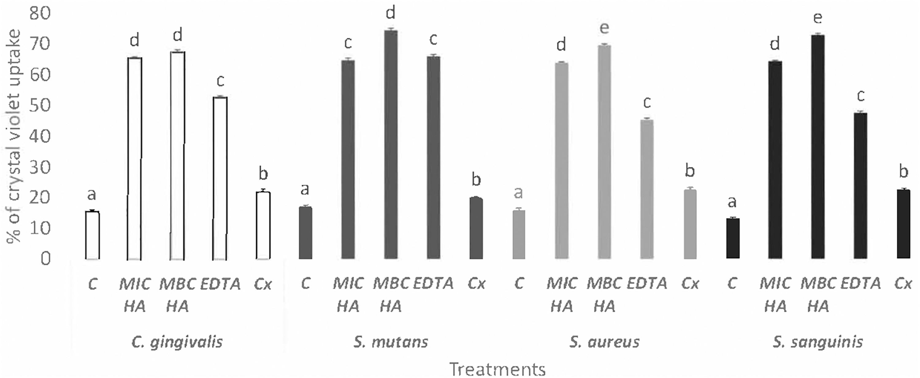
Change in bacterial membrane permeability of Capnocytophaga gingivalis, Streptococcus mutans, Staphylococcus aureus, and Streptococcus sanguinis (assayed by crystal violet uptake) in presence of different concentrations of hibiscus acid, 0.12% (w/v) chlorhexidine, and EDTA. Percentage of crystal violet uptake was plotted against the concentration of the treatment. The mean ± standard deviation for three replicates are illustrated. Values with different letters express significant difference at α = 0.05 by Tukey's test. C, control; Cx, chlorhexidine; EDTA, ethylenediaminetetraacetic acid; HA, hibiscus acid; MBC, minimum bactericidal concentration; MIC, minimum inhibitory concentration.
For S. aureus, there was an increase with respect to the negative control of 64% and 70% with the concentrations of MIC and MBC (Fig. 2). Also, for S. sanguinis, the MIC concentration increased to 64% and the MBC to 73% (Fig. 2). Finally, for the C. gingivalis strain, an increase compared with the control without treatment was observed: 65% for MIC and 67% for MBC (Fig. 2).
This shows that the effect of hibiscus acid on the permeability of the external membrane absorbs the CV dye. The statistically significant absorption of CV was observed in the strains S. mutans, S. aureus, S. sanguinis, and C. gingivalis treated with hibiscus acid compared with the negative control and the two positive controls (EDTA and 0.12% (w/v) chlorhexidine). This means that hibiscus acid permeates the bacterial membrane; likewise, the mechanism of action of chlorhexidine is carried out by damaging the bacterial membranes, so there is a subsequent leakage of cytoplasmic components. 17
Cytoplasmic membranes are the main sites of chlorhexidine action; the outer membrane of Gram-negative bacteria can act as a permeability barrier for chlorhexidine. Therefore, its antibacterial effect is limited. 18
This is the first report of an antimicrobial effect of hibiscus acid on oral pathogens and its comparison with chlorhexidine. Hibiscus acid has been shown to have an antimicrobial potential and a minimal toxicity, which may indicate that it could be used in natural therapeutics and with fewer expected adverse reactions for the control and reduction of dental biofilm; however, more studies are needed.
All experiments were replicated three times. An exploratory data analysis was performed to assess the assumptions of equality of variances and normal distribution of errors of the results obtained from the in vitro antimicrobial activity of hibiscus acid and chlorhexidine, which were analyzed by the Statgraphics Centurion XVI statistical program StatPoint Technologies USA software, 2009 for the one-way analysis of variance. Comparisons of means with the Tukey test were performed for each experimental section, with a significance level of P < .05. 19
Ethics Approval
The study was approved by the Laboratory Animal Research Ethics Committee of the Autonomous University of the Hidalgo State (UAEH2019-A1-S-8288).
Footnotes
Author Disclosure Statement
No competing financial interests exist.
Funding Information
The study was supported by the National Council for Science and Technology (CONACyT) for financial support to the project number A1-S-8288 “Antimicrobials from the Jamaican flower calyces alone and in combination with antibiotics: determination of the mechanisms of action on resistant and nonresistant pathogenic bacteria to antibiotics, the antimicrobial effect in vivo and adverse reactions in animals.”