
Abstract
Trigonella foenum graecum (Fenugreek) is used in traditional phytomedicine for its anti-inflammatory, antiseptic, antidiabetic, and several other therapeutic virtues. The current study was intended to investigate the protecting effects of fenugreek seeds' aqueous extract (FSAE) using experimentally ethanol (EtOH)-induced gastric peptic ulcer in rats, as immense alcohol consumption can lead to gastric ulcer. Sixty adult male Wistar rats were divided into 6 groups of 10 each: control, EtOH (4 g/kg body weight [b.w.]), EtOH + several doses of FSAE (50, 100, and 200 mg/kg b.w.), and EtOH + Omeprazole (OM, 20 mg/kg orally [p.o.]). Animals were p.o. pretreated with FSAE for 21 days and exposed to a single oral administration of EtOH (4 g/kg b.w.) for 2 h. Gastric ulcer in rats was induced with a single dose of EtOH. Ulcer index, malondialdehyde (MDA), hydrogen peroxide (H2O2), and thiol groups (−SH) content in stomach, and antioxidant enzymes superoxide dismutase (SOD), catalase (CAT), and glutathione peroxidase (GPx) were measured. Our recorded results showed that EtOH induced gastric damage, evidenced by the level of oxidative stress markers such as MDA and H2O2 in rats exposed to EtOH. However, significant increases in the activities of antioxidant enzymes were recorded, such as SOD, CAT, and GPx, and a decrease in nonenzymatic antioxidants, such as (−SH). Moreover, histopathological examinations showed the presence of lesions associated with severe tissue damage in the untreated rats. Interestingly, FSAE meaningfully protects against all gastric damages caused by EtOH. We propose that FSAE exhibits protective effects in EtOH-induced peptic ulcer in rats. This protection might be related to its antioxidant and anti-inflammatory properties as well as its opposite effects on some studied intracellular mediators.
Introduction
Although proton pump inhibitors were introduced into ulcer therapy more than 30 years ago, peptic ulcers remain a clinical challenge until today. 1 Naturally, it is known that gastric ulcers are known to benefit as a result of reduced protective factors (adherent mucus, bicarbonate secretion, antioxidant defenses, and blood flow) and by an amplification of aggressive agents, such as gastric acid, pepsin, and oxidative stress. 2 In addition to Helicobacter pylori, excessive alcohol consumption, long-term use of nonsteroidal anti-inflammatory drugs, physical inactivity, and stress are classic etiological factors inducing its appearance. 3
Numerous studies have demonstrated the side effects stimulated by prolonged antisecretory treatment, 4 as well as the recurrence of the ulcer after stopping this treatment. 5
In this context, herbal medicines have been an increasingly accepted choice by the population in the treatment of gastric ulcer. 6
Trigonella foenum graecum, also called fenugreek, has been widely cultivated in Mediterranean countries and in Asia for the manufacture of spices since ancient times. It has also been reported that its raw extract effectively prevents liver damage and acts as a natural hypoglycemic. The seeds of fenugreek are commonly used as a condiment in Tunisian homes. They have been reported to exhibit nutritive properties and to stimulate digestive processes. The seeds have been used to treat a number of gastrointestinal (GI) disorders in Tunisia and a lot of other countries system of medicine. 7
The antioxidant capacity is positively correlated with the richness of plants in phenolic compounds and particularly in total tannins that contribute to the formation of a protein-tannate complex leading to secretion decrease. These particles could prevent ulcer expansion through vasoconstrictor effects or precipitation of proteins into the site of ulceration, producing an impermeable coating on the mucosa that retains intestinal secretions and defends the underlying mucosa against lesions. 8,9 In contrast, flavonoids have been shown to inhibit prostaglandin release by inhibiting intestinal motility and the secretion of electrolytes known for their harmful effect on gastric mucosa. 10
Numerous studies have mentioned the beneficial effects of fenugreek seeds in diabetic and hypercholesterolemic states. 11 However, no experimental data are available concerning its gastric anti-ulcer potential.
Hence, this study aimed to promote this plant in the northwest of Tunisia, traditionally widely used in the treatment of gastrointestinal (GI) tract diseases, and to highlight the protective effect of fenugreek seeds against gastric injury induced by ethanol (EtOH) in rats. Omeprazole (OM), one of the proton pump inhibitors, which is a commonly prescribed drug for the treatment of gastric acid hypersecretion and gastric ulcers, was used as a reference to compare the data.
Materials and Methods
Reagents and chemicals
Bovine catalase, butylated hydroxyl toluene, epinephrine, gallic acid, hydrochloric acid, magnesium chloride, EtOH, methanol, sodium acetate, sodium hydroxide, 2-thio-barbituric acid, and trichloroacetic acid were from Sigma–Aldrich GmbH (Steinheim, Germany). Hydrogen peroxide (H2O2), sodium chloride, and OM were purchased from the Tunisia's Central Pharmacy. All the other chemicals used were of analytical grade.
Plant material
The fresh seeds of fenugreek (T. foenum graecum) were collected from North West Tunisia (Beja, Tunisia) during May 2018 and identified by Dr. Imen Bel Hadj Ali, Associate Professor at the University of Jendouba. Voucher specimens (No. TFG. 098) have been deposited at the Laboratory of Functional Physiology and Bio-resources Valorisation, Higher Institute of Biotechnology of Beja, University of Jendouba, Tunisia. The plant material was air-dried in the dark and powdered in an electric blender (Moulinex Ovatio 2, FR). The powder was then dissolved in bi-distilled water, and the mixture was incubated for 24 h in a shaking bath at room temperature. After centrifugation at 10,000 g for 10 min, the supernatant was freeze-dried to obtain the fenugreek seeds' aqueous extract (FSAE) in powder that was aliquoted and stored at −20°C until use.
Mineral determination
Evaluation of the mineral composition of the samples was determined using a flame atomic absorption spectrophotometer using 1 g of powder, which was placed in a crucible for calcination in a muffle furnace at 550°C for 4 h.
Total phenolic content
The rate of total phenols was determined by the colorimetric method as described by Haseeb et al. with a slight modification of the Folin–Ciocalteu method. 12,33
Total flavonoids determination
Total flavonoid content was evaluated as described by Haseeb et al. with a slight modification by the AlCl3 colorimetric method. 13,33
DPPH
•
radical scavenging activity
Antioxidant capacity of FSAE was determined as described by Selmi et al. with a slight modification using 2,2-diphenyl-1-picrylhydrazyl (DPPH) radical scavenging activity (RSA) as previously described by Grzegorczyk et al.
14
and expressed as a percentage. It was estimated utilizing the following formula:
Superoxide anion scavenging activity
The superoxide anion (O2
•−) scavenging action was performed according to Marklund and Marklund,
15
as described briefly by Selmi et al. The superoxide anion scavenging activity (SASA) is calculated using the following formula:
Anti-ulcerogenic activity of FSAE
Animals and treatment
Adult male Wistar rats (weighing 220–240 g; housed five per cage) were obtained from Pasteur Institute of Tunis and treated in agreement with the local ethic committee of Tunis University (TU2020-002) for use and care of animals, according to the National Institutes of Health (NIH) recommendations. They were provided with food (standard pellet diet; Badr Utique-TN) and water ad libitum and maintained in the animal house at controlled temperature (22 ± 2°C) with 12 h light–dark cycle. Experimental protocols were appropriate with the guidelines of the ethical committee of the Science Faculty of Tunis, Tunisia. The test was performed in compliance with the Commission Directive 2000/32/EC and the Organisation for Economic Co-operation and Development (OECD) Guideline 474.
Animals were separated into 6 groups of 10 animals each. Groups 1 and 2 were served as control and had bi-distilled water (5 mL/kg body weight [b.w.], orally [p.o.]). Groups 3, 4, and 5 were pretreated with several doses of FSAE (50, 100, and 200 mg/kg, b.w., p.o.), respectively. A preliminary study lasting 21 days proved that 50, 100, and 200 mg/kg Solid were the lowest doses that gave a significant protective effect. Finally, group 6 was pretreated with OM (20 mg/kg b.w., p.o.).
Animals were fasted for 24 h before the last administration of FSAE or reference molecule. After 2 h, each animal, except those of group 1, was exposed to a single dose of EtOH (4 g/kg b.w., p.o.). After 60 min, the animals were sacrificed for blood and tissue sampling. Blood was collected in heparinized tubes. After centrifugation at 3000 g for 15 min, the plasma was used for the determination of plasma scavenging activities (PSA) and metabolic and biochemical parameters.
Functional and metabolic parameters determination
Assessment of liver function tests, plasma alanine aminotransferase (ALT), alkaline phosphatase (ALP), aspartate aminotransferase (AST), total and direct bilirubin were measured using commercial diagnostic kits (Biomaghreb, Ariana, Tunisia).
Similarly, for the renal assessment, plasma analysis of urea, creatinine, uric acid, and albumin was also carried out using commercial diagnostic kits (Biomaghreb).
Cardiovascular indexes such as cholesterol, triglycerides, low-density lipoprotein (LDL), and high-density lipoprotein (HDL) were also carried out using commercial diagnostic kits (Biomaghreb).
Evaluation of gastric mucosal damage
The stomach sample from each animal was removed and opened along its greater curvature. Then, the tissues were gently rinsed in a 0.9% NaCl solution. The determination of the ulcer indices was calculated by determining the average of the sum of the lengths of all the gastric lesions (in square millimeters).
Histopathological analysis
Small pieces of stomach and duodenum were cut out and rinsed with ice-cold saline solution immediately after sacrifice. These tissue fragments were then fixed in 10% neutral buffered formaldehyde solution and embedded in paraffin for histopathological examination. Five-micrometer-thick sections were cut, deparaffinized, hydrated, and then subjected to hematoxylin–eosin (HE) staining. These were blinded to all treatments.
Stomach mucosa preparation
Gastric mucosa was excised and homogenized in a phosphate-buffered saline solution (KH2PO4/K2HPO4, 50 mM, pH 7.4) and then centrifuged at 10,000 g for 10 min at 4°C; the supernatants were used for the determination of the biochemical parameters and the assay of oxidative stress markers.
Lipid peroxidation measurement
The level of malondialdehyde (MDA), the end product of lipid peroxidation, is determined as described by Selmi et al. 16 with slight modifications.
Plasma scavenging activities
PSA were measured using the DPPH method according to Brand-Williams et al. 17 PSA are expressed in percentage and calculated according to the following equation:

Antioxidant enzyme activities
Superoxide dismutase (SOD) activity was assayed using modified epinephrine assays, 18 as described by Selmi et al. 16 with a slight modification.
Catalase (CAT) activity was determined by measurement of initial rate of H2O2 disappearance at 240 nm, 19 as described by Selmi et al. 16 with a slight modification.
Glutathione peroxidase (GPx) activity was quantified using the Flohé and Günzler method, 20 as described by Selmi et al. 16
Nonenzymatic antioxidants measurement
Thiol groups (−SH) determination
Total thiol groups (−SH) concentration was determined according to Ellman's method. 21
Glutathione was assessed in gastric tissue according to the Sedlak and Lindsay method. 22
H2O2 determination
H2O2 levels in gastric tissues were measured according to Dingeon et al. 23
Iron measurement
Gastric non-heme irons were assessed by the colorimetric method using ferrozine according to Leardi et al. 24
Calcium determination
Calcium levels in gastric tissues were assessed calorimetrically according to Stern and Lewis. 25
Protein determination
Proteins contents were evaluated according to Hartree, 26 which is a slight change of the Lowry method. 27
Statistical analyses
The data were analyzed by unpaired Student's t-test and are expressed in mean ± standard error of the mean. The data are representative of 10 independent experiments. All statistical tests were two-tailed, and a P-value of 0.05 or less was considered significant.
Results
Phytochemical composition
Our results showed that quantitative assessment of mineral contents and secondary metabolites in FSAE is presented in Table 1. Data first showed that the calcium was the most abundant component, followed by magnesium and manganese. The FSAE revealed high levels of total phenolics (169.94 ± 2.13 mg gallic acid equivalent/g dry matter [DM]), total flavonoids (87.48 ± 1.59 mg quercetin equivalent/g DM), as well as total tannins (74.83 ± 1.83 mg tannic acid equivalent/g DM).
Phytochemical Analysis and Half Maximal Inhibitory Concentration Values of DPPH Radical and Superoxide Anion Scavenging Activities of Fenugreek Seeds Aqueous Extract
Data are expressed as mean ± SEM (n = 3).
Ca, calcium; Cu, copper; DM, dry matter; DPPH, 2,2-diphenyl-1-picrylhydrazyl; Fe, iron; FSAE, fenugreek seeds aqueous extract; f.w., fresh weight; GAE, gallic acid equivalent; IC50, half maximal inhibitory concentration; K, potassium; Mg, magnesium; Mn, manganese; Na, sodium; O2 •−, superoxide anion; P, phosphorus; QtE, quercetin equivalent; SEM, standard error of the mean; TAE, tannic acid equivalent; Zn, zinc.
In vitro antioxidant capacity
Antioxidant capacity determination proved that RSA of FSAE against superoxide anions and DPPH radicals increased significantly and meaningfully dose dependent. The half maximal inhibitory concentration (IC50) values corresponding to the quantity of the fraction necessary to trap 50% of the DPPH and O2 − radicals are 188.49 and 198.4 μg/mL, respectively. However, for the reference molecule, quercetin, the IC50 values are 61.84 and 126.83 μg/mL, respectively, for DPPH and O2 − (Table 1).
Effect of EtoH and FSAE on liver and kidney functions and metabolic parameters
A single-dose administration of EtOH (4 mg/kg) was associated with liver and kidney disorders in rats, while a marked increase in certain hepatic biochemical parameters, in particular ALT, AST, alkaline phosphatase, and c-reactive protein (CRP) was noted in ulcered rats. The same effect was observed at the renal level of plasmatic biochemical parameters, whereas a significant increase in the plasma levels of creatinine and urea (P < .05), as well as a decrease in the uric acid content in the plasma, was observed in EtOH-ulcered rats (Table 2).
Effect of Fenugreek Seeds Aqueous Extracts and Omeprazole on Serum Metabolic and Biochemical Parameters in Ethanol-Induced Peptic Ulcer in Rats
Animals (n = 10). Data are expressed as mean ± SEM. Assays were carried out in triplicate.
P < .05 compared with the control group.
P < .05 compared with the EtOH group.
ALP, alkaline phosphatase; ALT, alanine aminotransferase; AST, aspartate aminotransferase; CRP, c-reactive protein; EtOH, ethanol; HDL, high-density lipoprotein; OM, omeprazole.
Furthermore, EtOH altered cardiovascular indices such as LDL, HDL, cholesterol, and triglycerides (Table 2).
All these parameters were significantly and dose dependently restored near to normal after pretreatment with FSAE. These effects were also observed for the reference molecule against gastric ulcer, OM (Table 2).
Effects on PSA, ulcer index, and percentage of protection
PSA was significantly decreased after EtOH administration compared with the control group (Fig. 1A). By contrast, PSA percentage was meaningfully and dose dependently augmented after the FSAE pretreatment. Analogous effects were also noted for OM. The gastroprotective effect of pretreatment with FSAE on EtOH-induced gastric lesions was determined. The pretreatment with FSAE (at 50, 100, and 200 mg/kg) and OM (at 20 mg/kg) (Table 3) significantly reduced the ulcer index of lesions compared with the ulcer control group. The reduction in the ulcer index was also expressed as a percentage of protection, being of 32.82%, 62.17%, 81.60%, and 80.42%, respectively, in 50, 100, and 200 mg/kg and OM (at 20 mg/kg) groups.

Effect of FSAE and OM on PSA
Effect of Trigonella foenum graecum Aqueous Extract and Omeprazole on Gastric Macroscopic Alterations Induced by Ethanol: Ulcer Index and Percentage of Protection
Animals (n = 10) were pretreated with various doses of FSAE (50, 100, and 200 mg/kg, p.o.) and OM (20 mg/kg b.w., p.o.).
P < .05 compared with the control group.
P < .05 compared with the EtOH group.
Effect of FSAE on EtOH-induced gastric lipoperoxidation and H2O2
To further investigate the effect of FSAE on oxidative stress, the gastric lipoperoxidation and H2O2 content were studied. EtOH intoxication severely improved the gastric MDA and H2O2 levels (Fig. 1B and C). FSAE pretreatment significantly and dose dependently overturned lipoperoxidation and H2O2 intensification induced by EtOH intoxication. The same results were observed with OM-pretreated rats.
Effect of FSAE on EtOH-induced gastric –SH groups decrease
We have observed a significant depletion in the gastric mucosa of EtOH-treated rats. Nevertheless, FSAE (50, 100, and 200 mg/kg b.w., p.o.) or OM (20 mg/kg b.w., p.o.) pretreatment meaningfully protected against this decrease compared with the EtOH group (Fig. 1C).
Effect of FSAE on EtOH-induced antioxidant enzyme activities depletion
The effect of FSAE on EtOH-induced antioxidant enzyme activities in the gastric mucosa was also assessed (Table 4). Ethanol improved antioxidant enzyme activities, such as SOD, CAT, and GPx. Nevertheless, subacute pretreatment with FSAE and OM significantly reduced the increase in antioxidant status to values near to control levels, particularly with the highest dose of FSAE (200 mg/kg b.w.).
Subacute Effect of Trigonella foenum graecum Aqueous Extract and Omeprazole on Ethanol-Induced Changes in Stomach Mucosa Antioxidant Enzyme Activities: Superoxide Dismutase, Catalase, And Glutathione Peroxidase in Rats
Animals were pretreated with various doses of FSAE (50, 100, and 200 mg/kg b.w., p.o.) and OM (20 mg/kg b.w., p.o.), challenged with a single oral administration of EtOH (4 g/kg b.w., p.o.) or NaCl 9‰ for 2 h. The data are expressed as mean ± SEM (n = 10).
P < .05 compared with the control group.
P < .05 compared with the EtOH group.
CAT, catalase; GPx, glutathione peroxidase; GSH; SOD, superoxide dismutase.
Quantitative macroscopic evaluation of FSAE anti-ulcer activities
In EtOH-intoxicated animals, macroscopic examinations of the stomach revealed the appearance of a broad, thick, elongated band, dark red and black, hemorrhagic lesions on the glandular part of the stomach. While quantitative reviews showed that FSAE or standard drug pretreatments significantly and dose dependently reduced ulcer index and improved percent protection against EtOH-induced gastric injury (Fig. 2a). The microscopic study of the control group (Fig. 2bA) showed a typical gastric histoarchitecture with intact epithelium and glands. The ulcer control group, however, showed several changes in the integrity of the gastric mucosa (Fig. 2bB), such as severe desquamation and loss of surface epithelial (mucosa) cells, necrosis, vacuolation, edema and dilated gastric glands, with infiltration of inflammatory cells (neutrophils and eosinophils).
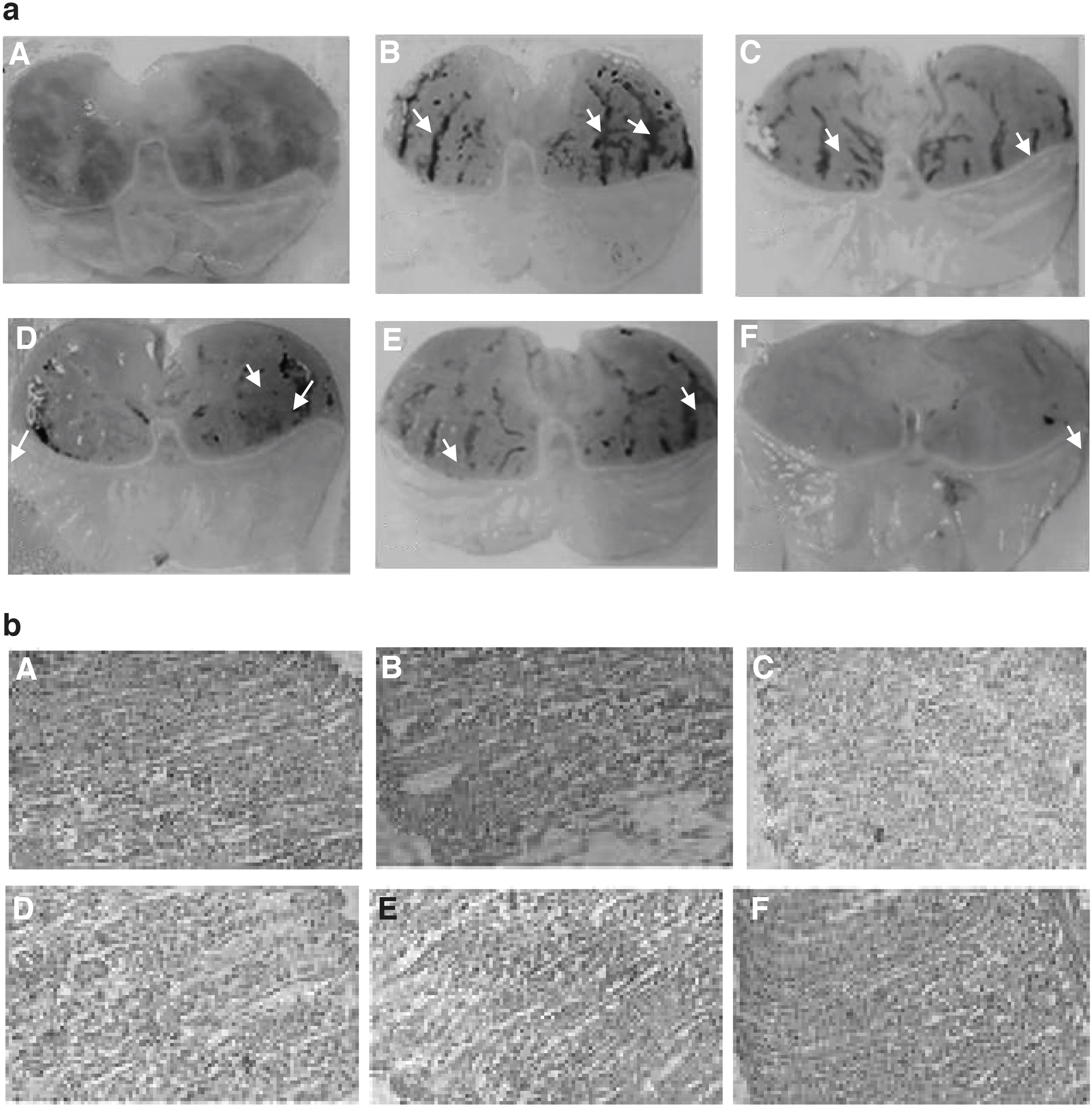
Effect of FSAE on ethanol-induced gastric lesions
Pretreatment with OM decreased gastric lesions compared with control (ulcer). The gastric mucosa exhibited focal loss of superficial gastric epithelium in the control group. The gastric glands were nearly ordinary in appearance in the same group. Pretreatment with many doses of FSAE caused gastric lacerations, characterized by focal areas of disruption in the mucosa, without a mucus layer in this zone. Nevertheless, the rest of the mucosa showed almost normal gastric glands, with mild edema and limited eosinophilic infiltration (Fig. 2bC–F) compared with the ulcer control.
Effects of FSAE and EtOH on free iron and calcium
The effects of EtOH and FSAE on intracellular mediators including free iron, magnesium, and calcium levels in gastric mucosa were studied (Table 2). The EtOH group showed a significant increase in free iron, magnesium, and ionizable calcium levels in gastric tissues when compared with the control group. Nevertheless, the FSAE treatment significantly and dose dependently reduced the EtOH-induced intracellular mediators' deregulating. A similar protective effect had been observed in OM-pretreated rats.
Discussion
T. foenum graecum, also termed fenugreek, is a plant extensively cultivated in the countries of the Mediterranean basin and in Asia for developed spices since very ancient times. 28 Its extract has been reported to effectively prevent liver toxicity 29 and type 2 diabetes effectively. 30,31
This study was aimed to determine anti-ulcer activity and antioxidant effects of FSAE. EtOH-induced gastric ulcer has been extensively used for the investigational evaluation of anti-ulcer activity. It has been reported that disorders in gastric secretion, damage to gastric mucosa, alterations in permeability, gastric mucus reduction, and extensive production of free-radical are correlated with the pathogenic effects of EtOH exposure. 32 The findings of the present study showed that FSAE p.o. administered provided gastric protection and fortification against the ulcerogenic effects of alcohol. An increase in gastric secretion, damage of the gastric mucosal barrier, modifications in permeability, gastric mucus decreasing, and free radical overproduction are proved to be the results of pathogenic effects of EtOH. 33
Three doses of FSAE were used to demonstrate the anti-ulcerogenic action of this plant. These doses of 50, 100, and 200 mg/kg b.w. were used for mucosal and antisecretory studies. Gastric mucus consists of mucin-like glycoproteins, which are the most important gelling constituents of mucus secretion and play a crucial role in the defense mechanisms against gastric ulceration. 34 Increased mucin activity reflects the efficient integrity of the mucosal barrier and serves as a consistent index of mucosal resistance.
The composition of glycoproteins and the amount of mucus were greater than those of animals treated with EtOH, suggesting a possible cytoprotective action of FSAE. This cytoprotective effect was also proven by histological evaluation showing the anticipation of hyperemia and mucosal edema. Furthermore, the antisecretory activity of the seed extract could be an important protector of the gastric mucosa. FSAE also seemed to influence the production of free radicals.
The lipid peroxidation index (MDA) was low in the FSAE-treated animals and was related with improved antioxidant enzyme activities. The increase in antioxidant enzyme activities such as CAT and GPx prevents the accumulation of H2O2, an inhibitor of prostaglandin synthase. This could be associated with the intensification of the mucosal barrier because prostaglandins are found throughout the gastric mucus and help an extensive variety of functions as well as maintenance of mucosal barrier, synthesis of mucus, regeneration of epithelial cells, and inhibition of acid secretion. 35
In this context, it is of interest to note that the administration of antioxidants inhibits EtOH-induced gastric injury in the rat. 36 In a previous work, we have earlier reported that the aqueous extract of the seeds possesses important antioxidant activity in vitro. 37 It is possible that the antioxidant potential of fenugreek seeds is linked to its gastroprotective effect given their richness in flavonoids that they contain. 38 It could be conceivable that the fenugreek seeds exert their anti-ulcer activity through the flavonoids, previously described to protect the mucosa by preventing the development of lesions by several necrotic agents. 39 In this sense, a pectin-like polysaccharide fraction from Bupleurum falcatum is reported to possess an anti-ulcer effect. 40
This study showed that EtOH intoxication induced an elevation of the end products of lipid peroxidation (MDA), a depletion of the levels of thiol groups, an increase in the content of H2O2, as well as an increase in antioxidant enzymatic activities (SOD, CAT, and GPx). Acute alcohol exposure-induced oxidative stress has been commonly documented in the gastric mucosa, 41 liver, 42 kidney, 43 heart, 44 and brain. 45 EtOH exposure induced oxidative imbalance through a number of pathways, including overproduction of reactive oxygen species (ROS). 46 Lipid peroxidation is an indicator of ROS generation in tissues. However, the role of SOD is to convert the reactive superoxide radical to H2O2, which has been diminished in the gastric mucosa, and if not scavenged by CAT, can on its own cause more severe lipid peroxidation by generation of hydroxyl radical. 47
More significantly, this study reveals that FSAE can abolish acute EtOH-induced oxidative stress in the gastric mucosa. These data entirely supported all earlier reported in vivo 48 and in vitro 49 antioxidant and anti-inflammatory effects of flavonoids and condensed tannins. 50 These latter molecules are the primal source of the antioxidant capacity of this plant, by scavenging free radicals, such as hydroxyl radical (OH•), which is the main cause of lipid peroxidation. 51 In addition, it is well known that sulfhydryls, found in fenugreek seeds, are involved in gastric cytoprotection. 52
These findings corroborate with the previous study dealing with the role of total polyphenolic compounds in FSAE in the maintenance of mucosal barrier integrity and the scavenging of free radicals formed due to the action of noxious agents. 53 Significant protective effect of FSAE against the increase in blood sugar caused by EtOH intoxication was also proved, confirming the antidiabetic effect of this plant. 8
Interestingly, our study revealed that there was a link between peptic ulcer, liver and kidney functions or lipid profile. We also noticed a high level of CRP and ALP for the groups that received EtOH. These results are in agreement with those published by Jedidi et al. 54 In this respect, FSAE significantly prevents EtOH-induced gastrotoxicity owing in part to its richness in phenolic compounds. However, the antioxidant properties of polyphenols unquestionably contribute to their anti-inflammatory roles by interrupting the ROS inflammation cycle. These bioactive molecules are known for their anti-inflammatory action by blocking many roots such as cyclo-oxygenase, lipoxygenase nitric oxide synthase, cytokines, NF-κB, and the matrix metalloproteinase. 55
Finally, the obtained results demonstrated an increase of intracellular mediators such as calcium and free iron in gastric mucosa in response to oxidative stress induced by EtOH administration. These data are in line with several previous studies. 56 However, FSAE exerted a beneficial effect by chelating free iron, scavenging H2O2, and regulating calcium homeostasis. Also, the pretreatment with FSAE showed a protective affect against overcharge of cells of the gastric mucosa by free iron and H2O2 induced by EtOH acute administration. In fact, these reactive substances are involved in the generation of hydroxyl radical, 57 which plays a major role in oxidative damage by affecting the molecular structures. In this respect, Jan et al. 58 concluded that living organisms create a complex endogenous and exogenous antioxidant defense system to restrict the production of this damaging radical.
In conclusion, the obtained results indicate that fenugreek could be considered a rich source of bioactive phenolic compounds, playing a potential role in the prevention from ulcer. In addition, due to the widely reported antioxidant activity of flavonoids, fenugreek seeds could be exploited as an important supplement in food manufacturing such as functional foods or other herbal preparations.
Footnotes
Authors' Contributions
S.S. contributed to the study concept and design, data acquisition and interpretation, and drafting of the article. K.R., D.G., and H.S. contributed to critical revision of the article. S.S. and L.M. contributed to the study concept and design, drafting of the article, critical revision, and final approval, and submission of the article. S.J., K.R., and H.S. designed and performed the study, and wrote the article. S.S., H.S., K.H., and L.M. performed the study. All authors approved the final version of the text.
Ethics Approval and Consent to Participate
Experimental protocols were approved with the guidelines of the Ethical Committee of Science Faculty of Tunis, Tunisia. The test was performed in compliance with the Commission Directive 2000/32/EC and the OECD Guideline 474.
Availability of Data and Materials
Data sharing not applicable to this article as no data sets were generated or analyzed during the current study.
Author Disclosure Statement
No competing financial interests exist.
Funding Information
This study was financially supported by the Tunisian Ministry of Higher Education and Scientific Research.