
Abstract
Allergic disorders, including atopic dermatitis (AD), are closely linked to the activation of type 2 helper T (Th2) cells. The aim of this study was to investigate the possibility of using Rosae multiflorae fructus extract (RMFE) for AD treatment in the AD-like mouse model induced by treatment with trimellitic anhydride (TMA). Oral treatment of RMFE reduced the increase in ear thickness and suppressed inflammatory cytokine expression (interleukin [IL]-1β and tumor necrosis factor [TNF]-α) and Th2-associated immune responses (immunoglobulin [Ig] E and IL-4) in mouse ears. Furthermore, messenger RNA (mRNA) expression levels such as IL-4, IL-5, and IL-13, in draining lymph nodes were decreased by RMFE. Furthermore, we found that RMFE increased the level of heme oxygenase-1 (HO-1) through ERK and p38 pathways, reducing IL-2 production and CD4+ T cell proliferation, and inhibited STAT6 phosphorylation. Therefore, this study suggested that RMFE could be an effective treatment of AD induced by Th2-mediated immune responses by suppressing proliferation of CD4+ T cells via increased HO-1.
INTRODUCTION
Atopic dermatitis (AD) is an inflammatory skin disease induced by allergic immune responses.
1
Patients with AD have a poor quality of life because it affects their social functioning and psychological well-being.
2
AD is linked to both an acute allergic reaction and a chronic inflammatory reaction. The acute phase of AD is mediated by type 2 helper T (Th2) cell-dominated immune responses, leading to a chronic inflammatory process of AD.
3,4
Interleukin (IL)-4, IL-5, and IL-13 are Th2 cytokines secrated by Th2 cells
5
and these Th2 cytokines lead to secretion of immunoglobulin (Ig) E by inducing B cells. The IgE receptor (FcɛRI)-IgE complex formed on the surface of mast cells binds the allergens, resulting in the release of histamines, tryptases, and leukotrienes.
6
These inflammatory factors can then increase tumor necrosis factor
Trimellitic anhydride (TMA), known as a hapten, produces immunogenic neoantigens via binding to host carrier protein, which then induce immune responses. 10 Furthermore, TMA can stimulate Th2- and relatively diminish Th1-related immune responses. 11 Thus, TMA-induced AD-like mouse model has been used as an alternative to other AD-like mouse models because of the similarity of symptoms observed between this mouse model and human AD patients. 12
Rosa multiflora
Thunberg, also called baby rose, belongs to the Rosaceae family and it is a sweet briar or an oriental wild rose. Importantly, its fruits are edible and have been traditionally used to treat various diseases, such as joint edema, inflammation, rheumatism, and beriberi in oriental countries. 13,14 In previous studies, we revealed that Rosae multiflorae fructus extract (RMFE) ameliorated symptoms of asthma, 15 allergic rhinitis, 16 and food allergy. 17 However, it is unknown whether RMFE has a beneficial effect on AD. Thus, to confirm the anti-AD effect of RMFE, we examined the effect of RMFE on the TMA-induced AD-like mouse model.
MATERIALS AND METHODS
Sample preparation
The RMFE was extracted twice using distilled water (85°C ± 5°C, 10 ± 2 × volumes) for 8 ± 2 h, and the extract was filtered by using a cartridge filter. Then, the filtrate was evaporated to obtain an over 20 brix extract. Next, the RMFE and dextrin were mixed in a ratio of 1:1 and then spray dried with an inlet air and outlet air temperatures of 180°C and 95°C, respectively. The RMFE (yield: 60% ± 10%) standardized to contain 1.34 mg of miquelianin (content range of ±20%) per gram of RMFE. The RMFE powder was stored at room temperature until use.
Animals
Animals were maintained by following established guidelines under specific pathogen-free conditions. Six-week-old female BALB/c mice used in this study were obtained from OrientBio, Inc. (Gyeonggi-do, Korea). Animals housed at 23°C ± 2°C in an air-conditioned room with a 12 h light/dark cycle and cared following the Korea Food Research Institute guidelines for animal care and use. The experimental protocol used in this study was approved by the Animal Care and Use Committee of Korea Food Research Institute (KFRI-M-14013).
Induction of AD-like symptoms by TMA
We used TMA (Sigma-Aldrich, St. Louis, MO, USA)-induced AD-like mouse model. 18 BALB/c mice (female, 7 weeks old) were divided as the following groups: naive group (TMA-non exposed group, naive, n = 8), sham group (TMA-exposed group, sham, n = 10), 200 mg/kg RMFE group (RMFE 200, n = 10), 400 mg/kg RMFE group (RMFE 400, n = 10), and 10 mg/kg prednisolone (PS) group (positive control, PS10, n = 8). Mice were sensitized by local application of 5% TMA (50 μL) solved in an acetone/isopropyl myristate mixture (4:1. v/v) on the shaved right flank on day 0. To induce inflammation, the surface of both ears was exposed, respectively of 2% TMA (20 μL) on days 5, 8, 11, 14, and 17 (Fig. 1A). RMFE or PS was dissolved in phosphate-buffered saline, and then each sample was orally administrated to mice using oral feeding needle from days 5 to 17 daily. The mice were sacrificed on day 18.
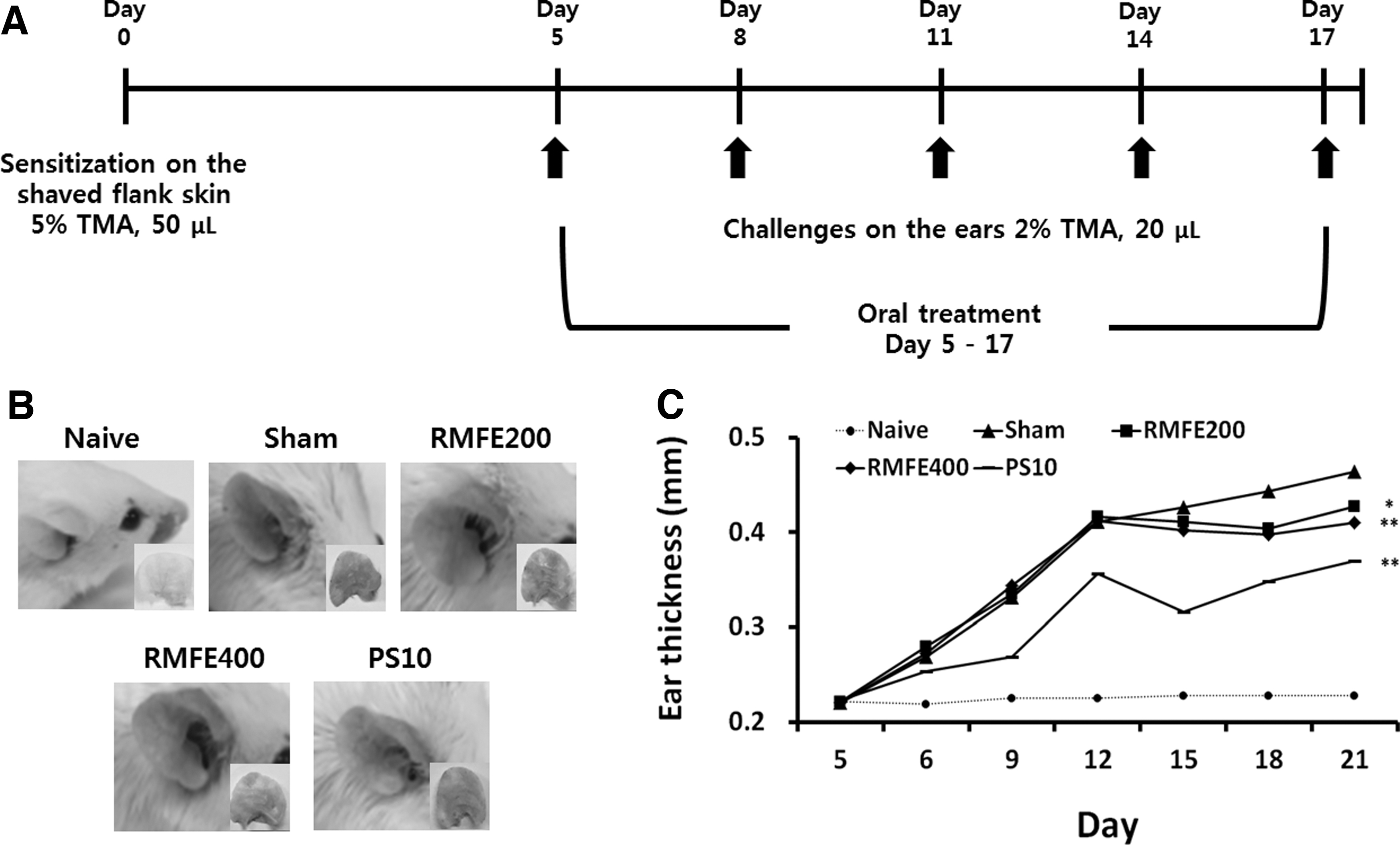
Effect of RMFE on ear inflammation and thickness in the TMA-treated AD-like mouse model.
Culture of draining lymph nodes and splenocytes
Draining lymph nodes (dLNs) isolated from TMA-induced AD-like mouse model were cultured in RPMI 1640 medium (10% fetal bovine serum, 100 U/mL penicillin, and 100 mg/mL streptomycin) with concanavalin A (2 μg/mL, Con A) for 48 h. Splenocytes isolated from BALB/c mice sensitized by OVA as described previously 18 were seeded in culture plate to 5 × 10⁶ cells/mL. The splenocytes were then treated with RMFE and OVA (100 μg/mL) and cultured for 72 h in RPMI 1640. Cells were maintained in a humidified incubator (37°C, 5% CO2, and 95% air).
CD4+ T cell isolation and culture for heme oxygenase-1 expression and proliferation assay
CD4+ T cells of BALB/c mouse splenocytes were isolated from by using the Magnisort™ mouse enrichment kit (Invitrogen, Carlsbad, CA, USA) and were cultured in RPMI 1640. CD4+ T cells (1 × 10⁶ cells/mL) were activated after seeding them in anti-CD3 antibody (1 μg/mL; Biolegend, San Diego, CA, USA) precoated 96-well plate with anti-CD28 antibody (1 μg/mLl; Biolegend) and RMFE. CD4+ T cells were cultured for 24 h to detect the expression of heme oxygenase-1 (HO-1) protein. MTT assay was used to measurement of CD4+ T cell proliferation after 48 h.
Measurement of cytokine and IgE
The levels of IL-2, IL-4, IL-5, IL-1β, TNF-α, total-IgE (BD Biosciences, San Diego, CA, USA), and IL-13 (R&D Systems, Minneapolis, MN, USA) were measured using enzyme-linked immunosorbent assay (ELISA) kits. The methods of all ELISA analyses were carried out according to the manufacturers' instructions.
Messenger RNA isolation and quantitative real-time reverse transcription-polymerase chain reaction analysis
To purify the total RNA in dLNs, we used an RNeasy Mini Kit (Qiagen), and synthesis of complementary DNA (cDNA) was carried out by using a QuantiTect Reverse Transcription Kit (Qiagen) following the method of the manufacturer's instructions. The real-time polymerase chain reaction was performed to dectect gene expression levels of samples by using SYBR Green master mix on a Rotor-Gene Q 2plex System (Qiagen). The fold change in relative gene expression levels was calculated compared to the GAPDH expression. The primers were obtained from Bioneer (Daejeon, Korea), and the sequence of primers is provided in Table 1.
List of Primer Sequence
IL, interleukin.
Western blotting analysis for STAT6, ERK, p38, and HO-1
To analyze the p-STAT6 (Abcam, Cambridge, United Kingdom) and total STAT6 (Santa Cruz, CA, USA), splenocytes were lysed and then the protein concentration was determined as previously described methods. 18
The HO-1 (Cell Signaling, Danvers, MA, USA) protein was detected in splenocytes or CD4+ T cells. To detect ERK and p38 (Cell Signaling) activation, CD4+ T cells were treated with RMFE for 2 h, and with U0126 (ERK inhibitor, 5 μM; Promega, Madison, WI, USA) and SB203580 (p38 inhibitor, 5 μmol/L; Sigma-Aldrich) for 1 h. The expression of protein levels was measured by an automated capillary-based size sorting system (WES; ProteinSimple, Santa Clara, CA, USA). All procedures were carried out following the method of the manufacturer's instructions, and the analysis of data was performd by using inbuilt Compass software (ProteinSimple).
Statistical analysis
All valuses are expressed as the mean ± standard deviation, significance of differences were assessed using one-way analysis of variance. A P value <.05 was considered as the minimum threshold for determining statistical significance.
RESULTS
Effects of RMFE on the symptoms of TMA-induced AD-like mouse model
We investigated whether RMFE could ameliorate the AD symptoms of mice induced by TMA exposure. RMFE and PS (positive control) treatments ameliorated the AD symptoms, such as erythema, edema, and hemorrhage (Fig. 1B), and ear thickness was significantly reduced by RMFE administration compared to that of the sham group (Fig. 1C).
Effects of RMFE on inflammatory parameters in ear tissues
Next, we observed the histological changes and epidermal thickness in mouse ear tissues by hematoxylin and eosin staining. Unlike TMA treatment, administration of RMFE attenuated both the infiltration of inflammatory immune cells and epidermal thickness increased by TMA treatment (Fig. 2A, B).

Effect of RMFE on TMA-induced inflammatory cell infiltration, epidermal thickness, and inflammatory cytokine levels in ear tissue.
We also investigated the IL-1β and TNF-α, AD-related inflammatory cytokines, produced by TMA in ear tissues. RMFE administration suppressed the IL-1β and TNF-α production increased by TMA exposure in ear tissues in a dose-dependent manner (Fig. 2C, D).
Effects of RMFE on TMA-induced allergic immune responses
According to previous studies, part of the etiopathogenesis of AD is an unbalanced immune response, which is predominantly mediated by Th2 immune responses. 3,19 IgE antibody was able to produce a Th2 immune response and increased levels in AD patients. 1 Thus, to confirm the effects of RMFE on Th2 immune response, we first investigated whether administration of RMFE suppressed serum IgE levels, a typical allergic marker in the TMA-induced AD-like mouse model. Serum IgE levels increased by TMA treatment, but decreased with RMFE administration dose-dependently (Fig. 3A). We also investigated the production of the AD-related allergic cytokine, IL-4, in ear tissues. RMFE administration significantly suppressed the TMA-induced IL-4 production (Fig. 3B).
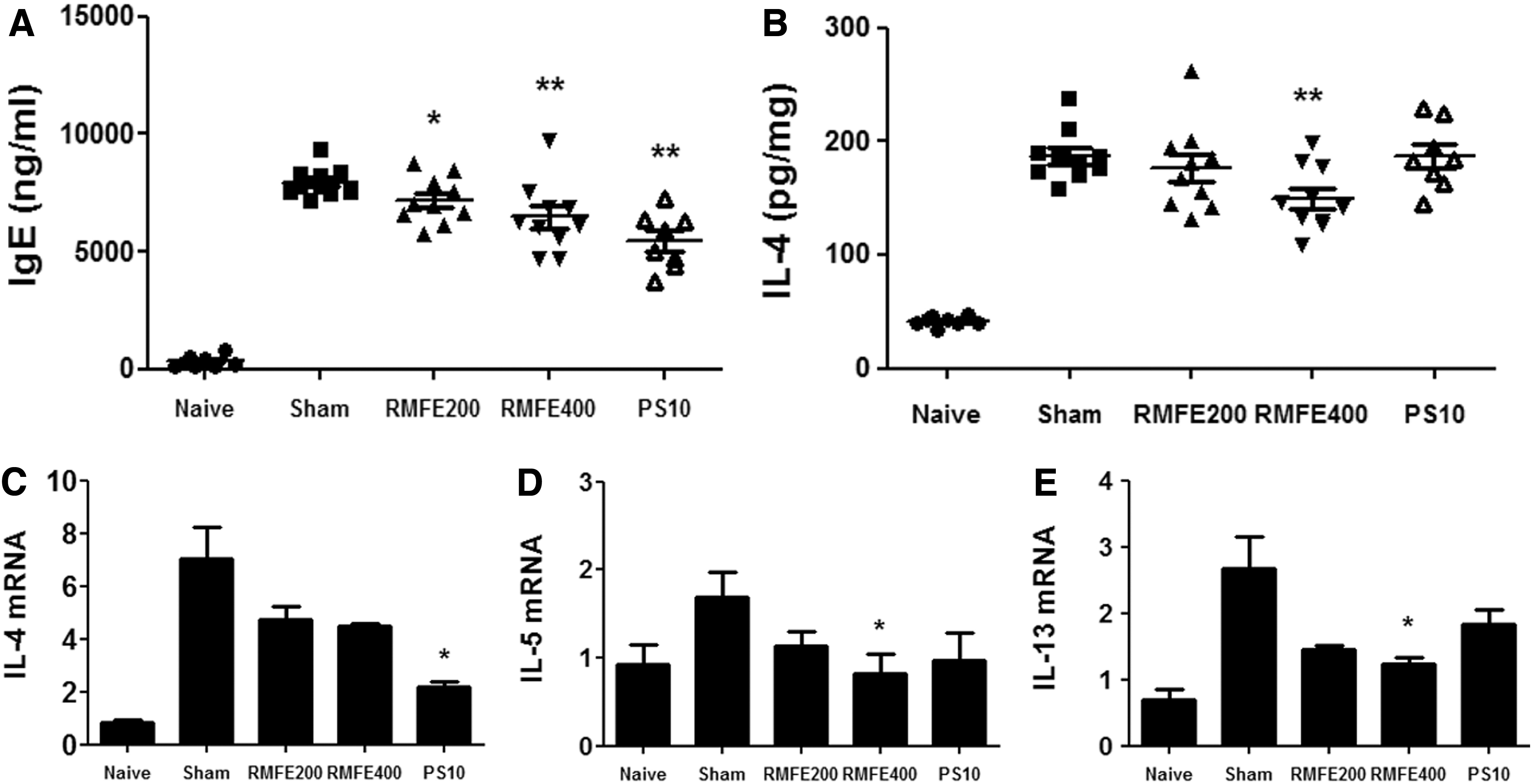
Effect of RMFE topical treatment of TMA-induced mice on IgE in serum, IL-4 production in ear tissue, and Th2 cytokine mRNA levels in dLNs. The production of
Also, dLNs play a central role in the adaptive immune responses by initiating the reaction of T cells and antigen-loaded antigen-presenting cells (APCs) reaction. Epidermal Langerhans cells (LCs) migrated into the T cell areas of dLNs after stimulation with microbial products and inflammatory cytokines, and dendritic cells (DCs) activated naive CD4+ T cells by presenting the antigen. 20,21 Therefore, we evaluated the effect of RMFE on Th2-related immune responses by measuring Th2 cytokine levels in dLNs. The messenger RNA (mRNA) levels of Th2-related cytokines in dLNs isolated from the TMA-induced AD-like mouse model indicated that IL-4, IL-5, and IL-13 were downregulated by RMFE (Fig. 3C–E).
Inhibitory effects of RMFE on OVA-induced Th2-associated immune responses in splenocytes
The TMA-induced AD-like mouse model is mainly characterized by the activation of Th2-dominant immune responses induced by allergen-specific effector T cells. 12 Those results were confirmed in our study, but Th2 immune responses were attenuated in the ear and dLNs after RMFE administration.
To confirm the observed effects after RMFE administration, we investigated the Th2 immune responses in splenocytes. OVA treatment strongly increased IL-4, IL-5, and IL-13 secretion in splenocytes, whereas RMFE treatment significantly suppressed those in a dose-dependent manner (Fig. 4A–C). Furthermore, we investigated whether RMFE inhibited the upstream signals associated with Th2 cytokine production. Phosphorylation of the STAT6, a Th2-related transcriptional factor, was suppressed by RMFE treatment in OVA-treated splenocytes (Fig. 4D). These results collectively demonstrated that RMFE treatment suppressed Th2 immune responses by inhibiting STAT6 phosphorylation, resulting in improvement of AD.
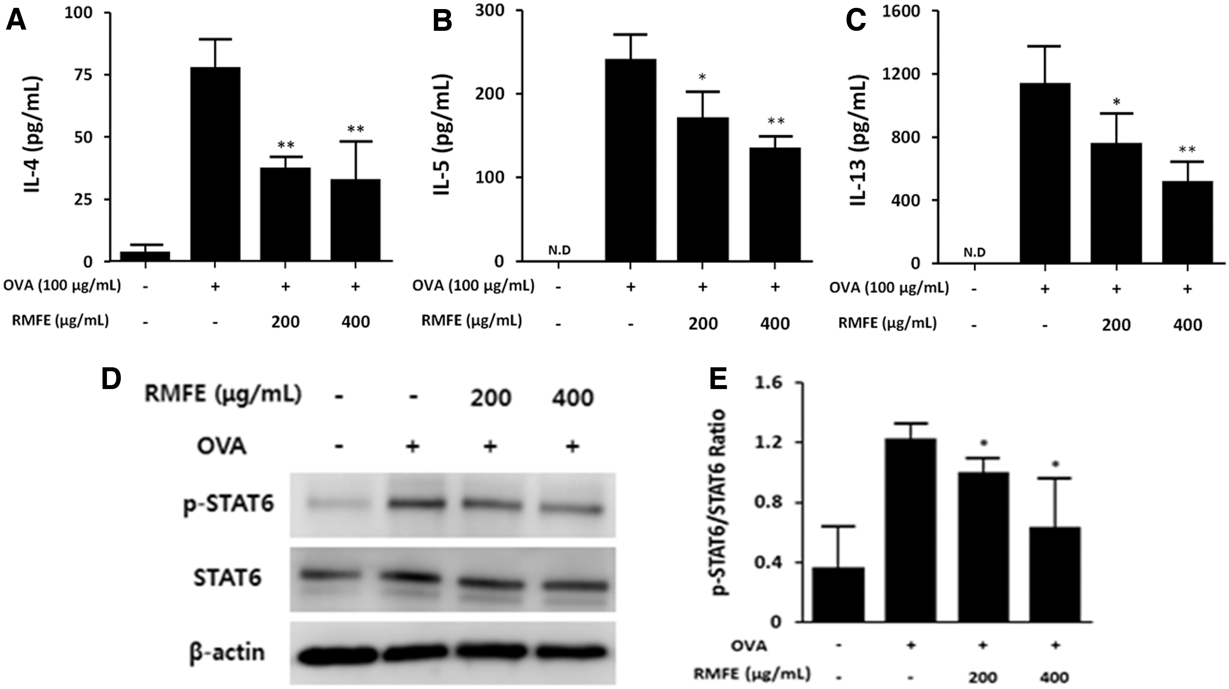
Effect of RMFE treatment on Th2 cytokine production and STAT6 phosphorylation induced by OVA-immunized splenocytes. The splenocytes of OVA-immunized BALB/c mice were seeded to 5 × 10⁶ cells/mL and cultured in the presence or absence of OVA (100 μg/mL) with RMFE (200 and 400 μg/mL). After 72 h, Th2 cytokines in the supernatant,
Inhibitory effects of RMFE on the proliferation of splenocytes
In previous studies, RMFE ameliorated asthma and rhinitis symptoms by suppressing Th2 immune responses and regulating Th1/Th2 balance. 15,16 In addition, Nguyen et al. revealed that RMFE elicited an improvement in food allergy symptoms via suppressing CD4+ T cells proliferation. 17
The effects of RMFE on reducing Th2 immune responses were confirmed in this study. Furthermore, to determine whether RMFE directly regulated Th1/Th2 balance of T cells in the splenocytes by increasing Th1 cytokines, we investigated the levels of IFN-γ in the culture supernatant of splenocytes cultured with OVA and RMFE. As a result, IFN-γ levels were reduced by treatment with RMFE (Fig. 5A). Thus, this result showed that RMFE attenuated Th1 as well as Th2 immune responses.
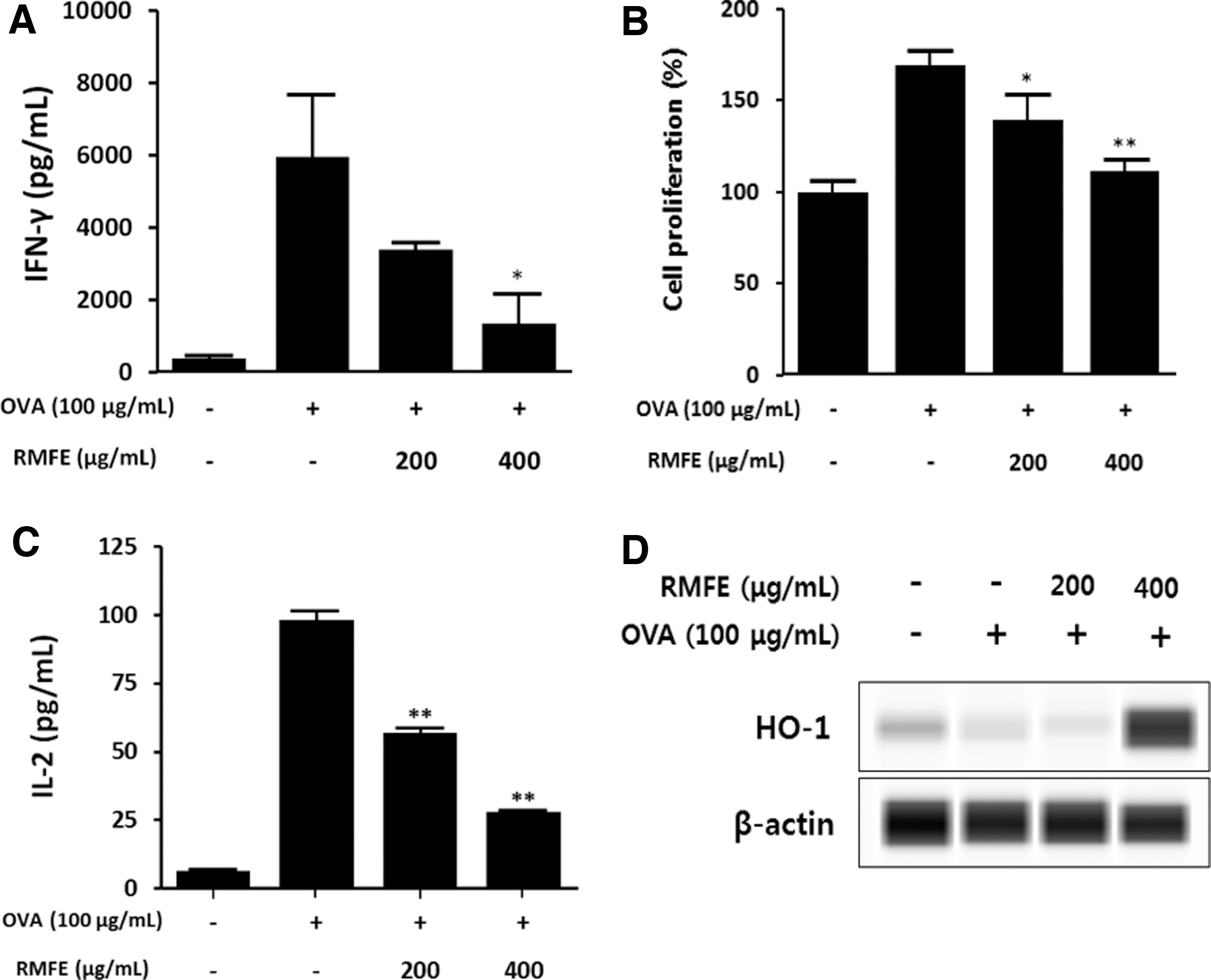
Effect of RMFE treatment on cell proliferation induced by OVA-specific immune responses and HO-1 expression in OVA-immunized splenocytes. The splenocytes of OVA-immunized BALB/c mice were seeded to 5 × 10⁶ cells/mL and cultured in the presence or absence of OVA (100 μg/mL) with RMFE (200 and 400 μg/mL). After 72 h,
Later, we hypothesized that suppressed Th1 and Th2 immune responses were because of reduced T cell proliferation by RMFE. To this end, RMFE suppressed cell proliferation of splenocytes, which were previously increased by OVA stimulation (Fig. 5B). Moreover, we investigated IL-2 levels, which is a T cell growth factor, 22 and confirmed that IL-2 cytokine production was also reduced by RMFE treatment (Fig. 5C). The study by Pae et al. revealed that HO-1 inhibited proliferation of CD4+ T cells via suppression of IL-2 secretion. 23 Like this report, we confirmed that HO-1 protein expression were increased in splenocytes by treatment with RMFE (Fig. 5D).
RMFE effects on proliferation of CD4+ T cell and HO-1 expression
We confirmed in an in vitro system that RMFE reduced IL-2 production and CD4+ T cell proliferation, which were previously activated by CD3 and CD28 antibodies, and increased HO-1 protein levels in CD4+ T cells (Fig. 6A–C).
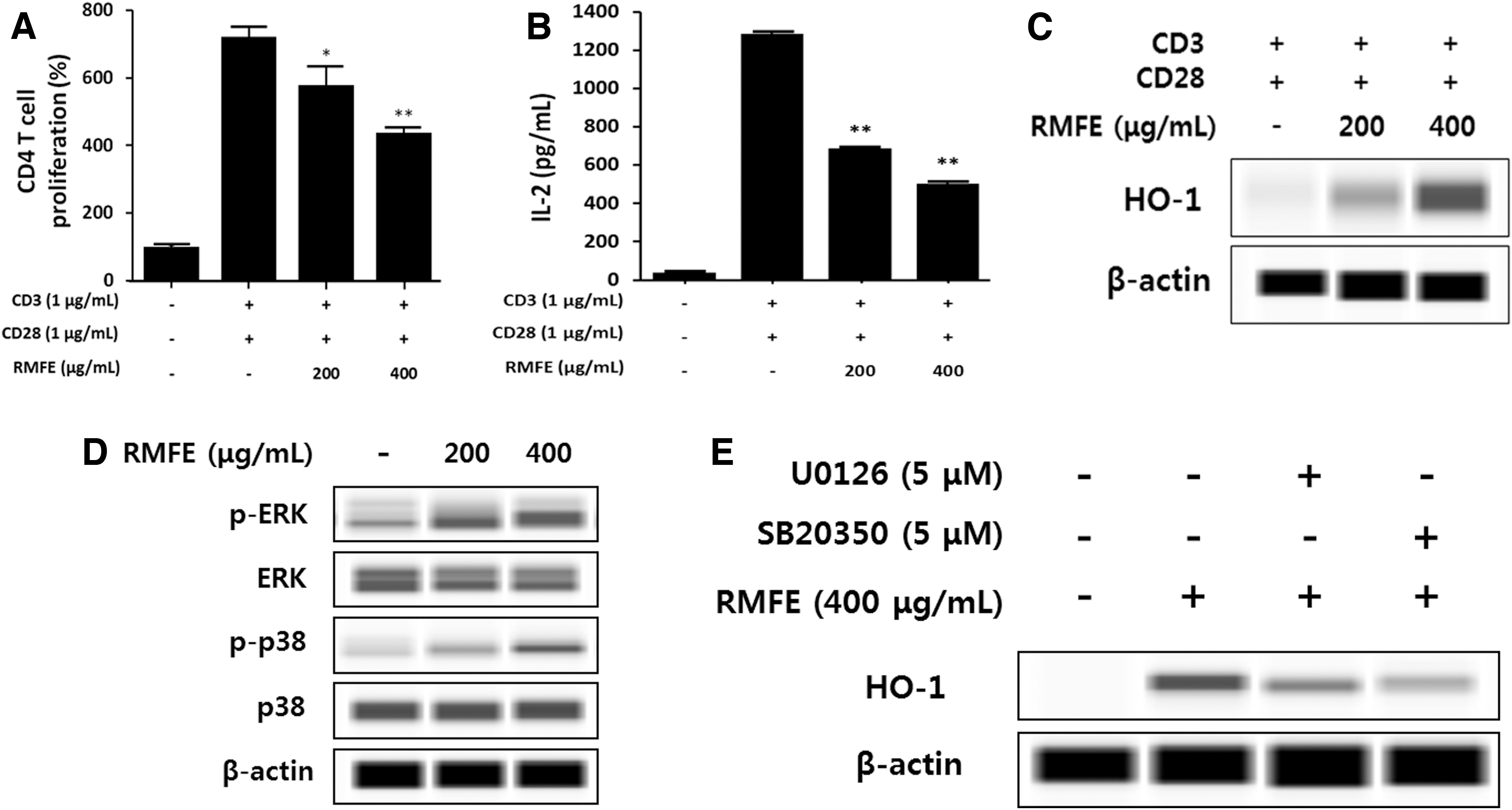
Effect of RMFE treatment on CD4+ T cell proliferation and HO-1 expression in CD4+ T cell isolated from BALB/c mouse splenocytes. Purified CD4+ T cell were seeded to 1 × 10⁶ cells/mL in anti-CD3 antibody (1 μg/mL) precoated plate and cultured in the presence or absence of RMFE (200 and 400 μg/mL) with anti-CD28 antibody (1 μg/mL).
Next, we investigated signaling pathways related to HO-1 protein expression. In a previously reported study, administration of quercetin showed an increase of HO-1 protein levels via phosphorylation of ERK and p38 in BV-2 microglial cells. 24 Like the previous study, in our system RMFE increased ERK and p38 phosphorylation in CD4+ T cells (Fig. 6D); and HO-1 protein levels, previously increased by RMFE treatment, were reduced in U0126- and SB203580-treated CD4+ T cells (Fig. 6E).
These results implied that RMFE treatment evoked HO-1 expression by activating ERK and p38 pathways in CD4+ T cells, which resulted in the suppression of IL-2 production and CD4+ T cell proliferation.
DISCUSSION
At least 85% of AD cases are reported to occur before 5 years of age. 25 A study in children with AD, aged <5 years, showed that Th1/Th2 balance was disrupted by increased CLA+ polarized Th2 cells and suppressed CLA+ Th1 cells, compared to the relative controls. 26 Another study performed on patients with acute-phase AD showed Th2 dominant immune responses and circulating Th2 cells in the peripheral blood, which resulted in increased serum IgE levels. 3 AD is divided into two types. extrinsic and intrinsic, that are correlated with IgE levels according to clinical phenotypes. 26 The extrinsic form was characterized by IgE-mediated sensitization, involving approximately 70–80% of the patients. 3,27 The patients showed high serum IgE levels, and their atopic lesions were infiltrated with highly activated T cells. Cutaneous T cells of extrinsic AD patients produced increased levels of Th2-associated cytokines, whereas intrinsic AD showed normal IgE levels and no specific IgE. Intrinsic AD is characterized by lower levels of Th2 cytokine expression, and higher levels of IFN-γ expression. 28,29 On the contrary, TMA elicited a Th2-dominant immune response, featuring increased IgE and Th2 cytokine levels. 12,30,31 Haptens like TMA have been known as small molecules inducing allergic skin inflammation after penetrating into the skin. The haptens that penetrate into the skin are able to form hapten–protein complex by conjugating with host protein, and the complex can induce adaptive immune responses through the process recognized as an antigen by LCs, which are skin-resident DCs. 10 Therefore, owning to previous evidence, which showed that TMA-induced AD-like mouse model expressed features of extrinsic AD immune response, we focused on studying IgE production and Th2 immune responses. As a result, this study showed that oral treatment of RMFE attenuated the AD symptoms and suppressed IgE- and Th2-related cytokine levels.
Naive helper T cells are activated after recognizing antigens presented by APCs via T cell antigen receptor, and in the presence of costimulation with CD28, resulting in expansion of effector cells. 32 In particular, the sensitization of OVA as antigen is able to induce the differentiation of naive CD4+ T cells forward to OVA-specific Th2 cells. In this study, the treatment with RMFE reduced not only Th2-dominant immune responses but also Th1-related immune responses in OVA-sensitized splenocytes. Furthermore, the RMFE suppressed cell proliferation induced by OVA treatment and IL-2, known as a T cell proliferation cytokine, was reduced. These results suggested that RMFE could suppress CD4+ T cell-mediated immune responses by reducing CD4+ cell proliferation, which was increased by antigen-stimulation, because RMFE did not affect the cell viability in nonsensitized splenocytes cultured without antigen (data not shown). Thus, we focused on HO-1 as a factor related to CD4+ T cell proliferation.
HO-1 is a rate-limiting enzyme critical for converting heme to bilirubin, carbon monoxide (CO), and iron, resulting in antiproliferation of CD4+ T cells. 33,34 Song et al. confirmed that CO reduced CD4+ T cell proliferation via a caspase-dependent pathway by increasing p21Cip1. 35 Also, HO-1 induced by cobalt protoporphyrin produced CO levels which inhibited IL-2 production and thus suppressed CD4+ T cell proliferation. 23 Various phytochemicals, 36 including quercetin and quercetin-3-O-β-D-glucuronopyranoside, increased the levels of HO-1 protein via the ERK and p38 pathways and ERK and PI3K-Akt pathways, respectively. 24,37 Thus, RMFE increased HO-1 protein levels through phosphorylation of ERK and p38, which resulted in inhibition of IL-2 production and the CD4+ T cell proliferation induced by stimulation of CD3 and CD28 antibodies, and thus ameliorated AD-like symptoms.
Many studies have identified various bioactive compounds in RMFE, including quercetin, rutin, isoquercitrin, multinoside A, multinoside B, multiflorin A, and multiflorin B. 38,39 However, various bioactive components are often not extracted or are extracted depending on different extraction methods. As the RMFE used in this study was extracted using hot water, it contained more glycosides than aglycon. We confirmed that the bioactive components of the extract, by using high performance liquid chromatography, were four glycosides: ellagic acid, isoquercitrin, hyperoside, and miquelianin, previously found in another study. 16
Ellagic acid, isoquercitrin, and hyperoside are naturally found in various fruits and plants, and are well-known for their antiallergic effects in disorders such as allergic asthma and allergic rhinitis. 40 –42 In addition, it was reported that the ellagic acid, hyperoside, and isoquercitrin have protective effects against oxidative stress by increasing HO-1 expression. 43 –47 However, in allergic immune responses, the physiological properties of quercetin 3-O-glucuronide (miquelianin) are unknown. Miquelianin was previously reported to exert antioxidant activities, neuroprotective effects, 48,49 and antiallergic activities inhibiting IL-4 production. 16 We thus suggested that miquelianin might help improve various allergic diseases. Therefore, we will conduct future studies to investigate the effect on variety of allergic diseases as well as the mechanisms.
In conclusion, our results demonstrated that RMFE significantly suppressed the symptoms of TMA-induced AD through suppression of antigen-specific Th2-associated immune responses and inhibition of inflammatory cytokine production. Therefore, we foresee that RMFE may be used as an ingredient in drugs or functional foods for the effective treatment of AD induced by Th2-mediated immune responses.
Footnotes
AUTHOR DISCLOSURE STATEMENT
No competing financial interests exist.
FUNDING INFORMATION
This study was supported by the research grant (E0170400-04) from the Korea Food Research Institute.