




Abstract
Chronic cough is very common in respiratory clinics, and no effective drugs are available. Schisandra chinensis (Turcz.) Baill. (S. chinensis), an important traditional Chinese medicine, has been extensively prescribed for patients with a persistent cough. Preliminary research indicated that 95% ethanol extracts (EE) of S. chinensis showed remarkable antitussive activity in guinea pigs exposed to cigarette smoke (CS). To find out the antitussive ingredients of S. chinensis, EE was divided into four fractions according to the polarity: petroleum ether extract (PEE), ethyl acetate extract (ECE), n-butyl alcohol extract, and residue extract. The antitussive, antioxidant, and anti-inflammatory effects of the four fractions were evaluated in a guinea pig model of cough hypersensitivity induced by CS exposure. Eighteen main constituents of the two effective fractions, PEE and ECE, were identified using ultra-high-pressure liquid chromatography electronic spray ion time-of-flight mass spectrometry. The cough inhibition activities of compound
Introduction
Chronic cough, broadly defined as a cough persisting for more than 8 weeks and without signs of imaging abnormalities, is very commonly seen in respiratory clinics. 1 The most important causes of chronic cough are upper airway cough syndrome, eosinophilic bronchitis, cough variant asthma, and gastroesophageal reflux–related cough. 2 The diagnosis and medical therapies of these four kinds of chronic cough are offered in the guideline. 3 However, about 10–46% of patients with chronic cough do not reveal any convincing etiology, and cough remains refractory. 4 This kind of chronic cough was defined as cough hypersensitivity syndrome (CHS). 5 Recently, the research on CHS has mostly focused on blockers of transient receptor potential ankyrin-1, transient receptor potential vanillic-1, and neuropeptides. 6,7 However, the antitussive effect and side effect of these blockers remain unknown due to the lack of clinical randomized controlled trials. 8
Schisandra chinensis (Turcz.) Baill. (S. chinensis), a traditional Chinese medicine, has been used for the treatment of chronic cough for thousands of years in China and Russia. 9,10 It was recorded as a herbal medicine for astringing lung and relieving cough in the Chinese Pharmacopoeia and legally listed as a health food by the Chinese Ministry of Health. However, the antitussive activity and potentially active compounds of S. chinensis are rarely reported. A previous study demonstrated that 95% ethanol extract (EE) of S. chinensis could reduce the frequency of cough and pulmonary inflammation in the cigarette smoke (CS)–induced cough hypersensitivity in guinea pigs, and lignans and polysaccharide may be the active antitussive components of EE and water-ethanol extract (EWE), respectively. At the same time, the antitussive activity of polysaccharides isolated from EWE was also demonstrated. 11
EE was divided into four fractions, and their antitussive effects were evaluated in this study to gain insight into the effects of the active compounds of S. chinensis EE on chronic cough. Moreover, the active fractions were chemically analyzed using ultra-high-pressure liquid chromatography electronic spray ion time-of-flight mass spectrometry (UHPLC-ESI-TOF-MS) to characterize the potential antitussive components of S. chinensis.
Materials And Methods
Animals
Male white Hartely guinea pigs with a body weight of 300 ± 50 g were obtained from Guangzhou Nan Fang Yi Da Laboratory Animal Science and Technology Development Limited Company [Guangzhou, China, SCXK(YUE)2016-0041]. Guinea pigs were raised in an animal facility of the Guangzhou Medical University [SYXK(YUE)2020-0227], with free access to food and water. The room temperature was controlled at 23°C ± 3°C, and the humidity was kept at 55% ± 15% on a 12-h light/12-h dark period. Guinea pigs were acclimatized for 1 week and used in experiments. All the animal experiments in this study were approved by the Committee for the Care and Use of Laboratory Animals in the First Affiliated Hospital of Guangzhou Medical University [LUN(SHEN)2018-60] and carried out according to the NIH and University Guidelines for the Care and Use of Laboratory Animals.
Materials
The following instruments were used for the experiment: PM-type unrestrained single-chamber animal plethysmography (1 L; Buxco Electronics Incorporated, Wilmington, USA), CS exposure box (0.6 × 0.6 × 1 m3, self-made; State Key Laboratory of Respiratory Disease), Anke TGL-16B centrifuge (Anting, China), and charge coupled device digital microscope camera system (Nikon, Japan).
The following reagents were used for the experiment: pentobarbital sodium (Merck, USA), codeine phosphate (30 mg/pill) (Sinopharm, China), and filtered cigarettes (Hongmei, 12-mg tar and 1.2-mg nicotine per cigarette; Guangdong Tobacco Industrial Limited Company, China); standards of schisandrin, schisandrol B, schisantherin A and B, schisanhenol, deoxyschizandrin, and schisandrin B (National Institutes for Food and Drug Control, China); and standards of gomisin J, gomisin G, and anwuligan (Shengyang Kaidi Science and Technology Ltd., China). Acetonitrile (CH3CN) and methanol (CH3OH) were of high-performance liquid chromatography (HPLC) grade. Deionized water was prepared using a Millipore water purification system. The other reagents were of analytical grade (Guangzhou Chemical Reagent Factory, China).
S. chinensis fruits, collected from Anshan County, Liaoning Province, China, were purchased from Shengyang Kaidi Science and Technology Limited Company in November 2019 and identified as Schisandra chinensis (Turcz.) Bail by Dr. Shan Zhong (Voucher Specimen No. 20191120S) according to the Chinese, Pharmacopoeia (2015).
Preparation of the four extracts from EE of S. chinensis
S. chinensis fruits were dried at 60°C for 24 h and ground into a powder before extraction. Then, 10 kg of the fruit powder was refluxed with 95% alcohol for three times, each for 2 h. The alcoholic extract was placed at room temperature for 12 h, and the supernatant was centrifuged at 3000 rpm for 20 min. The centrifuged supernatant was concentrated under reduced pressure and dried in vacuum to obtain EE (4256.5 g). Then, half of the EE was extracted with petroleum ether, ethyl acetate, and n-butyl alcohol, successively. Three extracts and the residue solution were collected, concentrated, and finally vacuum dried to obtain four parts: petroleum ether extract (PEE, 456.8 g), ethyl acetate extract (ECE, 956.2 g), n-butyl alcohol extract (BAE, 507.8 g), and residue extract (RE, 207.4 g).
Effect of the four extracts of EE on CS-induced cough hypersensitivity in guinea pigs
Model and intragastric administration
The effect of the four extracts of EE on chronic cough was evaluated on CS-induced cough hypersensitivity in guinea pigs as described in previous studies. 11,12
All guinea pigs underwent the following procedure after 1 week of adaption to screen the cough sensitivity: They were placed in a glass chamber (r = 12.5 cm, h = 18 cm) and nebulized with 0.8 mol/L citric acid for 1 min with a flow rate of 0.5 mL/min. A 5-min observation was followed, and the cough frequency was recorded during this 6-min period. The guinea pigs with a cough frequency between 10 and 30 were included in this study.
After 1 week of adaptation, 80 eligible guinea pigs were randomly and equally allotted to 8 groups: control group, CS exposure group, CS+solvent group (0.5% PEG-400), CS+codeine group, CS+PEE group, CS+ECE group, CS+AE group, and CS+RE group. All groups, except the control group, were exposed to CS (10 cigarettes) in a conscious and unrestrained state for 20 min, twice a day, 14 days successively. The period between two exposures was 6 h. The temperature inside the CS chamber was controlled at 23°C ± 3°C. During these 14 days, the animals of the four S. chinensis groups were intragastrically administered with 1 g/kg drugs daily for 3 h after the first smoke exposure. The dosage of the solvent group was 10 mL/kg. However, the codeine group was orally administered (30 mg/kg, qd) 1 h before the citric acid challenge on the 15th day.
Evaluation of cough sensitivity
After 14 days of oral administration and CS exposure, unrestrained guinea pigs were individually placed in a body plethysmograph (a chamber made of transparent plastic sheets). After 2 min of adaptation, guinea pigs were exposed to 0.4 mol/L citric acid aerosol produced by a nebulizer (REF AG-AL1000; Aerogen, Ireland) for 10 min with a flow rate of 0.5 mL/min. The cough frequency and airway resistance (Penh area under the curve [Penh-AUC]) were recorded for 20 min. The cough effort was defined as interruption of expiratory flow associated with a typical cough motion and sound observed by a trained staff and recorded using the Buxco system. 13
Infiltration of inflammatory cell and release of cytokines and chemokine
On the 15th day, guinea pigs were anesthetized using pentobarbital sodium (30 mg/kg), and blood was depleted from the heart as much as possible. Then the chest was surgically exposed, and the trachea was cannulated while the right main bronchus was ligated near the bifurcation. The left lung was lavaged thrice with 2 mL of ice-cold phosphate-buffered saline using the cannula. After a series of processing, the total cell counts were determined using a hemocytometer, and differential leukocyte counts were performed randomly in a minimum of 400 leukocytes.
Four segments of nonlavaged right lung tissue were homogenized with saline, and then the activities of superoxide dismutase (SOD) and glutathione orgotein peroxidase (GSH-px) and the content of malondialdehyde (MDA) were measured by colorimetric methods using the assay kits (JianCheng, Nanjing, China) and an ultraviolet-visible spectrophotometer (TU-1901; Purkinje General Instrument Limited Company, Beijing, China). The assay of tumor necrosis factor-α (TNF-α) and interleukin-8 (IL-8) in homogenizing lung tissue was performed using an Enzyme-Linked Immunosorbent Assay Kit (USCNK, Wuhan, China). The total protein of lung tissue was extracted from the lung tissue by radioimmunoprecipitation assay lysate and measured using a Bicinchoninic Assay Kit (Beyotime, Shanghai, China).
Histological evaluations
The nonlavaged end of trachea and a segment of right lung were removed and fixed in 10% formaldehyde. Then the tissues were embedded in paraffin, cut into 4-μm sections, and stained with hematoxylin and eosin (H&E) successively. Three different fields for each tissue were checked under light microscopy at a magnification of 200 × . Pulmonary inflammation, thickness of epithelium, and thickness of smooth muscle were semiscored by the blinded observer. The lung inflammation was graded into four categories, from 1 (normal) to 4 (severe). The thickness of epithelium and smooth muscle was directly measured and converted into relative values.
UHPLC-ESI-TOF-MS analysis of PEE, ECE, BAE, and RE
Sample preparation
The CH3OH solution containing certain amounts of the 10 lignan reference compounds (schisandrin, schisandrol B, schisantherin A and B, schisanhenol, deoxyschizandrin, schisandrin B, gomisin J, gomisin G, and anwuligan) was filtered through a 0.22-μm membrane filter before analysis. The PEE, ECE, BAE, and RE were dissolved in CH3OH at a concentration of 0.2 mg/mL and filtered through a 0.22-μm polytetrafluoroethylene filter into an Agilent amber sample vial for UHPLC analysis.
Personal database establishment
Compound information of 41 compounds, which were reported as constituents of S. chinensis, was downloaded from the Dictionary of Natural Products online (2014), compiled and saved as a “CSV” file. The file was then exported to Agilent MassHunter Personal Compound Database and Library (PCDL) software to build a personal database, in which the compound names, molecular formulas, and accurate masses were included. After an accurate MS screening, the database has the unique capability of adding LC retention times to increase the specificity of compound identification. 14 This personalized database combined with UHPLC-TOF-MS made it possible to identify the compounds in the extracts of S. chinensis.
UHPLC-ESI-TOF-MS analysis
Chemical analyses of PEE, ECE, BAE, and RE were conducted on an Agilent 1290 Infinity UHPLC system (Agilent Corporation, USA) with a Waters ACQUITY UPLC BEH C18 (2.1 × 100 mm2, 1.7 μm) column. The mobile phase consisted of (A) 0.1% formic acid in water and (B) 0.1% formic acid in CH3CN. The gradient was optimized as follows (flow rate, 0.35 mL/min): 0–12 min, 40–70% B; 12–15 min, 70–70% B; 15–17 min, 70–100% B; 17–19 min, 100–100% B; and 19.1–22 min, 40–40% B. The injection volume was 1 μL, and the column temperature was maintained at 40°C in each run. MS was performed on an Agilent 6230 TOF-MS equipped with a Jet Stream ESI source (Agilent Corporation). The ESI condition was set as follows: positive ion mode, sheath gas temperature 325°C and flow rate 11 L/min, capillary voltage −3.5 kV, nebulizer 40 psi, drying gas temperature 325°C and flow rate 11 L/min, nozzle voltage 1.5 kV, fragmentor 175 V, and recorded mass range m/z 100–1700. The MS data were processed using Agilent MassHunter Qualitative Analysis (version B.05.00).
Isolation and identification of compound 17
Compound
Antitussive activities of compound 1 , 3 , 9 , 10 , 17 on acute cough induced by citric acid in guinea pigs
After the cough sensitivities were screened, 108 qualified guinea pigs with a cough frequency between 10 and 30 were randomly divided into 18 groups (n = 6): control, vehicle, codeine (30 mg/kg), compound
Statistical analysis
The data were analyzed using statistical package for social science (SPSS) and expressed as mean ± standard deviation. Statistical significance of differences between different groups was assessed using one-way analysis of variance followed by the least significant difference test if all groups have constant variances, while the rank sum test was performed for unequal variance or abnormal distribution. For all experiments, P < .05 was taken as statistically significant. Animals showing signs of infection or who died during the experiment were excluded from the study.
Results
Effect of S. chinensis extracts in guinea pigs exposed to CS
Cough frequency and Penh
As shown in Figure 1, repeated exposure to CS doubled the citric acid–induced cough frequency in guinea pigs (P < .01). However, this could be significantly inhibited by oral administration of PEE and ECE with the inhibition rates of 35.7% (P < .05) and 49.7% (P < .01), respectively. BAE and RE showed no significant antitussive effect, and their inhibition rate was only 23.6% and 22.9%, respectively. No significant difference was observed between the CS and vehicle groups. The inhibition rate of codeine was 51.2%.

Effect of Schisandra chinensis extracts on CS induced cough hypersensitivity in guinea pigs. Animals were challenged with vaporized citric acid (0.4 mol/L) after 14 days of repeated CS exposure. Compared with control group, **P < .01; compared with model group, # P < .05, ## P < .01. CS, cigarette smoke.
As shown in Figure 1, the Penh-AUC of all groups showed no significant difference.
Infiltration of inflammatory cells and release of cytokines and chemokines in the lung tissue
Total cell counts and differential leukocyte counts in bronchoalveolar lavage fluid
The oral administration of ECE significantly decreased the total leukocyte cells (37.2%, P < .01) and the percentage of neutrophils in bronchoalveolar lavage fluid (BALF) (32.0%, P < .01) and increased the percentage of mastocyte (24.2%, P < .01) (Table 1). PEE, BAE, and RE showed no obvious significance on BALF.
Effect of Schisandra chinensis Extracts on Total Cell Counts and Differential Leukocyte Counts in Bronchoalveolar Lavage Fluid of Cigarette Smoke Exposure Guinea Pigs (Mean ± Standard Deviation)
× 106/mL; compare with Control group, ** P < .01; compare with CS group, # P < .05, ## P < .01.
BAE, n-butyl alcohol extract; CS, cigarette smoke; ECE, ethyl acetate extract; PEE, petroleum ether extract.
Release of IL-8 and TNF-α
As shown in Figure 2, 14 days exposure to CS significantly increased the contents of IL-8 (56.5%, P < .01) and TNF-α (34.0%, P < .05) in the lung tissue of guinea pigs, which could be slightly decreased by the oral administration of both PEE (29.0%, P < .05; 23.9%, P < .05) and ECE (46.4%, P < .01; 31.3%, P < .01). Yet BAE and RE could not show this kind of activity.
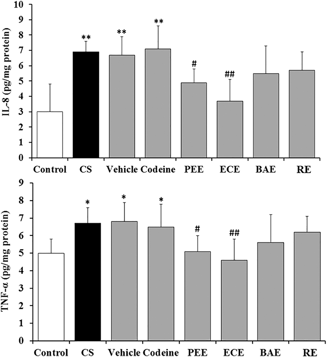
Effect of Schisandra chinensis extracts on release of IL-8 and TNF-α in lung tissue of CSE guinea pigs. Compared with control group, *P < .05, **P < .01; compared with model group, # P < .05, ## P < .01. IL-8, interleukin-8; TNF-α, tumor necrosis factor-α.
Contents of SOD, GSH-px, and MDA
As shown in Figure 3, 14 days of repeated CS exposure resulted in a significant decrease in both activities of SOD (9.0%, P < .05) and GSH-px (51.0%, P < .01) and a remarkable increase in the MDA (101.82%, P < .05) content in lung tissue. Oral administration of PEE and ECE significantly increased the activities of SOD (20.3%, P < .05; 24.6%, P < .01) and GSH-px (27.0%, P < .05; 41.8%, P < .01) and significantly decreased the content of MDA (39.2%, P < .05; 44.6%, P < .05) in lung tissue. BAE significantly decreased the content of MDA (47.3%, P < .05), but had no significant effect on SOD and GSH-px. However, daily treatment with RE showed no effect on the activities of SOD, GSH-px, or the content of MDA.
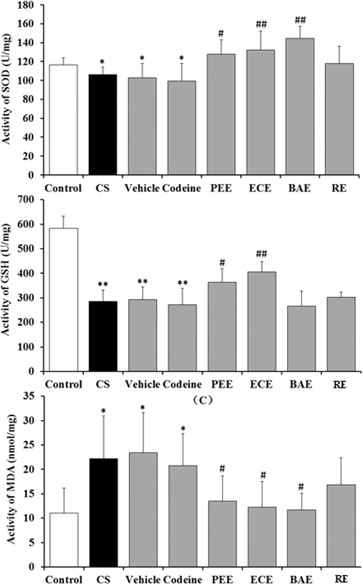
Effect of Schisandra chinensis extracts on content of SOD, GSH-px, and MDA in lung tissue of CSE guinea pigs. Compared with control group, *P < .05, **P < .01; compared with model group, # P < .05, ## P < .01. GSH-px, glutathione orgotein peroxidase; MDA, malondialdehyde; SOD, superoxide dismutase.
Histological changes
Semiquantitative scoring of H&E staining sections of lung and trachea showed that 14 days of CS exposure caused significant infiltration of inflammatory cells and thickening of the tracheal wall (Fig. 4). In the trachea sections, obvious hyperplasia of airway epithelial cells and smooth muscle cells could be found in the model group guinea pigs compared with the control group counterpart. Daily treatment with ECE significantly inhibited the hyperplasia of airway epithelial cells and smooth muscle cells induced by repeated CS exposure. Moreover, infiltration of inflammatory cells was obviously relieved in the lung tissue of guinea pigs daily treated with ECE or PEE. However, treatment with BAE, RE, or codeine (30 mg/kg, single dose) did not show a significant effect on the infiltration of inflammatory cells or thickening of tracheal wall.

Typically morphopathological changes in trachea and lung tissue (200 × ). Compared with control group, *P < 0.05, **P < 0.01; Compared with model group, # P < 0.05, ## P < 0.01. Color images are available online.
UHPLC-ESI-TOF-MS analysis of PEE, ECE, BAE, and RE
The 16 peaks in PEE and 3 peaks in ECE were identified by comparing the accurate mass, isotope ratios, and retention times. Ten available lignan standard compounds and one isolate of the major peak (No. 17) from ECE were used to verify the identities (Fig. 5). The 16 major peaks in PEE were characterized as 15 lignans (compounds
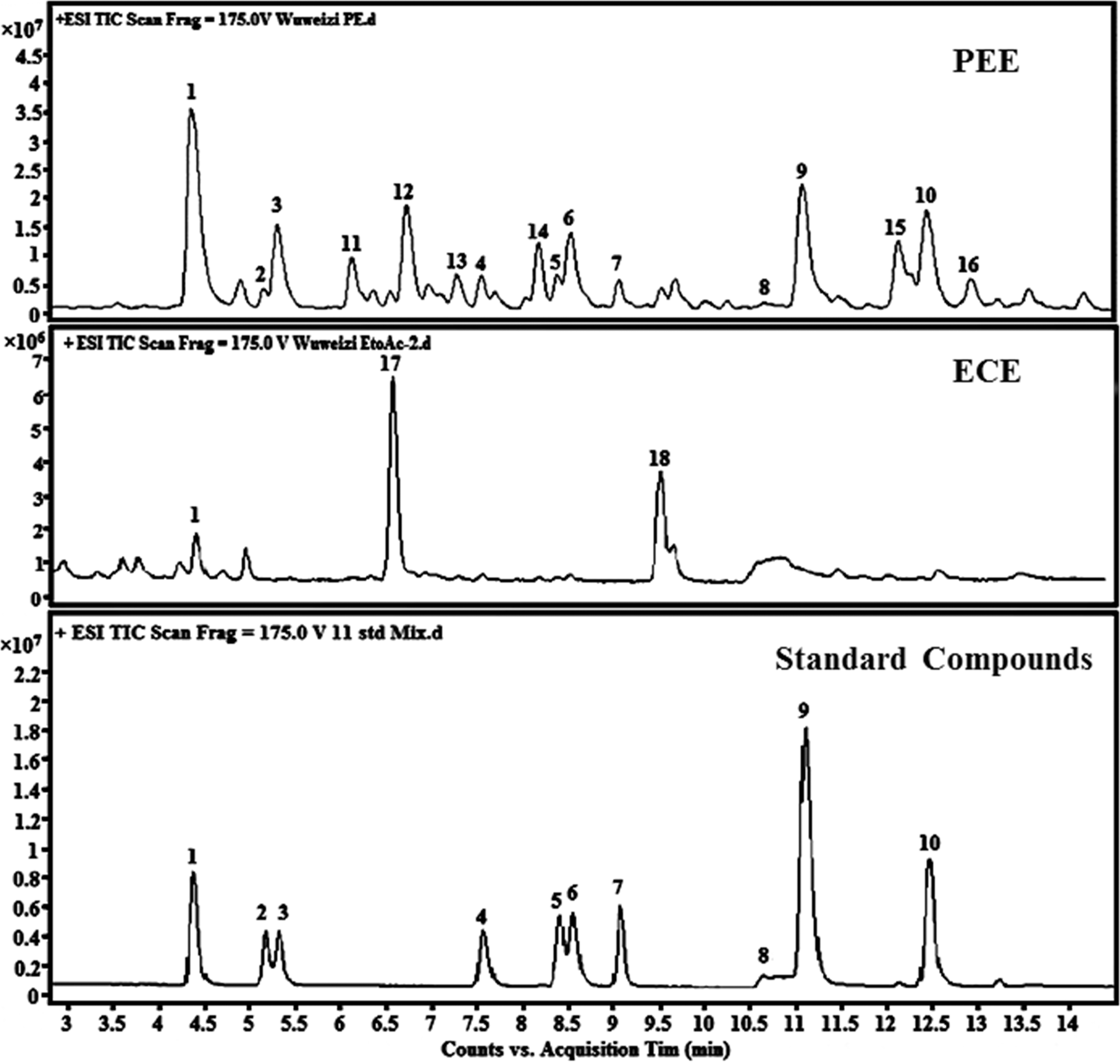
UHPLC-ESI-TOF-MS chromatography of PEE, ECE, and standard compounds. Lignan standard compounds (peaks 1–10) were Schisandrin, Gomisin J, Schisandrol B, Gomisin G, Schisantherin A, Schisantherin B, Schisanhenol, Anwuligan, Deoxyschizandrin, and γ-Schizandrin B, respectively. ECE, ethyl acetate extract; PEE, petroleum ether extract; UHPLC-ESI-TOF-MS, ultra-high-pressure liquid chromatography electronic spray ion time-of-flight mass spectrometry.
Identification of Compounds in Petroleum Ether Extract and Ethyl Acetate Extract of Schisandra chinensis by High-Resolution Mass Spectrometry
Denotes that either one is propinquanin F, the other is methylschisantherin F;
Denotes that the compound is either schisanwilsonin A or schisanwilsonin B.
Analysis of the chemical structure of compound 17
Compound

The structure of compound
Antitussive activities of compound 1 , 3 , 9 , 10 , 17 on acute cough induced by citric acid in guinea pigs
The effect of compound
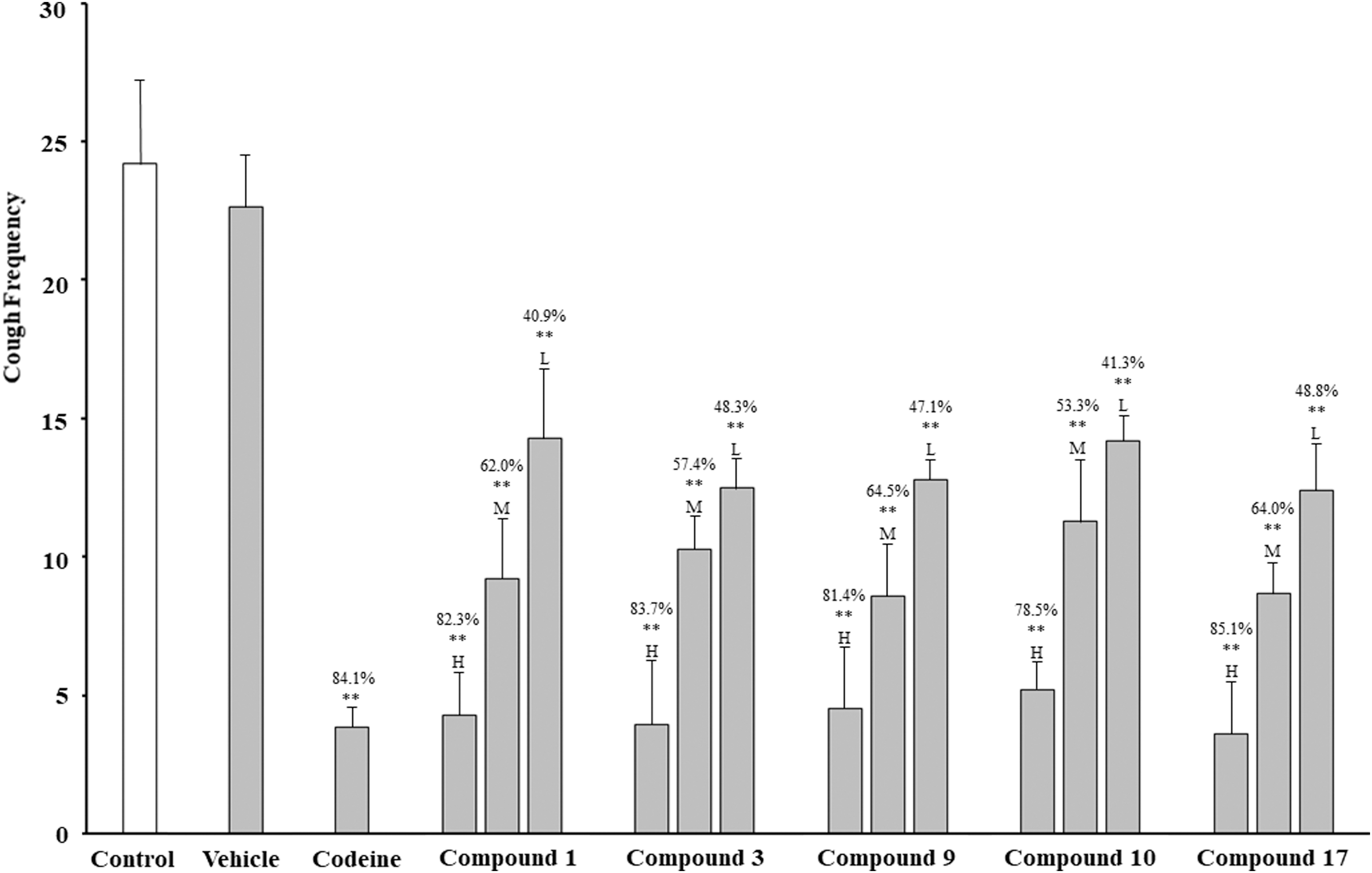
Antitussive activities of compound
Discussion
CHS has a complex pathogenesis and is very difficult to treat. So far, no effective drugs targeting CHS have been found, whose typical characteristics are increased cough sensitivity and airway inflammation. Moreover, the pathogenic mechanism of CHS is also related to oxidative stress injury. 16 –18
A previous study that established a guinea pig model of CHS by CS exposure demonstrated that the EE of S. chinensis could decrease cough sensitivity and airway inflammation. In the present study, EE was separated into the following four extractive fractions to investigate the antitussive compounds of S. chinensis: PEE, ECE, BAE, and RE. The pharmacological results revealed that pretreatment with PEE and ECE could remarkably reduce the CS-induced cough sensitivity in guinea pigs with inhibition rates of 35.7% and 49.7%, respectively, which were almost close to that of codeine (51.2%). PEE and ECE could also reduce the airway inflammation and the proportion of inflammatory cells in BALF and evidently affect the content of IL-8, TNF-α, SOD, GSH-px, and MDA in pulmonary tissue.
The results of UHPLC-ESI-TOF-MS showed that the main compounds of PEE and ECE, 2 antitussive parts in EE, were mostly lignans: 16 compounds, including 15 lignans and 1 triterpene, were identified in PEE, and 3 lignans were characterized in ECE. None of these lignans was detected in the other two high-polarity parts without antitussive activity (BAE and RE). The compound
Lignans are a large group of concentrated compounds in S. chinensis. They are also acknowledged as active substances of S. chinensis. Although no reports about the antitussive activity of lignans in S. chinensis are available, their strong anti-inflammatory and antioxidative activities have been demonstrated in previous studies.
19
–21
On the basis of pathological characteristics and mechanisms of CHS, pharmacological experiment results, and UHPLC-ESI-TOF-MS analysis, lignans were speculated to be the antitussive substance compounds of S. chinensis. To verify the antitussive activities of lignas, we isolated and purchased compound
This study provided sufficient scientific evidence for the clinical application of S. chinensis on CHS and benefits of the usage of S. chinensis and laid the foundation of developing new drugs for CHS.
In conclusion, lignans, with low chemical polarity, are the main antitussive substances of EE of S. chinensis, and they could be concentrated from the extracts by further extraction with petroleum ether and ethyl acetate.
Footnotes
Author Disclosure Statement
No competing financial interests exist.
Funding Information
This study was supported by the project of National Natural Science Foundation of China (No. 81573575), the project of Natural Science Foundation of Guangdong Province (No. 2016A020217023), the Incubative Project for Innovation Team of Guangzhou Medical University (2017-159), and the project for clinical application and transformation of the First Affiliated Hospital of Guangzhou Medical University (No. 201511-gyfyy).
Supplementary Material
Supplementary Data S1
References
Supplementary Material
Please find the following supplemental material available below.
For Open Access articles published under a Creative Commons License, all supplemental material carries the same license as the article it is associated with.
For non-Open Access articles published, all supplemental material carries a non-exclusive license, and permission requests for re-use of supplemental material or any part of supplemental material shall be sent directly to the copyright owner as specified in the copyright notice associated with the article.
