
Abstract
Nitraria tangutorun Bobr. has been used for thousands of years as a native folk medicine to alleviate dizziness and neurasthenia due to oxygen. In our previous study, natural antioxidant components (namely, NJBE) were isolated from industrial N. tangutorun Bobr. juice byproducts (NJBE) from the Qinghai-Tibet plateau. The current investigation assessed the effects of NJBE on ischemic stroke in mice and the potential mechanisms. C57BL/6 mice received NJBE (25, 50, or 100 mg/Kg) by gavage for 14 days and then stroke was induced by the middle cerebral artery occlusion (MCAO) model, followed by reperfusion for 72 h. The evaluation of brain infarct size, behavioral tests, and functional assessments was conducted to assess the effects of NJBE after MCAO. Our results suggested that NJBE significantly decreases infarct size, improves neurological deficits, as well as reduces the number of GFAP+ and Iba-1+ cells after MCAO. NJBE inhibited nitric oxide and malondialdehyde production in the ischemic brain. Meanwhile, it attenuated the expressions of superoxide dismutase (SOD), catalase (CAT), and glutathione peroxidase (GPx). Also, NJBE significantly attenuated the expression levels of proinflammatory indicators, including TNF-α, IL-1β, IL-6, and IL-12. This process was accompanied by the downregulation of TLR4, TRAF6, pIκB/pIκB, and MMP9 expression and the upregulation of claudin-5 expression. NJBE induced improvements in brain injury. The neuroprotective effect of NJBE provides evidence for its potential application in stroke treatment.
Introduction
Ischemic stroke is a dynamic and complex disease caused by multifactorial and multisegmented injuries. 1 Stroke is a major factor associated with disability, and stroke treatment is an important issue worldwide. Current therapies for ischemic stroke remain unsatisfactory. 2,3 More attention has been given to the prophylactic effects of neuroprotectants against ischemic stroke. Due to enhanced therapeutic efficacy and reduced adverse effects, herbal therapies and traditional food and medicine may be effective alternatives in the neuroprotective treatment of ischemic stroke. 4 –6
Nitraria tangutorun Bobr. (N. tangutorun) is a high-altitude-growing plant (>3000 m) in the Tibetan Plateau, and it has been widely used as a traditional medicine to alleviate dizziness and neurasthenia due to oxygen deficiency for the past millennia. N. tangutorun fruits and seeds contain plentiful flavones, alkaloids, quercetin, and polysaccharides, which exhibit strong antioxidant and antifatigue properties. 7 –10 N. tangutorun fruit can be processed into a functional health drink, 11 and the reconstituted juice byproducts are of great interest because the fruit may serve as a low-cost source of some unique bioactive ingredients. A previous study isolated NJBE from N. tangutorun juice byproducts using a microwave/ultrasonic-assisted enzymatic extraction method. 12 Chemical composition assays showed that the phenol, flavonoid, and anthocyanin contents in NJBE were 157.54 ± 5.42 mg GAE/g, 101.28 ± 6.84 mg QE/g, and 82.26 ± 4.79 mg CGE/g, respectively. NJBE could protect cells against UVB-induced oxidative phototoxicity and doxorubicin-induced oxidative cardiotoxicity, 12 and NJBE had a cardioprotective effect against doxorubicin-induced injury in H9c2 cardiomyocytes by directly regulating the reactive oxygen species (ROS) system. 13
The current study evaluated the potential effect of NJBE on ischemic injury and the possible mechanisms using the middle cerebral artery occlusion (MCAO) mouse model. Ischemic brain infarct size, neurological deficits, the serum levels of oxidative stress indicators, and other inflammatory-related indicators were simultaneously examined.
Materials and Methods
Materials
Fresh N. tangutorun Bobr. fruits were collected from Dongshangen of Dulan County, Haixi, which is a national municipality of Mongol and Tibetan, Qinghai, China. The plant name was checked using The Plant List website (TRO. record 50126119). Amberlite XAD-4 was purchased from Dow Chemical Co. (MI, USA).
Preparation of N. tangutorun juice byproduct extract
NJBE was prepared according to the procedure described in our previous study. 12 Briefly, fresh N. tangutorun fruits were juiced using a screw juice extractor. After centrifugation, the natural juice byproducts (NJBs) were freeze dried and ground. A simultaneous microwave/ultrasonic-assisted enzymatic method was used to extract the ground NJB powder according to the optimal antioxidant conditions as follows: extraction solvent of 70% ethanol with 0.6% cellulase; ultrasonic power, 800 W; the ratio of solvent to raw material, 20:1; pH value, 4.5; extraction temperature, 66°C; extraction time, 43 min; and microwave power, 420 W. After that, the extraction mixture was centrifuged, and the supernatant was collected and purified using an Amberlite XAD-4 macroporous resin column to obtain the NJBE.
Animals
Male C57BL/6 mice, 8–10 weeks old (weighing 22–25 g) were procured from the Pharmacology Experimental Center of Jilin University (Changchun, China). The mice were freely fed a standard laboratory chow diet and water. Ambient temperature was maintained at 22°C ± 2°C, and relative humidity was 55% ± 5%, respectively. The mice acclimatized for 3 days before studies. Animal handling procedures were conducted with the permission of the Ethics Committee of Jilin University (No. 202013) under the National Institutes of Health Animal Care and Use Guidelines (NIH publication No. 85–23, revised in 1985).
Mouse experimental design
Group I: Sham group; mice underwent a sham operation.
Group II: Vehicle group; animals were subjected to the MCAO model procedure.
Groups III, IV, and V: NJBE plus vehicle group; animals received NJBE (25, 50, and 100 mg/kg) for 14 days and then were subjected to MCAO model procedure followed by reperfusion for 72 h. A schematic of the experimental design is shown in Figure 2A.
MCAO surgical procedure
MCAO is a focal cerebral ischemia model and was conducted as previously described. 14 Briefly, the procedure was conducted under inhalation anesthesia with 5% isoflurane and maintenance with 1.5% to 2% isoflurane in medical air using a face mask. A silicon rubber-coated size 6-0 nylon filament (Doccol, MA, USA) was gently inserted into the left middle cerebral artery (MCA) to induce focal cerebral ischemia. Successful occlusion was verified (cerebral blood flow rate reduced to 20% of preischemic levels) by using laser Doppler flowmetry. After 45 min of occlusion, reperfusion was initiated by gently withdrawing the occluding filament. The body temperature of each mouse was controlled around 37°C. Mice in the sham group were subjected to the same surgical procedure without the occlusion. A schematic of the experimental design is presented in Figure 2A. The mice were euthanized 3 days after surgery, and then the ischemic brains were collected. The spleen, heart, and liver of mice were weighed, and we calculated the relative weights of each organ to the final body weight (organ index).
Behavioral tests
Modified neurological severity score
The modified neurological severity score (mNSS) is a comprehensive evaluation of exercise, sensory, balance, and reflex assessments in experimental animals, and the highest possible score is 18. 15 The scores were determined as follows: no neurological deficit was 0 to 6 points, moderate neurological impairment was 7 to 12 points, and severe neurological impairment was 13 to 18 points. Neurological deficits of mice were evaluated in a blinded fashion.
Corner test
Each tested mouse was allowed to advance into a 30° angle corner as previously described. 16 The mouse would turn left or right to quit the corner, and only turns made when the mouse was completely standing on its hind legs were counted. The test was repeated for each mouse 10 times per experiment, and the percentage of left turns was calculated. This test was repeated 10 times, allowing 30 sec between trials, and the investigators recorded the numbers of right and left turns.
Forelimb placing test
A vibrissae-elicited forelimb placing test was performed to evaluate sensory motor function in mice. 17 Briefly, the mice were torso handled, allowing the forelimbs to move at will. The mice were positioned parallel to the top of the table and gently moved up and down. Forelimb movements were induced by vibrissae touching of the edge of the tabletop. Movements of the corresponding impaired (left) forelimb were recorded. Each tentacle-induced forelimb placement was investigated, and the number of successful placements in 10 consecutive trials was recorded.
Cerebral infarction volume
After brain removal, 2,3,5-triphenyl tetrazolium chloride (TTC) (Sigma, St. Louis, USA) staining was used to evaluate cerebral infarct size. The area of infarction was calculated by measuring the white regions of the cerebral sections, and noninfarct areas that possessed normal mitochondrial enzymatic activity were stained deep red.
Immunofluorescence staining
The mice were perfused with 4% paraformaldehyde followed by dehydration with 30% sucrose. Then the mouse brains were collected and embedded with OCT to prepare frozen sections. The brain sections were cut into 10 μm coronally using a cryostat (Leica, Wetzlar, Germany). All sections were first blocked with 10% BSA for 1 h at room temperature. Thereafter, primary antibodies, such as rabbit anti-mouse GFAP (1:1000; Abcam, Cambridge, MA, USA) and rabbit anti-mouse Iba-1 (1:100; Abcam), were added to each slide, which was then incubated overnight at 4°C. Then, the slides were incubated with secondary antibody, Alexa 594-conjugated goat anti-rabbit (Invitrogen, Carlsbad, CA, USA) for 1 h at room temperature. After washing, nuclei were costained with DAPI (Abcam).
Detection of oxidative stress markers
The mouse anterior optic chiasma was rapidly isolated and washed with cold saline. Samples were homogenized in 10-times (w/v) phosphate buffer (0.1 M, pH 7.4). The supernatant was collected through centrifugation at 4°C, 1200 g for 10 min. The levels of superoxide dismutase (SOD), malondialdehyde (MDA), catalase (CAT), and glutathione peroxidase (GPx) were determined using commercially available kits (Sigma).
Detection of nitric oxide
Nitric oxide (NO) levels within the supernatants prepared as described above were determined using an NO Assay Kit (Beyotime, Nantong, Jiangsu, China). The absorbance was measured by a microplate reader at 540 nm (Bio-Rad, West Berkeley, CA, USA). Nitrite concentration was determined based on a standard curve, and a stock solution of sodium nitrite was used to create the standard curve by serial dilutions. All the protocols were according to the manufacturer's instructions.
Cytokine assay
Cytokines in the ischemic brain were measured by the Multi-Analyte ELISArray Kit (Qiagen, Inc., Germany) according to the manufacturer's instructions, and included multiple cytokines, such as IL-1α, IL-1β, and so on. The mice were sacrificed on day 3 after MCAO. Each ischemic hemisphere was dissected and homogenized in normal cold saline (w/v = 1:9). The supernatants were collected after being centrifuged for 10 min. The absorbance of each well was measured with a microplate reader (Bio-Rad, West Berkeley, CA, USA) at 450 nm. After that, we used ELISA Kits (eBioscience, San Diego, CA, USA) to analyze the significantly changed markers in the Multi-Analyte ELISArray. All the measurements were repeated three times on individual ischemic brains (Fig. 4A).
Real-time PCR
Total RNA was collected from the ischemic hemispheres on day 3 after MCAO using TRIzol reagent (Invitrogen). cDNA was transcribed using the PrimeScript™ RT Reagent Kit (TaKaRa, Shiga, Japan). Then, we performed PCR on an Opticon 2 Real-Time PCR Detection System (Bio-Rad, Hercules, CA, USA) by using SYBR Green PCR Master Mix (Roche, Indianapolis, IN, USA). All the protocols were according to the manufacturer's instructions. Samples were analyzed in duplicate and normalized to GAPDH. The primer sequences are shown in Table 1. Target mRNAs expressions are reported as fold changes relative to the control.
Primer Sequences and Reaction Parameters Used for Real-Time Reverse Transcription-PCR Analysis
Western blotting
Ischemic hemisphere samples were collected on day 3 after ischemia and subjected to Western blot analysis. The cold lysis buffer (10 mmol/L 4 to 2-hydroxyethyl-1-piperazine-ethanesulfonic acid [pH 7.9], 10 mmol/L KCl, 1.5 mmol/L MgCl2, and 1 mmol/L dithiothreitol) was used to prepare the brain tissue homogenate. Proteins were separated using sodium dodecyl sulfate–polyacrylamide gel electrophoresis (SDS-PAGE) and transferred to a PVDF membrane (Millipore, Billerica, MA, USA), and then blocked with 5% nonfat milk solution for 1 h at room temperature, then incubated overnight at 4°C with the corresponding primary antibodies. The following antibodies were used in this study: anti-TLR4, anti-TRAF6, anti-pIκB, anti-IκB, anti-MMP9, and anti-claudin-5 (Cell Signaling Technology, Danvers, MA, USA). GAPDH was used as an internal control.
Blood biochemical analysis
Fossa orbitalis blood samples were collected from the normal mice that were treated with NJBE for 2 and 4 weeks. Thereafter, we used an automatic biochemical analyzer (Beckman, CA, USA) to measure the serum levels of AST (aspartate aminotransferase, AST), ALT (alanine aminotransferase, ALT), and BUN (blood urea nitrogen, BUN).
Statistical analysis
Data are shown as the mean ± SEM. Statistical significance was performed by GraphPad Prism 6.0 software (GraphPad, CA, USA). The differences between each group were assessed, as appropriate, by one-way ANOVA followed by Tukey's HSD. A P value <.05 (<.05) was considered statistically significant.
Results
MCAO is a classical animal model that has been widely used to research the mechanisms of ischemia. 18,19 We investigated the neuroprotective effects of NJBE pretreatment using the MCAO model.
NJBE has no effect on body/organ weight or liver injury
Body weight was recorded at three time points, including before the experiment, and at 14 and 17 days. As shown in Figure 1A, there was no significant effect on body weight between these groups, although the body weights of MCAO mice were slightly decreased. The ratios of the liver (Fig. 1B), heart, and spleen weight to body weight did not change significantly between the sham and vehicle groups. These results exclude NJBE toxicity or effects on mouse organs or body weight.
The AST, ALT, and BUN in blood were evaluated for the safety issue after normal mice were treated with NJBE (25, 50, 100 mg/kg, i.g.) at 2 and 4 weeks. The results showed (Supplementary Data) that when comparing with the vehicle group, the mice injected with NJBE (25, 50, 100 mg/kg, i.g.) exhibited no significant differences.

NJBE has no effect on body or organ weight
NJBE protects against brain injury after MCAO
The mNSS fully reflects the neurological deficit levels of the nervous system, and it is widely used to assess cerebral ischemic animal models. NJBE significantly reduced the mNSS scores in a dose-dependent manner (25, 50, 100 mg/kg) (Fig. 2B). The corner test results showed that the vehicle group exhibited significantly worse scores than the sham group (Fig. 2C), and NJBE reversed this effect to some extent. Mice in the vehicle group showed significant moderate acute deficits in the forelimb placing test, and the administration of NJBE restored these deficits to presurgery levels (Fig. 2D). These behavioral results showed that NJBE significantly improved neurological deficits after MCAO.

NJBE protects against brain injury after MCAO.
The infarct size was evaluated using TTC assays. TTC is a lipid-soluble, light-sensitive complex that reacts with dehydrogenases in tissues, and it shows the sizes of ischemic infarct areas in animal tissues by staining. Significant cerebral infarction was evident in the vehicle group after ischemic injury (Fig. 2E, F), which was consistent with previous reports. 20,21 The TTC results confirmed the neuroprotective effect of NJBE on ischemic stroke. Figure 2 shows that NJBE (25, 50 and 100 mg/kg) treatment significantly reduced the infarct size, and 50 mg/kg NJBE showed the highest neuroprotective effect and was the minimum dose that decreased the infarct size.
NJBE decreases the number of GFAP- and Iba-1-stained cells
Resident astrocytes and microglia are two primary cell types in the brain. The activation of astrocytes and microglia plays a crucial role in ischemic injury. The number of those two cell types was detected by immunostaining in this study. We found that the number of GFAP- and Iba-1-positive cells in the peri-infarct cortex was significantly decreased compared with those in the vehicle group (Fig. 3). This result suggests that NJBE could inhibit the proliferation of microglia and their migration toward damaged tissue.
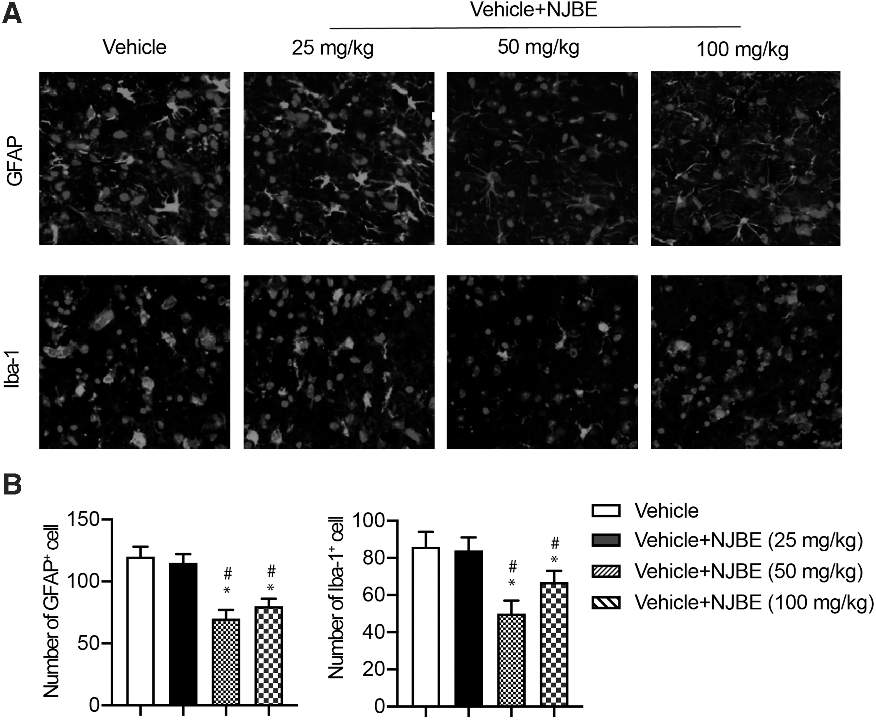
NJBE reduces the number of microglia after ischemic stroke. Immunofluorescent staining of astrocytes and microglia in cortical brain sections. Brain slices from MCAO mice stained with anti-GFAP or anti-Iba-1 antibody.
NJBE inhibits oxidative stress
MDA, GPx, SOD, and NO levels in the ischemic core and ischemic brain homogenates 3 days after ischemia/reperfusion are shown in Figure 4. In brain tissue, MDA and NOS levels were significantly increased in MCAO mice, and GPx, CAT, and SOD levels were significantly decreased. However, NJBE significantly reversed the reduction in antioxidant enzymes in brain tissue compared with the MCAO group, based on the partial recoveries of GPx, CAT, and SOD levels. NJBE inhibited lipid peroxidation and reduced oxidative stress after ischemia/reperfusion, as shown by the MDA and NO levels, respectively. These results support the beneficial effects of NJBE on neurological deficit recovery and brain infarct size.

NJBE inhibits oxidative stress after MCAO. Pretreatment with NJBE (25, 50, and 100 mg/kg, i.g.) was performed for 14 days followed by 2 days of administration after MCAO. MDA content
NJBE maintains the integrity of blood-brain barrier
Blood-brain barrier (BBB) dysfunction after MCAO contributes to vasogenic cerebral edema. Therefore, we examined the impact of NJBE on the integrity of the BBB after MCAO. Western blot analysis results indicated that NJBE preserved the tight junction protein claudin-5 and MMP9 after MCAO (Fig. 5). These results suggest that NJBE preserves BBB integrity after MCAO.
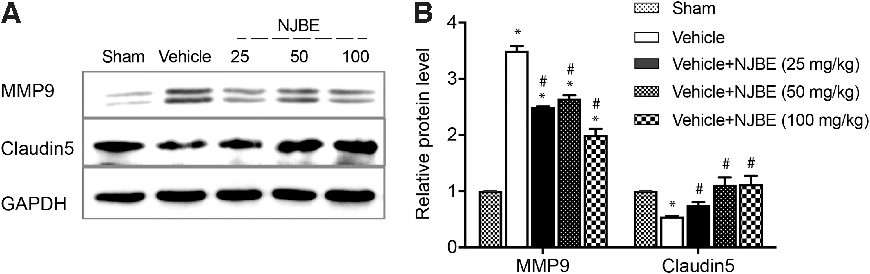
NJBE preserves the integrity of the blood-brain barrier after MCAO.
NJBE inhibits the inflammatory response in the ischemic brain
Several trials have shown that ischemic stroke induces a prominent inflammatory response in the periphery during the acute phase. 22 Cerebral ischemia injury leads to increases in circulatory proinflammatory cytokine and chemokine levels. Therefore, a potential therapeutic strategy is to inhibit the inflammatory responses after ischemic stroke. However, the anti-inflammatory effect of N. tangutorun Bobr. on ischemic stroke is still unknown.
The current study examined several inflammatory cytokines using ELISArray Kits and significantly changed cytokines were further confirmed using single ELISA Kits. The levels of TNF-α and IL-1β, which are indicators of inflammation, were significantly increased after cerebral ischemia in the vehicle group, which was consistent with the literature. However, NJBE attenuated the ischemia-induced increases in TNF-α and IL-1β to some extent (Fig. 6 A, B). NJBE treatment also upregulated IL-10 expression. These data suggest that NJBE exerts an anti-inflammatory effect after MCAO.

NJBE inhibits the inflammatory response in ischemic brain after MCAO. Pretreatment with NJBE (25, 50, and 100 mg/kg, i.g.) was performed for 14 days followed by 2 days of administration after MCAO. Samples prepared from ischemic hemispheres were assayed for inflammatory cytokines using ELISArray Kits
NJBE upregulates M2 markers and downregulates M1 markers
M1-to-M2 polarization of microglia/macrophages is associated with pro- and anti-inflammatory activities, as indicated by differential expression of surface markers. Our findings suggest that the polarization of microglia changes after NJBE treatment. Real-time PCR revealed that the mRNA expression levels of M1-type gene markers, such as monocyte chemotactic protein (MCP)-1, IL-1β, CCL-2, and TNF-α, were decreased by NJBE treatment after cerebral ischemia. In contrast, the mRNA expression of the tested M2 gene markers, such as Arg-1, CD206, and YM-1, was dramatically induced (Fig. 7). In combination with the ELISA results, these results showed that the anti-inflammatory effect of NJBE was associated with a shift in the polarization of microglia/macrophages in the MCAO model.
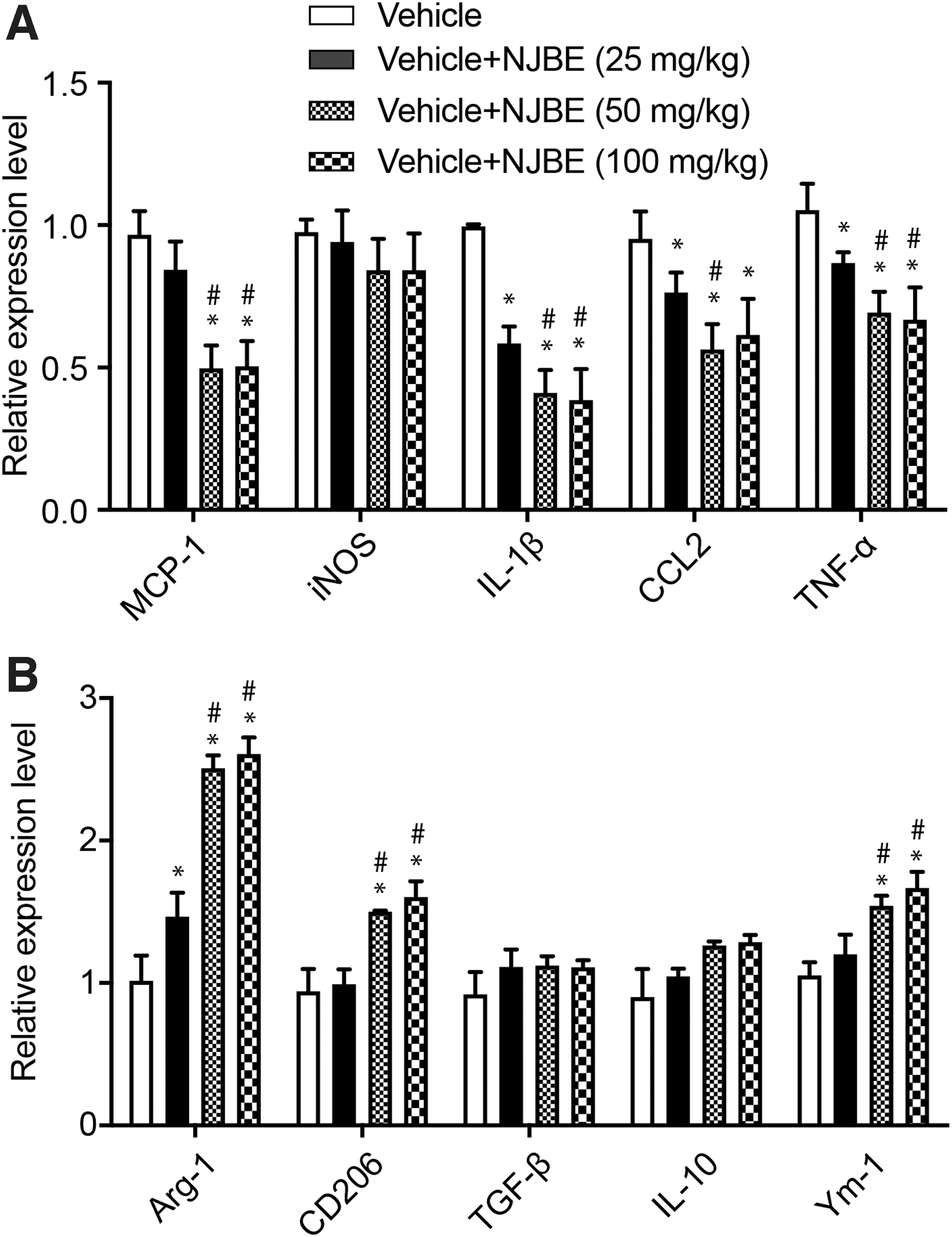
NJBE promotes M2 marker expression and inhibits M1 marker expression in the ischemic brain.
NJBE inhibits the activation of TLR4-TRAF6 signaling
Recent studies have revealed that the inflammatory mediator TLR4 participates in the process of ischemic damage. 18,23 TLRs primarily signal through the MyD88-dependent and the TRIF-dependent pathways to initiate immune responses. MyD88 and TRIF recruit TRAF6, which activates NF-κB or IRF3 to promote inflammation. Therefore, TRAF6 plays an essential role in TLR-mediated inflammation in ischemic injury. 18
To further reveal the mechanism of the anti-inflammatory effects of NJBE, we investigated the signal transduction associated with NJBE-induced neuroprotection in MCAO mice. TLR4, TRAF6, and pIκB/IκB were measured using Western blotting. As shown in Figure 8, compared with the sham group, TLR4 and TRAF6 expression significantly increased in the MCAO group. Consistent with previous reports, these results indicated that ischemic injury activates TLR4, TRAF6, and pIκB/IκB signaling. However, NJBE decreased TLR4, TRAF6, and pIκB/IκB expression. These results suggested that suppression of the TLR4-TRAF6-initiated inflammatory reaction is the possible mechanism associated with the neuroprotective effects of N. tangutorun Bobr. during ischemic stroke.
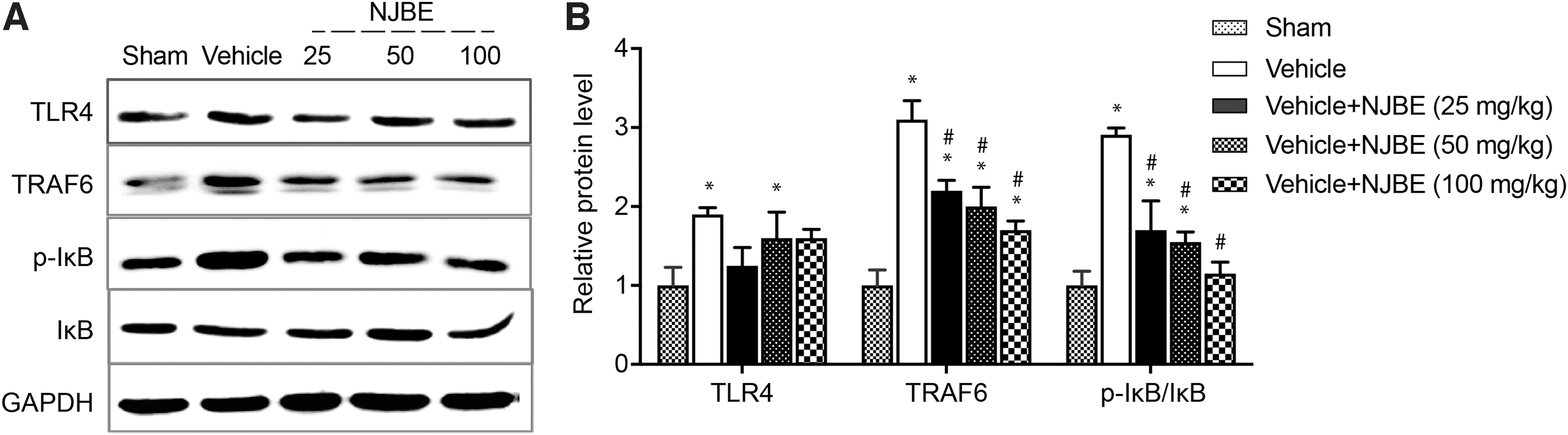
NJBE inhibits the activation of TLR4/TRAF6 signaling.
Discussion
In our previous study, NJBE was isolated from N. tangutorun and primarily consisted of phenols, flavonoids, and anthocyanins. NJBE shows antioxidant activity, as it protects cells from UVB-induced oxidative phototoxicity and doxorubicin-induced oxidation in vitro; NJBE exerts cardioprotection against doxorubicin-induced injury in H9c2 cardiomyocytes in vitro. 13 Several studies have reported that phenolics affect brain cognition through mechanisms that include decreasing neuroinflammation and scavenging oxidative stress factors. 24 –29 Based on these considerations, the current study evaluated the potential effect of NJBE on ischemic injury and the possible mechanisms by using an MCAO mouse model. We examined the effects of NJBE after MCAO utilizing a series of behavioral tests to evaluate the neurological deficits after ischemic stroke. The results indicated that NJBE significantly reduced infarct size, alleviated neurological deficits, and decreased the number of GFAP+ and Iba-1+ cells after MCAO.
Several studies showed that supplementing with plant-derived compounds, including flavonoids, effectively reduced weight gain in animals fed a high-fat/high-sucrose diet. 30 However, the flavonoid contents of NJBE exhibited no effects on weight loss. This difference may be due to the other components of NJBE or the different structures of the flavonoids derived from these plants.
Resident astrocytes and microglia are the primary cells in the central nervous system. As we know, astrocytes could express several kinds of inflammatory mediators, resulting in increased GFAP expression when they are activated after ischemia. The activation of microglia is the first step of inflammatory response after acute cerebral injuries, such as infection, inflammation, trauma, ischemia, and neurodegeneration. Microglia are rapidly activated within minutes after ischemia and develop an amoeboid morphology in response to insult, resulting in the secretion of various molecules. Iba-1, regarded as the surface marker of microglia, is also upregulated in the ischemic injured brain. In our study, GFAP and Iba-1 in the NJBE groups decreased compared with the vehicle group. These results showed that NJBE involved in the activation of astrocytes and microglia might signal its role as a critical regulator of ischemic stroke.
Oxidative stress is induced through cerebral ischemia/reperfusion injury, which is a pivotal factor that exacerbates brain damage. 4,23,31 Ischemia/reperfusion leads to increased production of ROS and other harmful chemical groups, which results in energy metabolism disorders in the brain, mitochondrial dysfunction, and increased secondary inflammation and apoptosis, and may lead to irreversible brain damage. ROS and hypoxia overwhelm endogenous antioxidant enzymes, such as SOD, GPx, and CAT. SOD is the critical enzyme for the scavenging of free radicals; CAT decomposes hydrogen peroxide and reduces inflammation. MDA indirectly reflects the level of ROS production and tissue damage. 4 NO plays a dual role in the pathological process of ischemic stroke by promoting the recovery of blood supply to the ischemic penumbra area and promoting the formation of ROS and proinflammatory effects that exacerbate brain tissue injury. 24 NJBE reduced oxidative stress injury and stimulated endogenous antioxidation in the ischemic brain tissue. Our results demonstrated that NJBE has a neuroprotective effect on acute cerebral ischemia in mice, regulating the oxidative/antioxidant balance in the ischemic brain.
Mechanical brain damage causes pathological changes, such as ischemic hypoxia and inflammatory reactions, in brain tissue, which result in the destruction of BBB. Therefore, treatment of BBB dysfunction may be a breakthrough in improving the efficacy of brain injury treatments. MMP9 and claudin-5 are critical indicators involved in tight junctions sealed and preserving the integrity of BBB. Several studies have reported that ischemic injury significantly enhances MMP9 and reduces claudin-5 expression. 19 Also, our results suggested that ischemic injury reduced claudin-5 expression and enhanced MMP9 expression. NJBE partially preserved the expression of claudin-5 and decreased the expression of MMP9. These results suggest that NJBE restored the disruptions in the integrity and function of BBB.
The inflammatory response induced by brain ischemia may not be locally restricted to the cerebral parenchyma and is an important factor that affects stroke patients. Several reports have shown that proinflammatory molecules and PMNs increase the severity of brain damage. Shifting the peripheral immune response from a proinflammatory pattern to an anti-inflammatory pattern helps repair the ischemic brain. 22 Our results suggested that NJBE has a potent anti-inflammatory activity.
In the ischemic brain, our results indicated a phenotypic shift in microglia/macrophages in response to NJBE treatment. The expression levels of inflammatory M1 cytokines (MCP-1, IL-1β, CCL-2, and TNF-α) significantly reduced, and the expression of anti-inflammatory M2 cytokines (Arg-1, CD206, and YM-1) increased compared with the vehicle group. This process was accompanied by the downregulation of TLR4 and TRAF6 and the ratio of pIκB/IκB. TLR4 triggers immune responses through MyD88- and TRIF-dependent pathways. MyD88 and TRIF recruit TRAF6, which activates NF-κB and leads to proinflammatory cytokine release. Studies have demonstrated that TRAF6 may promote macrophage differentiation into the M1 phenotype. 8 Our results also demonstrate that there is a potential link between the inhibition of M1 polarization and the suppression of the TLR4-TRAF6-NFκB signaling pathway in ischemic injury.
The neuroprotective effect is reflected in behavioral effects and reductions in infarct size in the mouse, which may be achieved by multiple factors, including anti-inflammatory and antioxidative factors. In our study, the results of the corner test, forelimb placement test, and infarct size analysis showed a certain concentration-dependent effect. The effects in the 50- and 100 mg/kg-treatment groups were relatively close. Thus, we hypothesize that 50 mg/kg is the optimal dose for this protective effect in MCAO mice. The optimal dose for clinical application still needs further exploration.
The World Stroke Organization emphasizes that one of the most important indicators for evaluating the final clinical efficacy of drugs is neurobehavioral functional recovery, which is an important issue that affects the quality of life of patients, but it also relates to preclinical research and the clinical treatment of stroke patients. 32 Traditional medicine improves the prognosis of clinical neurological function primarily by repairing the nervous tissue in the ischemic penumbra area. 33 –35
Recent reports have shown that dietary anthocyanin intake may play a pivotal role in the gut/brain axis because the gut microbiota converts anthocyanins to active compounds that influence brain functions, such as improving neuroinflammation and brain cognitive processes, through the gut/brain axis. 30 Our results demonstrate that NJBE derived from N. tangutorun Bobr. has great potential as a neuroprotective agent for clinical use in prophylactic protection against stroke.
In conclusion, our study evaluated the neuroprotective effect and underlying mechanism of NJBE using the MCAO mouse model. The neuroprotective effects were evidenced by significant improvements in neurological deficits and cerebral infarction sizes attributed to the antioxidant, BBB-protective, anti-inflammatory characteristics of NJBE. Our findings also support the hypothesis that NJBE mediates the microglia/macrophage phenotype modulation and suppresses the immune response in ischemic stroke through inhibiting the TLR4-TRAF6 signaling pathway. These results provide insight into the underlying mechanisms and provide valuable information for facilitating the design of more efficient adjuvants for ischemic stroke therapeutics. Further preclinical and clinical studies are needed to study the possible neuroprotective effects of NJBE and its therapeutic function in treating ischemic stroke patients.
Footnotes
Author Disclosure Statement
No competing financial interests exist.
Funding Information
This work was funded by the Department of Science and Technology of Jilin Province (No. 20200201376JC, No. 20200201144JC, and No. 20190701060GH), the Science and Technology Project of the Jilin Province Education Department (JJKH20201045KJ), the National Natural Science Foundation of China (No. 31300291, No. 81701193), and the Natural Science Fund of Tianjin Medical University (2015KYZM06).
Supplementary Material
Supplementary Data
References
Supplementary Material
Please find the following supplemental material available below.
For Open Access articles published under a Creative Commons License, all supplemental material carries the same license as the article it is associated with.
For non-Open Access articles published, all supplemental material carries a non-exclusive license, and permission requests for re-use of supplemental material or any part of supplemental material shall be sent directly to the copyright owner as specified in the copyright notice associated with the article.