




Abstract
Ankaferd Blood Stopper (ABS) is a medicinal plant extract that has anti-inflammatory effect. Inflammatory bowel disease is a pathological condition that directly affects colon health and increases the risk of colon cancer. Especially inflammation is an important factor in the formation and progression of this disease. The aim of the study was to investigate the protective effect of ABS on colonic inflammation. Caco-2 and RAW 264.7 cells were used as a model of in vitro colonic inflammation. RAW 264.7 cells were treated with lipopolysaccharide for 12 h to induce inflammation, and an inflammatory medium (IM) was obtained. Caco-2 cells were treated with 15 μL/mL ABS for 4 h, then incubated with IM. The cells also were incubated with 15 μL/mL ABS and IM together for 12 h. Tumor necrosis factor alpha (TNF-α) protein levels were targeted in testing inflammatory condition and cyclooxygenase-2 (COX-2) mRNA level was used as a marker gene to show the possible anti-inflammatory effect of ABS in Caco-2 cells. TNF-α level was 26.1-fold higher than the control group. IM caused 3.2-fold increase in COX-2 expression in Caco-2 cells. Pretreatment of Caco-2 cells with ABS resulted in 3.3-fold decrease in COX-2 mRNA levels relative to IM group. Furthermore, COX-2 mRNA level reduced 4.7-fold when ABS and conditional medium were given at the same time. ABS has suppressive effect on COX-2 mRNA expression in Caco-2 cells. These results suggest that ABS might have protective and therapeutic effect for colonic inflammation.
Introduction
Ankaferd Blood Stopper (ABS) is a medicinal standardized plant extract that comprises five various plant extracts: Thymus vulgaris, Glycyrrhiza glabra, Vitis vinifera, Alpinia officinarum, and Urtica dioica that have historically been used as hemostatic agents in Turkish medicine. 1 Each of these plant extracts shows antibacterial and cellular proliferation activity as well as hemostatic, antithrombotic, antineoplastic, and wound healing effects. 2,3 The basic mechanism of ABS is based on the formation of a protein network that provides red blood cell aggregation through spectin and ankyrin receptors. 2,3 It is used to stop bleeding caused by injuries and surgical interventions, also involved in wound healing of intestinal mucosal damage due to oxidative stress and inflammation. 4 It has been shown that ABS has an in vitro antimicrobial effect against both gram negative and gram positive bacteria. 2 In addition, an in vitro study demonstrated its antioxidant and antimutagenic activity. 5 Furthermore, a recent study suggested that ABS influenced regulation of cancer-related proteins and pathways in Caco-2 cells. 6 Previous studies have reported that plant extracts have an effective role against chronic inflammation and may contribute to reduction of inflammatory-related diseases. 7
Inflammatory bowel disease (IBD), including Crohn's disease and ulcerative colitis, is a common chronic disorder of the gastrointestinal tract that causes abdominal pain, diarrhea, gastrointestinal bleeding, and increases the risk of colon cancer. 8 All these conditions cause elevated levels of cytokines such as interleukin-6 (IL-6), interleukin-8 (IL-8), and tumor necrosis factor alpha (TNF-α), which are key signals of intestinal immune response. 9 Cyclooxygenase-2 (COX-2) is an important gene whose production is enhanced by proinflammatory cytokines at the sites of inflammation. 10 Previous research has also demonstrated that COX-2 is induced in large intestinal epithelium in active human IBD. 11 Several studies have been focused on the physiological effects of ABS; however, ABS-dependent mechanism on colonic inflammation has not been elucidated. Therefore, the aim of the study was to investigate the effect of ABS on colonic inflammation. Caco-2 and RAW 264.7 cells were used to form in vitro colonic inflammation model. Lipopolysaccharide (LPS) was used as an inflammation inducer on RAW 264.7 cells and an inflammatory medium (IM) was formed. Caco-2 cells were pretreated with ABS, then incubated with IM. The cells also were incubated with ABS and IM together. Finally, the effects of ABS treatments were determined in the formed colonic inflammation model.
Materials and Methods
Reagents
ABS was taken from local pharmacy as 2 mL ampoule form. This standardized plant mixture composition is shown in Table 1. Dulbecco modified Eagle's medium (DMEM, Cat. No. D5546), RPMI 1640 (Cat. No. R2405), LPS from Escherichia coli O11:B4 (Cat. No. L2630), Penicillin-Streptomycin (Cat. No. P4333), and 1-(4,5-dimethylthiazol-2-yl)-3,5-diphenylformazan (M2003; MTT) were purchased from Sigma. Fetal bovine serum (FBS; Cat. No. 10500; Gibco, NY, USA) was purchased from ThermoFisher. Mouse TNF-α enzyme-linked immunosorbent assay (ELISA) Kit (Cat. No. EKO527) was purchased from Boster Bio. Folin & Ciocalteu's phenol reagent for phenolic measurements (Cat. No. F9252) was purchased from Sigma. All other chemicals were analytical grade. Unless stated otherwise, all chemicals were prepared in Milli-Q® water.
Dried root extract.
Dried leaf extract.
Dried grass extract.
Cell culture
The RAW 264.7 murine macrophage cell line were cultured in RPMI 1640 medium supplemented with 10% FBS, 1% penicillin and streptomycin. Caco-2 cells were maintained in DMEM with 10% FBS, 1% nonessential amino acids, 1% sodium pyruvate, and 1% penicillin and streptomycin. Cells were cultured in 5% CO2 incubator at 37°C. Passage numbers of the cells were kept between 20 and 35.
ABS toxicity on Caco-2 cells
MTT assay was applied to Caco-2 cells for determining the toxicity levels. As the colonic inflammation study has been conducted to confluent cells, the cytotoxicity analysis of the ABS on Caco-2 cell line was also determined in the same conditions. The cells were seeded in 96-well plates and waited to adhere completely to the well. After differentiation, the cells were treated with 15 μL/mL ABS for 12 h, which is the maximum contact period. Then, the medium was discarded and 100 μL of fresh medium was added into each well. Next, 10 μL of MTT reagent was applied to each well and incubated for 4 h. At the end of this period, the medium was discarded. To terminate the reaction, the cells were lysed with 100 μL dimethyl sulfoxide and the absorbances were recorded in 450 nm.
Measurements of TNF-α secretion from RAW 264.7 macrophages
LPS-dependent release of TNF-α cytokine was determined by ELISA method. The liquid growth medium from LPS-treated RAW 264.7 cells were centrifuged in 1000 g at 5 min and placed in 96-well plates coated with antibody specific to TNF-α proteins. The procedure was applied according to the kit instructions. The amount of TNF-α released from the samples will be determined by spectrophotometric measurement.
Establishment of colonic inflammation model
Caco-2 cells were seeded at 9 × 10⁴ cells/cm2 density onto 12-well plates and grown for 21 days. At day 20, RAW 264.7 cells grown in 75 cm2 flask and treated with 0.5 μL/mL LPS for 12 h to induce the inflammation, and IM was obtained. The Caco-2 cells were treated with 15 μL/mL ABS for 4 h, then incubated with IM for additional 12 h to determine the preventive effect of ABS. In addition, the cells were also incubated with 15 μL/mL ABS and IM together for 12 h to determine the therapeutic effect of ABS on simulated colonic inflammation model (Fig. 1).
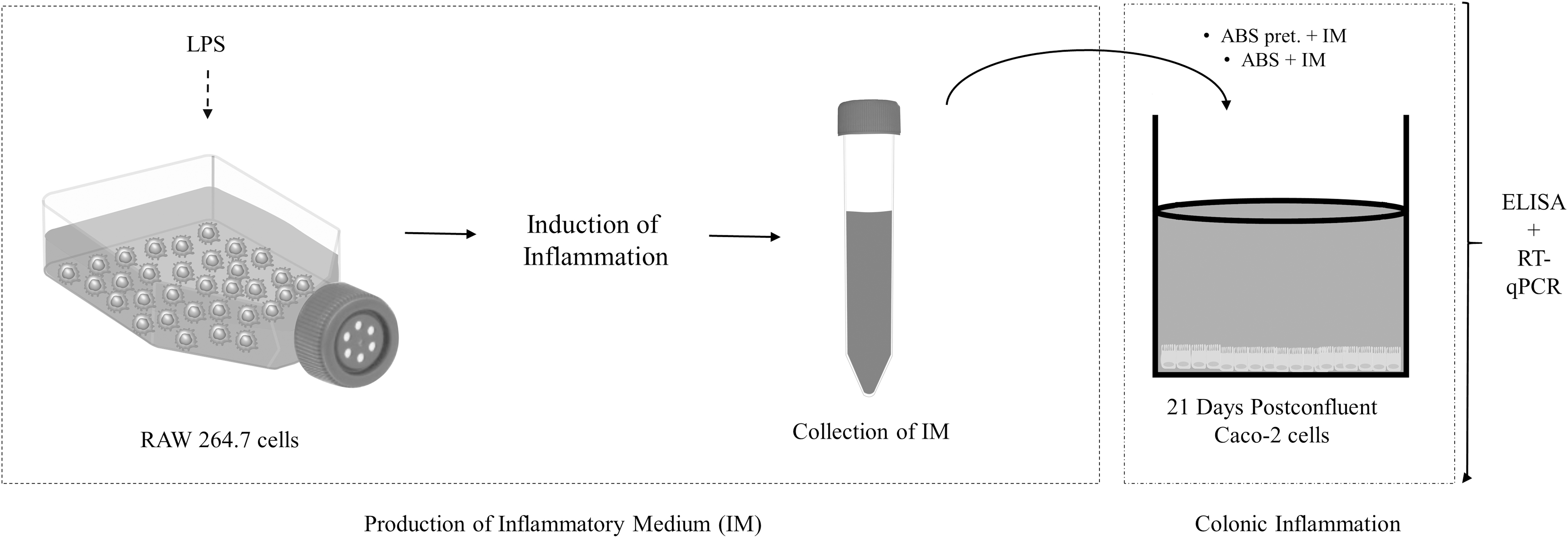
The overall experimental procedure by using in vitro colonic inflammation model.
RNA isolation and quantitative reverse transcription polymerase chain reaction
RNA isolation procedure was performed by using RNAzol® reagent (Cat. No. RN190; MRC, Inc., Cincinnati, OH) according to the company's protocol. One milligram total RNA sample was used to convert cDNA using cDNA synthesis kit (Cat. No. 4368814; Life Technologies, WG, UK). To determine the mRNA expression levels of genes, reverse transcription quantitative polymerase chain reaction (RT-qPCR) was performed on an ABI StepOnePlus instrument (Lifetech, WDL, Singapore) by SYBR-Green mix (Cat. No. 4367659; Lifetech, WG, UK). Human cyclophilin A (CYPA, F:tacgggtcctggcatcttg and R:cgagttgtccacagtcagca) was used as a housekeeping gene for normalization of mRNA expression results and primers were used for target mRNAs: COX-2, F:cttcacgcatcatttttcaag and R:tcaccgtaaatatgatttaagtccac; IL-6, F:caggagcccagctatgaact and R:gaaggcagcaggcaacac; IL-8, F:tggctctcttggcagccttc and R:tgcacccagttttccttggg). The RT-qPCR data were converted to gene expression levels through 2ΔCt calculation methods. 12
Statistical analysis
All results were expressed as mean ± standard error with at least three replicates. In the comparison of the experimental groups, one-way analysis of variance with Tukey's post hoc test was performed for all experiments. GraphPad Prism (version 8.2.1) was used in the preparation of all analyses and figures.
Results and Discussion
Establishment of inflammatory condition using RAW 264.7 cells stimulated with LPS
RAW 264.7 cells were treated with LPS to induce inflammation and the released cytokines were collected from these macrophage cells. TNF-α is an important cytokine that mediates inflammation. 13 In this study, it is used as a marker to confirm inflammatory conditions. LPS concentration was determined based on the measurement of the TNF-α protein levels from RAW264.7 cells where the treatment caused most significant difference when compared with the control group. 0.5 and 1 μL/mL of LPS treatments for 12 h caused 7.5- and 6.8-fold increase in TNF-α proteins, respectively (Fig. 2). Since the TNF-α production was higher in 0.5 μL/mL LPS experimental group, the incubation concentration is fixed at 0.5 μL/mL for RAW 264.7 cells. In addition, the result is in agreement with the previous report that showed induction of TNF-α levels upon stimulation of macrophages by LPS. 14

Concentration profile of LPS and relative TNF-α production in RAW264.7 cell line. The cells treated with 0.5 and 1 μg/mL LPS for 12 h. TNF-α protein levels were determined by ELISA. Different letters indicate statistical significance between groups, P < .05 (1-factor ANOVA and Tukey's post hoc test). Values represent the means ± SE (n = 3). CTRL, control group. ANOVA, analysis of variance; ELISA, enzyme-linked immunosorbent assay; LPS, lipopolysaccharide; SE, standard error; TNF-α, tumor necrosis factor alpha.
Responses of the colonic inflammation model toward LPS
In vivo and in vitro studies showed the hemostatic, antimicrobial, and proliferative effects of ABS. 2,15,16 It was indicated that ABS given Caco-2 cells induced protein levels of TNF receptor-associated factor 3 (CAP1) and heat shock 70 kDa protein 4 (HSPA4), which are involved in immune system response. 6 Another study also suggested that the anti-inflammatory effects of ABS on a rat model of necrotizing enterocolitis by causing reduction of TNF-α and IL-6 levels. 17 However, much uncertainty still exists about the relation between ABS and colonic inflammation. Within the scope of this project, Caco-2 and RAW 264.7 cell lines were used as a combined model of colonic inflammation. Caco-2 cells were grown for 21 days to postconfluency to form physiological similarities with enterocyte cells. 12 We measured TNF-α levels for every new experiment to determine the inflammatory condition in RAW 264.7 cells. Thus, at day 20, RAW 264.7 cells treated with 0.5 μL/mL LPS for 12 h and IM was obtained. In LPS-treated groups, 26.1-fold induction in TNF-α was observed (Fig. 3A). A statistically significant increase in TNF-α production in the 0.5 μL/mL LPS-treated group was accepted as an evidence of successful inflammatory condition. After that, Caco-2 cells were treated with IM and inflammatory gene expressions were measured by RT-qPCR. IL-6, IL-8, and COX-2 genes were used as marker genes to evaluate whether IM treatment induced the mRNA expression of inflammatory cytokines. Induction of proinflammatory cytokines (interleukin-1β, IL-6, prostaglandin E2, and TNF-α) in immune cells are generally observed in IBD. 18 Results showed that IM treatments significantly increased IL-6 (1.97-fold), IL-8 (2.05-fold), and COX-2 (3.24-fold) mRNA levels (Fig. 3B). It has been indicated that LPS treatments in Caco-2 cells lead to elevated production of IL-6, IL-8, and COX-2 genes, and this is consistent with the current findings. 18,19 When the results were compared among themselves, COX-2 genes were expressed more significantly than other marker genes, thus it was used as a marker to determine whether the protective or therapeutic effects of ABS on colonic inflammation. According to the literature, COX-2 is known as an indicator for inflammatory response. 20 A recent study showed that various plant extracts were used as a treatment of IBD by using different mechanisms such as antioxidant, antiulcer, and inhibiting production of COX-2 cytokine. 21 Cyclooxygenase enzymes play role in conversion of arachidonate to prostaglandin H2, which is a precursor for several cell-specific prostaglandin and thromboxane synthases. Prostaglandins involve in several biological processes such as immune function and gastrointestinal integrity. 22
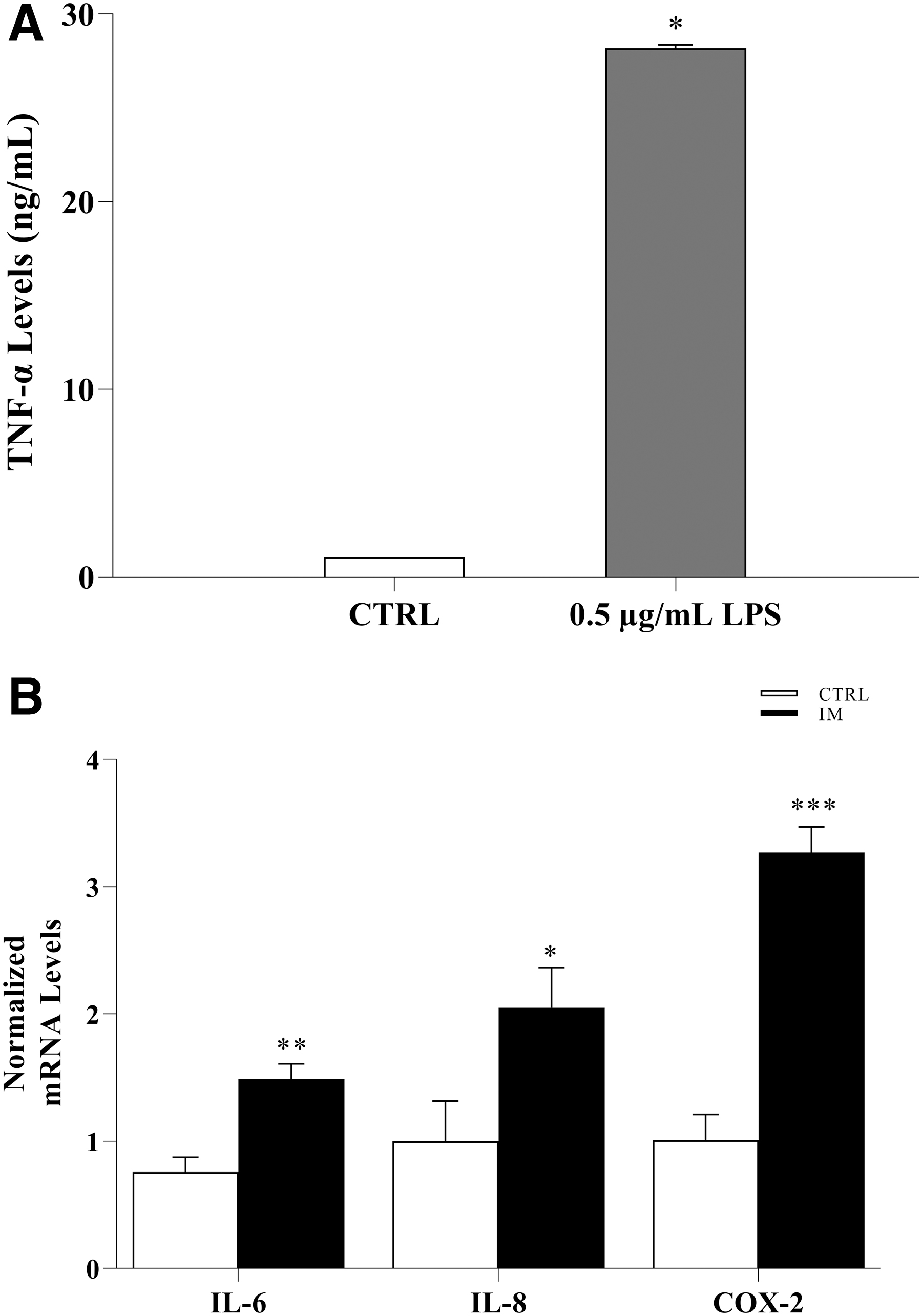
The inducible effect of LPS and IM on TNF-α production, IL-6, IL-8, and COX-2 mRNA expression in vitro colonic inflammation model.
Protective and therapeutic effect of ABS treatments on colonic inflammation model
A cytotoxicity test was performed by using MTT reagent to evaluate the cytotoxic effect of ABS on Caco-2 cells. When the cells were treated with 15 μL/mL ABS for 12 h, the cell viability was not affected by the treatment (P > .05) (Fig. 4A). Thus, 15 μL/mL was selected as the treatment concentration on Caco-2 cells. At day 20, Caco-2 cells were pretreated with ABS for 4 h and then IM was introduced as an inflammation inducer for 12 h time period. In addition, ABS and IM were given at the same time to determine the therapeutic effect. To determine the potential anti-inflammatory properties of ABS, expressions of COX-2 were analyzed (Fig. 4B). Treatment of IM induced COX-2 gene expression (3.2-fold), whereas both pretreatment (3.3-fold) and treatment (4.7-fold) of ABS resulted in the downregulation of LPS-induced expression of COX-2 mRNA similar to the nontreated control group, suggesting that ABS has both preventive and therapeutic effects on inflammation in human colon system.
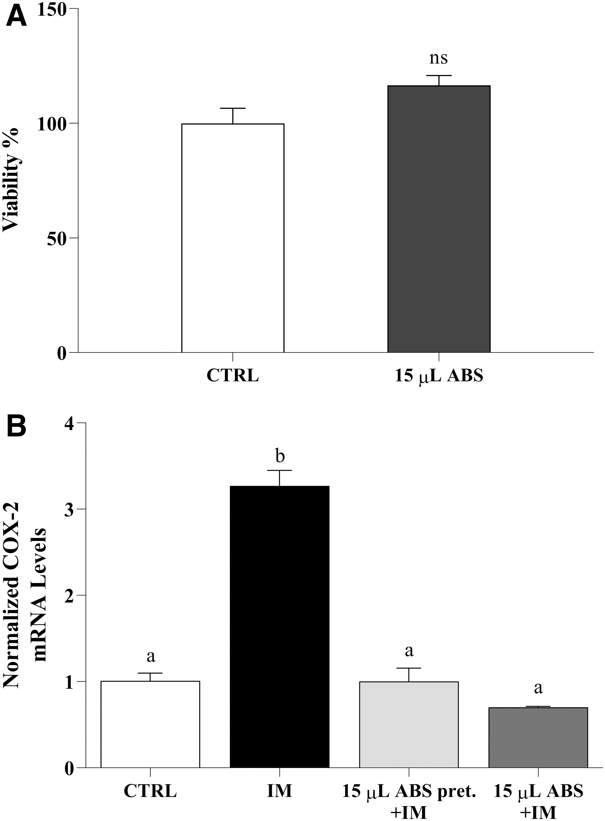
Cytotoxicity analysis and suppressive effect of ABS on COX-2 mRNA gene expression in Caco-2 cells. The cells were treated with 15 μL/mL ABS for 12 h and cell viability was determined by using an MTT assay.
ABS treatments cause downregulation in COX-2 mRNA levels compared with IM experimental group in vitro colonic inflammation model. The findings of this study showed that ABS has significant effects on the inflammation that may occur in the colon and it might be an alternative approach for controlling of IBD or in individuals at risk of this disease. This is the first in vitro study to investigate the effect of ABS on intestinal inflammation. However, in vivo and clinical studies are necessary to clarify the effect of ABS on colonic inflammation.
Footnotes
Authors' Contributions
All authors designed the investigation, performed the experiments, wrote the article, prepared figures, and approved the final article.
Author Disclosure Statement
No competing financial interests exist.
Funding Information
This project was funded by İzmir Institute of Technology (IZTECH) Scientific Research.