
Abstract
Quercus ilex fruit is widely used in the treatment of various gastrointestinal disorders, including diarrhea, for its bioactive compounds and astringent property. The current study focuses on the phytochemical characterization of the Q. ilex-aqueous extract (QIAE) and its protective effect against gastroduodenal (GD) ulcer (GDU) produced by absolute ethanol (EtOH) intoxication in adult male Wistar rats. Experimental rats were divided into six groups (n = 6): control, EtOH [95%, 4 g/kg body weight (b.w.)], EtOH + different doses of QIAE (100, 200, and 400 mg/kg, b.w.), and EtOH + Famotidine (FAM, 10 mg/kg, b.w.). Animals were orally pretreated (p.o.) with QIAE for 15 days and intoxicated with a single oral administration of EtOH for 2 h. The findings showed that the QIAE is rich in phenolic-astringent compounds and fibers, and it exhibited a significant scavenging activity on DPPH/ABTS free-radicals with half maximal inhibitory concentration (IC50) values of 177.00 ± 5.11 and 203.9 ± 2.23 μg/mL, respectively. In vivo part, QIAE significantly reduced the GD mucosal injury revealed by edema and leukocyte infiltration of the submucosal layer. GD mucosal homogenates revealed a remarkable increase in endogenous antioxidant enzyme activities (catalase, superoxide dismutase, and glutathione peroxidase) and a decrease in the lipid peroxidation levels (malondialdehyde) in animals pretreated with QIAE compared with the ulcer control group. QIAE exerted significant and dose-dependent anti-GDU protection in the rat model with a more effective action than FAM. The GD protective effect of the QIAE might be related to a direct radical scavenging activity, increased antioxidant enzymes, and depression of lipid peroxidation.
Introduction
Apeptic ulcer represents a collective term for a group of chronic events that affect the integrity of the lining of the stomach and/or duodenum. It is marked by pain, perforation, bloating, nausea, blood in the stool or vomiting, loss of appetite, and weight loss. 1 It is now known that the majority of peptic ulcer cases are associated with Helicobacter pylori infection and/or the use of nonsteroidal anti-inflammatory drugs. 2 The incidence of this disease increases with age, with gastric ulcers peaking between the fifth and seventh decade, in contrast to duodenal ulcers, which typically occur 10–20 years earlier and both sexes are affected equally. 3
This disease is often defined as a mucosal break greater than 3–5 mm in the stomach or duodenum with a visible depth. Also, it is the result of an imbalance between the protective (mucus secretion, cell regeneration secretion, blood flow, and epithelial barrier) and damaging factors (acid-pepsin secretion and reactive oxygen species [ROS]) of the stomach and duodenum lining. 4,5 ROS, especially the hydroxyl radical (OH•), are responsible for the oxidative degradation of mucous membranes in all types of ulcers. 6 As a result, the gastric mucosa provides a dynamic barrier against the effects of harmful agents through a series of endogenous antioxidant defense systems. 7,8
Nowadays, antisecretory drugs, including histamine type 2 receptor antagonists and irreversible proton pump inhibitors, for example, omeprazole and famotidine (FAM), are the main pharmacological treatment for this disease. Despite its efficacy, long-term treatment with these drugs is associated with several side effects and poor gastric healing, leading to the recurrence of ulcers. 9 This pathology has led researchers to develop many pharmaceutical products and they have already tested several natural compounds to prevent or treat gastric ulcers such as quercetin 10 and curcumin. 11 Phytotherapy is used to treat various pathologies such as gastrointestinal diseases, including peptic ulcers without any side effects. 12
Quercus ilex L. (the green oak) belongs to the family of Fagaceae, and is distributed in several countries such as France, Italy, Croatia, Greece, and Maghreb, including Morocco, Algeria and Tunisia. 13 In north Tunisia, large areas of unexploited holm oak forests have attracted the attention of scientists, mainly because of their high availability and resistance to drought. 14 To their content of carbohydrates, amino acids, proteins, lipids, and several sterols, the fruit of oak tree, have long been used as a source of tannin, oil, and especially food. 15 Oak acorns were traditionally used in medicine, especially roasted grains as astringents and antidiarrheal agents. 16,17
However, no scientific data exist to our knowledge regarding the effect of the Q. ilex fruit on the pathology of digestive ulcers. Therefore, this study was conducted to assess the gastroduodenal (GD) protective role of the Q. ilex-aqueous extract (QIAE) against gastroduodenal ulcer (GDU) and oxidative stress induced as a result of acute exposure to ethanol (EtOH) and the mechanism involved in this protection.
Materials and Methods
Chemicals
Methanol, EtOH, sodium chloride (NaCl), aluminum chloride (AlCl3), phosphate-buffered saline, Folin–Ciocalteu, hematoxylin, eosin, trichloroacetic acid (TCA), 2,2-diphenyl-1-picrylhydrazyl (DPPH), 2,2′-azino-bis (3-ethylbenzothiazoline-6-sulfonic acid) (ABTS), hydrochloric acid (HCl), butylated hydroxytoluene (BHT), 2-thiobarbituric (TBA), 2,4-dinitrophenylhydrazine, epinephrine, bovine catalase, tris, catechin, quercetin, and gallic acid were procured from Sigma-Aldrich (GmbH, Steinheim, Germany).
Preparation of QIAE
Green oak acorns were collected from the area of Fernana (northwest of Tunisia) during the month of December 2018 and identified by the botanic coordinator, Institute of Biotechnology of Beja, University of Jendouba. Thereafter, the plant material was dried at 40°C for a duration of 72 h with air circulation and later rigorously crushed in a traditional metallic mortar and then with an electric blender. The powder was dissolved in doubly distilled water. The water extracts were prepared under magnetic conditions for 24 h and the homogenate was filtered under pressure. Finally, the QIAE was immediately used for in vitro and in vivo experiments.
Physicochemical characterization of QIAE
The physicochemical analyses are carried out according to the standards described by the Association of Official Analytical Chemists. 18 All the analyses were realized in a minimum of three tests. The dry matter was determined by drying 0.5 g of the powder in a ventilated oven at 105°C until a constant weight was obtained. The difference in weight corresponds to the loss of moisture (water content) and the residue characterizes the dry matter content of the sample. 18
The ash and mineral content of green oak acorns can be calculated from this analysis. To do this, 2 g of the powder was placed in a porcelain crucible, and tared and then incinerated in a muffle furnace at 550°C for 3 h. The weight loss observed during calcination corresponds to the organic matter and the weight of the residue to the mineral matter. 18
QIAE crude fiber content
The fiber contents are analyzed using enzymatic gravimetric analysis according to Association of Analytical Chemists (AOAC) International Official Method 991.42. 18
QIAE polyphenol content
The overall phenolic content was quantified by the colorimetric method, the Folin–Ciocalteu method. 19 Briefly, 500 μL of the extract was added to 10 mL of water and 0.5 mL of Folin–Ciocalteu Reagent. After 5 min, 8 mL of 7.5% sodium of the carbonate solution was added. The reaction was kept in the dark for 2 h and was measured at 765 nm using a UV-visible detector spectrophotometer. Gallic acid was applied as a standard, and the results were expressed in milligram gallic acid equivalent per gram dry weight (mg GAE/g DW).
QIAE flavonoid content
The total flavonoid content was detected by the AlCl3 colorimetric method. 20 In a brief step, 1 mL of the sample was mixed with 1 mL of 2% AlCl3 solution. After a 15-min incubation at room temperature, the optical density of the reaction mixture was evaluated at 430 nm. Quercetin was used as a reference standard and the total flavonoid content was expressed as milligram quercetin equivalent per gram dry weight (mg QE/g DW).
QIAE-condensed tannin content
The condensed tannin content was analyzed using colorimetric methods demonstrated by Mahmoudi et al. 21 Concentrated HCl (750 μL) was added to 1.5 mL of vanillin solution (4%) prepared in methanol and 250 μL of sample. The aliquots obtained were incubated at room temperature for 15 min. Absorbance was measured at 500 nm using a UV-visible spectrophotometer. Catechin was used as standard and the results were expressed in milligram catechin equivalent per gram dry weight (mg CE/g DW).
Free radical-scavenging activity on DPPH and ABTS
The antioxidant capacity of the QIAE was realized with the help of DPPH. 22 In brief, different concentrations of QIAE (0, 20, 50, 100, 150, 200, 250, and 300 μg/mL) were added to 1 mL of 0.1 mM methanol solution of DPPH and incubated at 27°C for 30 min. The optical density of the sample was measured at 517 nm. DPPH radical scavenging (RSA), expressed as a percentage, was estimated using the following formula:

Ascorbic acid was used as the reference molecule in the same concentration as the extract tested. All analyses were carried out in triplicate. Efficacy 50 was determined as the value of concentration (μg/mL) of the compound needed to recover 50% of the DPPH radical.
The antioxidant capacity of the QIAE was evaluated using the extract of ABTS. 23 In brief, 1 mL of diluted extract was mixed with 3 mL of 7 mM ABTS radical solution (ABTS•+) and was kept in the dark at room temperature for 60 min. Absorbance was determined at 734 nm. The scanning capability was calculated as ([1 – Ab/A0] × 100%), Ab and A0 are the absorbance of samples as well as the ABTS•+ solution at 734 nm.
Animals and treatment
Male Wistar rats about 15 weeks old (weighing 200–220 g, housed six per cage)) were obtained from the Central Society of Pharmaceutical Industries of Tunisia (SIPHAT, Ben-Arous, Tunisia). They received feed (standard pellet-based diet—Badr Utique, Tunisia) and water at will and were kept in temperature-controlled barns (22 ± 2°C) with a 12-h light–dark cycle.
All the animals were divided into six groups of six rats each. The control groups 1 and 2 were treated with bidistilled water (5 mL/kg b.w./day). However, groups 3–5 were treated with various doses of QIAE (100, 200, and 400 mg/kg b.w.), while group 6 was treated with FAM (10 mg/kg b.w.) for a period of 15 days. Rats were fasted for 24 h before the last administration of QIAE or FAM. One hour later, each animal, except Group 1, received EtOH (95%, 4 g/kg b.w.) by oral administration. After 2 h, the rats were sacrificed. The gastric mucosa was immediately collected and homogenized in Tris-NaCl buffer (50mM, pH 7.6). The resulting supernatants were used for biochemical assays.
Evaluation of GD mucosal damage
The stomach of each animal was removed and opened at its greatest curvature. The tissues were carefully rinsed in 0.9% NaCl. The lesions of the gastric and duodenal mucosa were macroscopically examined and photographed. The indices of ulcer were determined as the total sum of the lengths of all gastric lesions (in square millimeter). 24 Two independent blind observers made the measurements of lesion length.
Gastric volume juice determination
Gastric juices were extracted and centrifuged at 3000 g for 5 min to eliminate insoluble matter. The supernatant was later measured using graduated tubes. 25
Histopathological analysis
Directly after the sacrifice, small pieces of stomach were collected and washed with ice-cold saline solution. The fragments of tissue were then fixed in 10% neutral buffered formalin solution, embedded in kerosene, and used for further histopathologic examination. Sections 5 μm thick were cut, deparaffinized, hydrated, and stained with hematoxylin and eosin (H&E). Gastric sections were examined in a blinded manner for all treatments.
GD mucosal homogenate preparation
The mucosa of both organs was subsequently homogenized using a T-18 digital ultra-turrax homogenizer in Tris-buffered saline (TBS) buffer (50 mM, pH 7.6). The homogenates were then centrifuged at 3000 g at 4°C for 15 min. The resulting supernatants are used to determine malondialdehyde (MDA) and protein levels as well as the activity of antioxidant enzymes such as superoxide dismutase (SOD), catalase (CAT), and glutathione peroxidase (GPx).
Lipid peroxidation measurement of GD mucosal homogenates
The peroxidation of lipids in the gastric mucosa was determined by measuring MDA using the double heating method. 26 In brief, aliquots of gastric mucosal homogenates were mixed with a BHT-TCA solution that contained 10 mg/mL BHT (w/v) dissolved in 200 mg/mL TCA (w/v) and centrifuged at 1000 g for 5 min at 4°C. The supernatant was mixed with 0.5 N HCl and 120 mM TBA within 26 mM Tris and heated at 80°C for a duration of 10 min. The absorbance of the resulting chromophore was determined at 532 nm after cooling. The levels of MDA were determined using an extinction coefficient for the MDA-TBA complex of 1.56 × 10⁵ M−1 cm−1.
Antioxidant enzyme activities of GD mucosal homogenates
The SOD activity was obtained using modified epinephrine assays. 27 At alkaline pH, the superoxide anion (O2 –) causes the auto-oxidation of epinephrine to adrenochrome; while competing with this reaction, SOD decreased the formation of adrenochrome. An SOD unit is the quantity of the extract that inhibits the rate of adrenochrome formation by 50%. The enzyme extract was added to 2 mL reaction mixture containing 10 μL bovine catalase (0.4 U/μL), 20 μL epinephrine (5 mg/mL) and 62.5 mM sodium carbonate/bicarbonate buffer pH 10.2. Absorbance changes were observed at 480 nm.
The CAT activity was evaluated by measuring the initial rate of hydrogen peroxide (H2O2) disappearance at 240 nm. 28 The reaction mixture contained 33 mM H2O2 in 50 mM phosphate buffer at pH 7.0 and the activity of CAT was calculated using the extinction coefficient of 40 mM−1 cm−1 for H2O2. The GPx activity was quantified by the Flohé and Günzler method. 29 In brief, 1 mL of reaction mixture containing 0.2 mL gastric mucosal supernatant, 0.2 mL 0.1 M phosphate buffer pH 7.4, 0.2 mL glutathione (GSH) (4 mM), and 0.4 mL H2O2 (5 mM) was incubated at 37°C for 1 min and the reaction was stopped by addition of 0.5 mL TCA (50 mg/mL, w/v).
After centrifugation at 1500 g for 5 min, aliquot (0.2 mL) of the supernatant was combined with 0.5 mL of 0.1 M phosphate buffer pH 7.4 and 0.5 mL of 5,5-dithio-bis-(2-nitrobenzoic acid) (DTNB) (10 mM) and the absorbance was read at 412 nm. The GPx activity was expressed in nanomolar of GSH consumed per minute per milligram of protein.
Statistical analyses
Statistical analyses were calculated using SAS Statistical Analysis System statistical software. Differences were stated as mean ± standard error of the mean and designated significant when the values of P were <.05.
Results
Physicochemical characteristics and bioactive compounds of QIAE
The variation of physicochemical characteristics of QIAE was determined and the results were expressed as a percentage (Table 1). QIAE contains 10.95% of water and 2.12% of ash. Furthermore, the results proved that the QIAE is rich in phenolic compounds, flavonoids, astringent compounds, and fibers. The secondary metabolites of QIAE in GAE, QE, and CE were found to be 52.60 ± 0.75 mg GAE per g dry matter (DM), 33.00 ± 1.70 mg QE per g DM, and 1.76 ± 0.10 mg CE per g DM, respectively. Moreover, the rate of fiber was 6.86 ± 0.82 mg/g DM.
Physicochemical Characteristics, Bioactive Compounds, and DPPH/ABTS Free-Radical Scavenging Activity of Quercus ilex aqueous Extract
ABTS, 2,2′-azino-bis (3-ethylbenzothiazoline-6-sulfonic acid); CE, catechin equivalent; DM, dry matter; DPPH, 2,2-diphenyl-1-picrylhydrazyl; GAE, gallic acid equivalents; IC50, half maximal inhibitory concentration; QE, quercetin equivalent; QIAE, Quercus ilex aqueous extract.
DPPH/ABTS free-RSA activity of QIAE
Antioxidant activity and the capability of QIAE to scavenge free radicals in vitro were evaluated by the DPPH/ABTS assays. The RSA activity of QIAE radicals increased significantly with the graduated doses. The percentage inhibition of DPPH/ABTS free-RSA activity of QIAE to be significant with IC50 values of 177.00 ± 5.11 and 203.9 ± 2.23 μg/mL, respectively. These results were compared with the standard ascorbic acid. The percentages of inhibition levels of the DPPH/ABTS free-RSA activity of the standard were 190.47 ± 1.22 and 175.13 ± 0.91 μg/mL for the DPPH and ABTS radicals, respectively (Table 1).
Macroscopic appearance examination of the QIAE or FAM effects on EtOH-induced GD mucosal injuries
The effect of a single oral administration of EtOH (4 g/kg, b.w.) shows the installation of ulcerous areas in the GD regions (Fig. 1). The animals in the lesion EtOH-group showed extensive and visible hemorrhagic injury in the gastric mucosa with lesion area 53.83 ± 2.02 mm2. Pretreatment of rats with QIAE (groups 3–5) or the reference drug group (FAM) before exposure to EtOH significantly (P < .05) reduced areas of gastric lesion from 40.66 ± 2.08 mm2 (24.46% of protection) to 4.66 ± 0.11 mm2 (91.34% of protection), which was more effective than FAM (10.00 ± 0.12 mm2 or 81.42% of protection).

Macroscopic appearance evaluation of gastroduodenal mucosa. Animals were pretreated with various doses of QIAE (100, 200, and 400 mg/kg, b.w., p.o.), FAM (10 mg/kg, b.w., p.o.), or bidistilled water, challenged with a single oral administration of EtOH (4 g/kg, b.w., p.o.) or NaCl 9‰ for 2 h.
These ulcerous areas were accompanied by a decrease in mucus volume (Table 2). GD ulcerated animals yielded the lowest mucus content of the gastric mucosa, while animal groups pretreated with various doses of QIAE or FAM showed significantly increased mucus content compared with the GD group.
Effects of Subacute (15-Day) Pretreatment with Quercus ilex Aqueous Extract (100, 200, and 400 mg/kg, b.w.) or Famotidine (10 mg/kg, b.w.) on Different Gastric Parameters of Ethanol (4 g/kg, b.w.)-Induced Rats
P < .05 compared with control group.
P < .05 compared with EtOH group.
EtOH, ethanol; FAM, famotidine.
Histopathological evaluation of the QIAE or FAM effects on EtOH-induced GD mucosal injuries
The stomach and the duodenum of the control section showed normal histologic features, while following EtOH administration it revealed for the stomach prominent and severe gastric mucosal damage, presence of edema, very important submucosal congestion, alteration of the surface coating, and leukocyte infiltration of the mucosal and submucosal layer. For the duodenum it revealed loss of surface coating and congestion of the intestinal villi (Fig. 2).
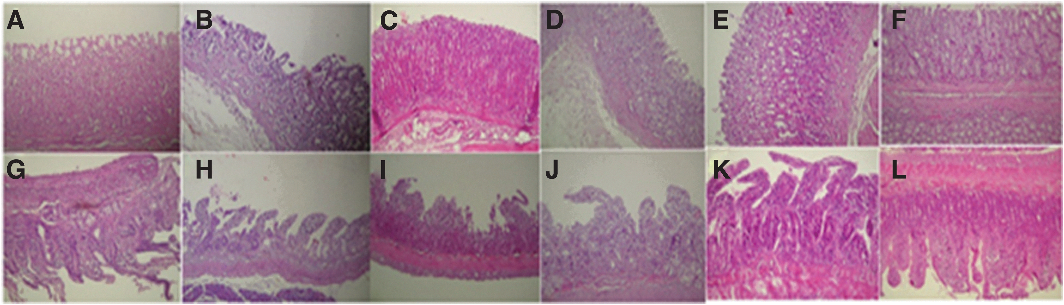
Subacute effect of QIAE and FAM on histopathological changes of the stomachs of rats with EtOH-induced gastritis. Animals were pretreated with various doses of QIAE (100, 200, and 400 mg/kg, b.w., p.o.), FAM (10 mg/kg, b.w., p.o.), or bidistilled water, challenged with a single oral administration of EtOH (4 g/kg, b.w., p.o.) or NaCl 9‰ for 2 h.
Pretreatment with QIAE presents a clear dose-dependent protection of the gastric and duodenal mucosa as seen by reduction of lesions, mucosal and submucosal edema, as well as leukocyte infiltration. A similar protective effect was also observed in FAM-pretreated rats.
Effect of QIAE or FAM on EtOH-induced GD mucosal lipoperoxidation
The single oral administration of EtOH at the dose (4 g/kg, b.w.) induces an increase in the level of MDA in both gastric mucosa and duodenal mucosa. Subacute pretreatment with QIAE at increasing doses (100, 200, and 400 mg/kg, b.w.) provides significant dose-dependent protection against lipid peroxidation induced by ethyl poisoning. The same results were observed in the FAM-pretreated group (Fig. 3).
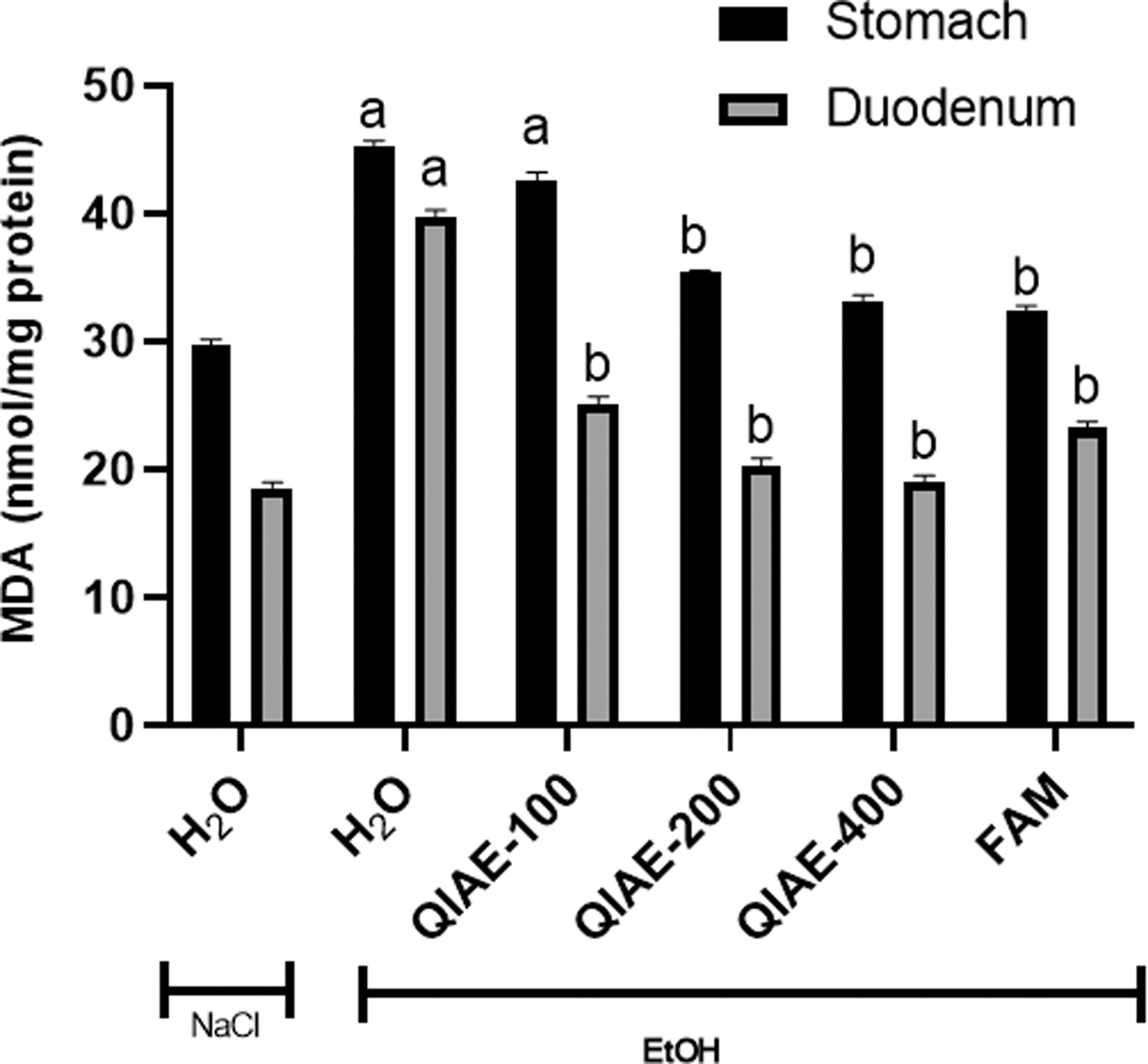
Subacute effect of QIAE and FAM on EtOH-induced changes in stomach and duodenum mucosa MDA levels in rats. Animals were pretreated with various doses of QIAE (100, 200, and 400 mg/kg, b.w., p.o.), FAM (10 mg/kg, b.w., p.o.), or bidistilled water, challenged with a single oral administration of EtOH (4 g/kg, b.w., p.o.) or NaCl 9‰ for 2 h.
Effect of QIAE or FAM on EtOH-induced GD mucosal antioxidant enzyme activity depletion
The EtOH intoxication induces a depletion of GD endogenous antioxidant enzyme activities such as SOD (Fig. 4), CAT (Fig. 5), and GPx (Fig. 6). However, animals pretreated with QIAE or FAM show a marked increase in these enzymes in a concentration-dependent manner. At a dose of 100 mg/kg, compared with the lesion control group, QIAE-pretreatment resulted in an evident augmentation in enzyme levels. Upon increasing the concentration to 400 mg/kg, the SOD activity was significantly elevated in comparison with the control group in both GD mucosal homogenates (P < .05).
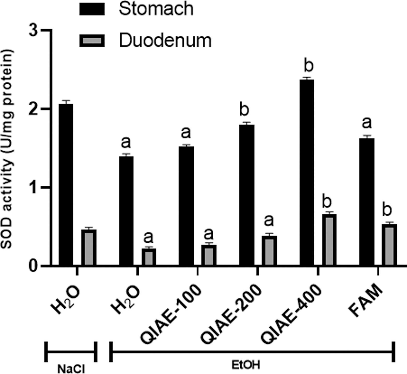
Subacute effect of QIAE and FAM on EtOH-induced changes in stomach and duodenal mucosa SOD activity in rats. Animals were pretreated with various doses of QIAE (100, 200, and 400 mg/kg, b.w., p.o.), FAM (10 mg/kg, b.w., p.o.), or bidistilled water, challenged with a single oral administration of EtOH (4 g/kg, b.w., p.o.) or NaCl 9‰ for 2 h.
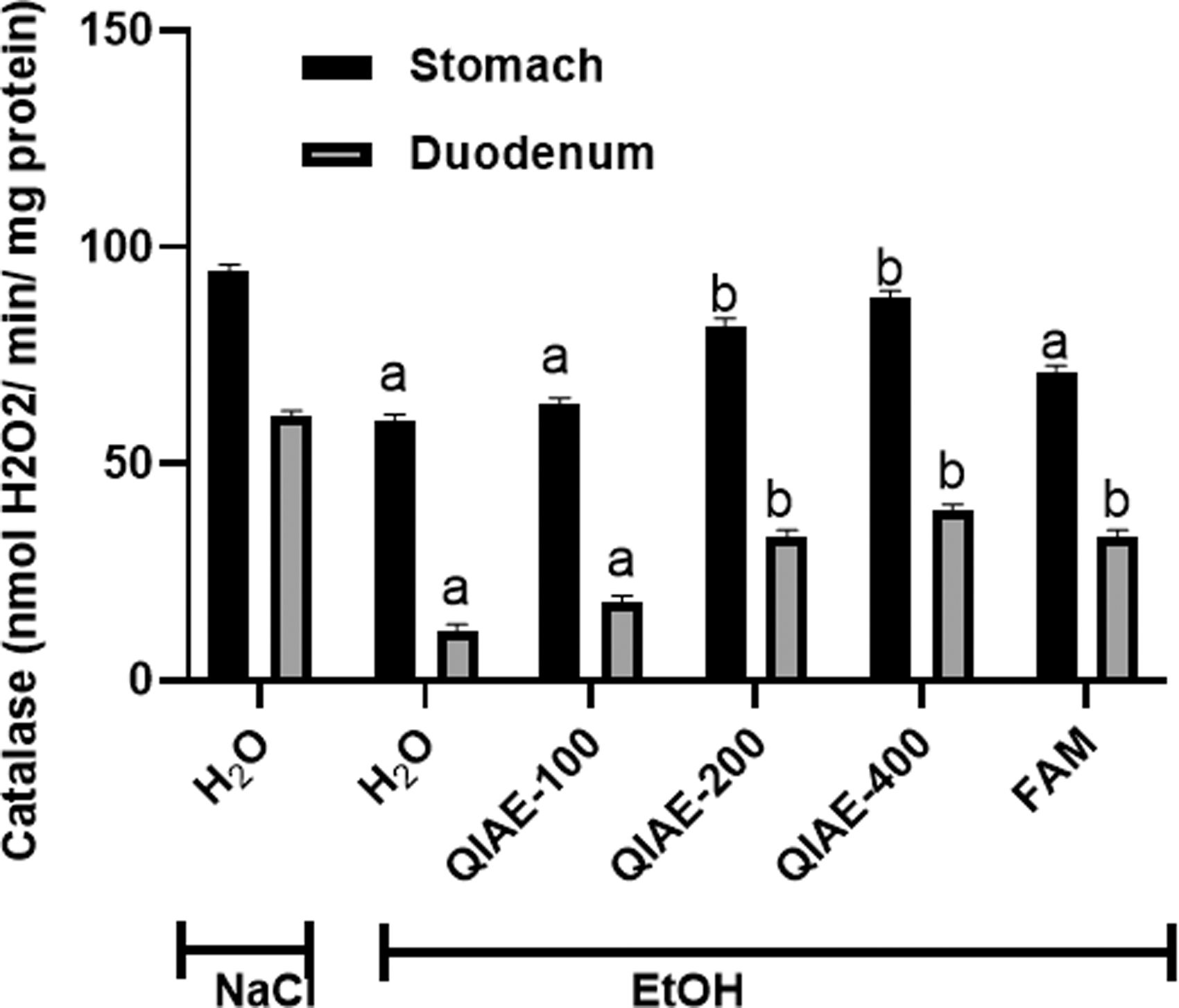
Subacute effect of QIAE and FAM on EtOH-induced changes in stomach and duodenal mucosa CAT activity in rats. Animals were pretreated with various doses of QIAE (100, 200, and 400 mg/kg, b.w., p.o.), FAM (10 mg/kg, b.w., p.o.), or bidistilled water, challenged with a single oral administration of EtOH (4 g/kg, b.w., p.o.) or NaCl 9‰ for 2 h.

Subacute effect of QIAE and FAM on EtOH-induced changes in stomach and duodenal mucosa GPx activity in rats. Animals were pretreated with various doses of QIAE (100, 200, and 400 mg/kg, b.w., p.o.), FAM (10 mg/kg, b.w., p.o.), or bidistilled water, challenged with a single oral administration of EtOH (4 g/kg, b.w., p.o.) or NaCl 9‰ for 2 h.
Discussion
The recent research was designed for the first time, to study the gastro- and duodenoprotective effects of QIAE against EtOH-induced GD mucosal ulcer 30 in comparison with FAM, which is widely medically used for gastric ulcer treatment. This study is based on our phytochemical screening of QIAE, which revealed the presence of phenolic acids, flavonoids, and tannins. These phytoconstituents were previously established to be among the possible cytoprotective agents implicated in decreasing GD ulcer. 31
Regarding the physicochemical characterization, our results showed that QIAE is very rich in dry matter (98.15%). The water, organic matter, and mineral matter contents are 10.95%, 97.88%, and 2.12%, respectively. These results can give us an idea of the climatic conditions of the sampling area “northwest Tunisia,” a soft and humid region characterized by high rainfall. 32
The phytochemical study showed the richness of QIAE in total polyphenols (52.60 ± 0.75 mg gallic acid equivalent [EAG]/g DM), total flavonoids (33.00 ± 1.70 mg quercetin equivalent [EQt (QE)]/g DM), condensed tannins (1.76 ± 0.10 mg catechin equivalent [Ect (CE)]/mL), and fiber (6.86%). On the contrary, the study of the antiradical activity of QIAE against DPPH and ABTS radicals has shown that the latter is endowed with a good antioxidant power, which is manifested by low IC50 (177.00 ± 5.11 and 203.9 ± 2.23 μg/mL, respectively, for DPPH and ABTS) close to that of ascorbic acid, used as a reference antioxidant molecule (190.47 ± 1.22 and 175.13 ± 0.91 μg/mL, respectively). However, this richness in phenolic compounds, as well as this very important antioxidant power, means that this food probably participates in good dietary balance and especially the proper functioning of the body.
The interest in green oak is partly due to its phenolic compounds, which are beneficial secondary metabolites to defend against several pathologies. Due to its high content of phenolic compounds, this plant has a number of beneficial effects on health and it is well used as an antidiabetic 14 and antidiarrheal. 33 The phytochemical investigation by high-performance liquid chromatography-photodiode array detector/(-)electrospray ionization-tandem mass spectrometry (HPLC-PDA/ESI-MS) showed that the main phenolic compounds of QIAE are gallic acid, quinic acid, protocatechuic acid, p-coumaric acid, syringic acid, and naringin (Table 3). However, the antioxidant activity of QIAE could be attributed, in part, to its high levels of phenolic compounds and, in particular, gallic acid and quinic acid. 13
High-Resolution Liquid Chromatography/Electrospray Ionization Identification of Quercus ilex Aqueous Extract
The compounds are suggested according to the dictionary of natural products and the characteristic fragmentation pattern
In vivo part, the acute oral administration of EtOH at a dose of 4 g/kg showed the development of lesions in the gastric and duodenal mucosa resulting in the appearance of elongated bands of dark red coloration. These lesions are parallel to the longitudinal axis of the stomach. They are accompanied by edema, alteration of the surface coating, leukocyte infiltration of the mucosal and submucosal layer, loss of surface coating, and congestion of the intestinal villi. These lesions are the result of the penetration of EtOH into the gastroduodenal mucosa, the same observations have been reported by several other studies. 31,34 –36
However, several mechanisms are involved in the development of alcohol-induced mucosal injury. 37 Alcohol is one of the few elements absorbed by the gastric mucosa. H+ ions are potent inducers of ulcers because they pass through mucus-producing cells and reach deep layers. 38 In addition, alcohol has a triple ulcer-inducing action; it erodes the mucous membrane and causes congestion and cellular necrosis. 39,40 EtOH is responsible for the decrease in gastroprotective mechanisms, in particular, the decrease in gastric mucosal secretion and blood flow and the increase in acidity in the stomach lead to the initiation of the inflammatory reaction.
This is followed by an increase in the formation of ROS such as O2 –, H2O2, and OH•, which promote lipid peroxidation and the formation of gastric lesions. 41 –43
In this context, we have also shown that lesions of the gastric and duodenal mucosa are accompanied by a disturbance of redox balance, which is reflected in an increase in the level of MDA, reflecting membrane lipoperoxidation, and a decrease in the activity of antioxidant enzymes such as CAT, SOD, and GPx, which is in agreement with previous data observed in rats 35 and in mouse 44 as well as in macrophages in culture. 45 However, many recent studies have shown the involvement of oxidative stress in the development of certain gastrointestinal disorders, including ulcers, 36 constipation, 46 and diarrhea. 14
Subacute treatment with QIAE protects against alcohol-induced marked morphological and structural changes in gastric and duodenal mucosa. However, it has been well established that there are several medicinal plants that can protect the body from alcohol-induced gastric injury such as sour orange, 47 prickly pear, 48 carob, 35 and chamomile. 49
Subacute administration of QIAE at increasing doses for 15 days corrects gastric and duodenal lipoperoxidation and restores CAT, SOD, and GPx activities modified by EtOH administration. These results are consistent with previous studies demonstrating the antioxidant and protective properties of green oak acorns against several toxic molecules such as 2,4,6- trinitrobenzenesulfonic acid (C6H3N3O9S) 50 and castor oil. 14
The antioxidant capacity of QIAE is mainly linked to its richness in phenolic compounds such as total polyphenols, total flavonoids, and condensed tannins. The latter is well known for the ability to trap free radicals such as the superoxide radical (O2 •–), OH•, and other ROS. 51,52 However, a positive correlation between phenolic compounds and antioxidant capacity is observed in most natural extracts. 53
In addition, it has been shown that the main antioxidant constituents are phenolic compounds and the positions of the active groups play an important role. Thus, their antioxidant activity seems to be linked to their molecular structure, or more precisely to the presence and number of hydroxyl groups, as well as to the conjugation of double bonds and resonance properties. 54 However, antioxidant supplements modulate endogenous mechanisms by reducing ROS production or increasing enzymatic activities that break down ROS. 55
In this context, Li et al. 56 showed that pretreatment with cinnamic acid could lead to some reduction in lipid peroxidation and rapid stress tolerance by enhancing the activities of antioxidants such as SOD, CAT, GPx, and GSH. Also, gallic acid acts as an antiulcer through its effect on gastric acid secretion, protection of mucous membranes by endogenous factors (NO, PGE2, and TNF-α), and inhibition of apoptosis due to oxidative stress. 57 Protocatechuic acid has been well shown to have a strong scavenging and antioxidant activity by decreasing lipid peroxidation, lowering low-density lipoprotein (LDL) levels, reducing the production of H2O2 and O2 –, and restoring glutathione-related enzymes (GSH). 58
Duodenal ulcer is associated with a large acid load to the duodenum caused by several factors, including increased acid secretion by the stomach. Fiber, especially soluble fiber, slows down the release of digested food materials from the stomach to the duodenum, thereby reducing the exposure of the duodenum to gastric acid. This action of dietary fiber makes high-fiber foods essential for protecting the lining of the duodenum from gastric acid-induced sores and ulceration. 59
As a conclusion, the data clearly demonstrate that QIAE has protective effects against EtOH-induced acute ulceration in the gastric and duodenal mucosa of rats, due, in part, to its direct RSA activity, increased antioxidant enzymes, and depression of lipid peroxidation. We intend to further investigate the chemical components of QIAE and combine it with pharmacodynamic experiments to identify the protective effect of this fruit on GD mucosa. Therefore, there is still a great need to realize other deep studies that offer the opportunity to develop future therapeutic strategies for GDU-management.
Animal Welfare Statement
The authors confirm that they have followed EU standards for the protection of animals used for scientific purposes.
Footnotes
Author Disclosure Statement
No competing financial interests exist.
Funding Information
Financial support of Tunisian Ministry of Higher Education and Scientific Research.