




Abstract
We aimed to analyze the effects and explore the molecular mechanisms of a natural herb mixture extract (NME) on osteoblasts during differentiation in human bone marrow-derived mesenchymal stem cells (hBMSCs). We tried to confirm the regulation of osteogenic differentiation during NME treatment. Alkaline phosphatase assay and Alizarin red S staining were performed to evaluate the regulation of osteogenic differentiation. Real-time polymerase chain reaction was performed to analyze the expression of osteoblast maker genes, and Western blot was used to verify the signaling pathway. Signaling pathway conformation, selective bone morphogenetic protein receptor inhibitor, and dorsomorphin homolog 1 were used as pretreatments before inducing osteogenic differentiation. We determined that MME (natural herb mixture extract) was a safe material and significantly increased osteoblast differentiation and that SMAD phosphorylation is a key signaling pathway that regulates osteogenic differentiation in hBMSCs.
Introduction
Mesenchymal stem cells (MSCs) represent an effective model for studying cellular therapy or regenerative medicine. 1 –3 MSCs are multipotent stromal cells that can be differentiated into a variety of cell types, including osteoblasts (bone cells), chondrocytes (cartilage cells), myocytes (muscle cells), and adipocytes (fat cells, which give rise to marrow adipose tissue). 4 Osteoblast differentiation regulation plays an important role in maintaining bone density and strength. 5 Several signaling pathways and transcription factors are important for musculoskeletal development. 6 –8 For example, transforming growth factor (TGF)-β and bone morphogenetic protein (BMP) are well-known osteocyte-inducing factors in MSCs. 8,9
Medicines originating from natural herbs are much easier to use for treatment of human diseases than chemical-based medicines. Therefore, people try to identify the medically effective components of natural herbs because they have fewer adverse reactions and are more suitable for long-term use compared with chemically synthesized medicines. 10,11
Achyranthes (Achyranthes aspera) has occupied a pivotal position in Indian culture and folk medicine. Since ancient times, the rural people of India commonly use this herb for various disorders. 12 Achyranthes aspera, an herb in the Amaranthaceous family, is a native medicinal plant found in Asia, South America, and Africa. It is commonly used as a traditional cure for treatment of malarial fever, dysentery, asthma, hypertension, and diabetes. 13,14 Especially, the roots of Achyranthes aspera are reported to have efficacy in a variety of disease types, including infantile diarrhea and common cold, and the leaves are used to treat asthma. The seeds are regarded as having emetic and hydrophobic properties. Achyranthes aspera contains a variety of triterpenoid saponins and is an effective cardiac stimulant and diuretic.
Safflower seed (Carthamus tinctorius) is commercially cultivated for vegetable oil extracted from the seeds. It is rich in unsaturated fatty acids and α-linoleic acid and is a commonly used vegetable oil in the United States and Europe, and it is also used for clinical treatment of osteoporosis and rheumatism in Korea. 15,16
The Acanthopanax (Eleutherococcus senticosus) species is a small woody shrub in the Araliaceae family native to northeastern Asia, Korea, Japan, and the Far Eastern region of Russia. The root extracts of Eleutherococcus senticosus have a reputation in traditional medicine for providing beneficial health effects, but such claims are not supported by medical evidence. 17 Acanthopanax is generally known to relieve palsy and extravasated blood, improve bone strength, support sexual function, and alleviate tingling sensations. In addition, it has been used to treat lung diseases such as asthma and pulmonary tuberculosis. In particular, β-sitosterol, which is abundant in the stem and bark, is known to be effective for weight loss and improvement of blood circulation because it breaks down bad cholesterol and fat. 18 Kim et al. 19 reported that Acanthopanax extracts promote osteoblast differentiation, and Moon et al. 20 reported that Acanthopanax extracts with local herbs inhibit osteoclast differentiation and promote chondrocyte differentiation.
In this study, we investigated natural herbal candidates and their composition related to bone regeneration. Achyranthes (Achyranthes aspera), safflower seed (Carthamus tinctorius), and Acanthopanax (Eleutherococcus senticosus) were selected as candidates, and two types of enzymes were used to increase the extraction rate of the active ingredients. As a result of analyzing the effect of the extract by evaluating the expression of the osteoblast marker gene, it was confirmed that osteoblast differentiation was enhanced, and this result occurred through the SMAD signaling pathway.
Materials and Methods
Natural herb mixture extract extraction
The herbal mixture extract for bone regeneration used in the experiment was provided by Gagopa Healing Food (Changwon, Korea) as a mixture containing Achyranthes (Achyranthes aspera), safflower seed (Carthamus tinctorius), Acanthopanax (Eleutherococcus senticosus), and other herbs that are blended and then chopped for dissolution. The composition of the natural herb mixture is listed in Table 1. The extraction rate was increased by adding 0.3% of protamex and 0.2% of flavourzyme after boiling for 48 h in a pressure cooker at 100°C for 2 days by adding water to the listed natural herb mixture at 1:10. After extraction, the solution was filtered using a 0.2-μm filter paper, lyophilized, and then used for experiments. The name of this extract is natural herb mixture extract (NME).
Composition of the Natural Herb Mixture Extract
Cell culture
Human bone marrow MSCs (human bone marrow-derived mesenchymal stem cells [hBMSCs], PromoCell, Heidelberg, Germany) were purchased, and the cells were cultured in MSC Growth Medium 2 (PromoCell, Heidelberg, Germany) and 1% antibiotic–antimycotic (Biowest, Nuaillé, France) in a humidified 5% CO2 incubator at 37°C. The cells were used at passages 3–6 in this study.
Cell proliferation assay
To assess the effects of NME on cell proliferation, cells were seeded in a 96-well plate at a density of 3 × 10³ cells per well. The cells were incubated in growth medium for the indicated duration at various concentrations of NME (1, 10, and 100 μg/mL). They were cultured for 3 days. Next, the medium was removed, cells were washed twice with phosphate-buffered saline, CCK-8 assay solution was added following the procedure, and absorbance was measured at 450 nm using a TECAN microplate reader (Sunrise, Australia).
Alkaline phosphatase activity assay
Four days after inducing osteogenic differentiation, the alkaline phosphatase (ALP) activity assay was performed using the QuantiChrom™ Alkaline Phosphatase Assay Kit (DALP-250; BioAssay Systems, USA) according to manufacturer's instructions. Absorbance was recorded at 405 nm at time 0 and again after 4 min on a plate reader while the samples were maintained at 37°C between readings. The quantity of p-nitrophenylphosphate was normalized to the total protein content. ALP activity was recorded in terms of μmol/mg protein.
Osteogenic differentiation induction and Alizarin red S staining
Osteogenic differentiation was induced through culturing of cells for 10 days to 2 weeks in osteogenic medium (10% fetal bovine serum, 0.1 mM dexamethasone, 10 mM β-glycerophosphate, and 50 mM ascorbic acid in α-MEM), and extracellular matrix calcification was estimated by using 2% Alizarin red S with a pH of 4.3 (Sigma-Aldrich, St. Louis, MO, USA) for 15 min. For obtaining quantitative data, 300 μL of 10% sodium phospate solution was used to dissociate CPC powder. Cetylpyridinium chloride (Sigma- Aldrich) solution were added to stain dishes and absorbance of the extracted dye was determined at 570 nm using a Sunrise microplate reader (TECAN, Mannedorf, Switzerland).
Quantitative real-time polymerase chain reaction
Total RNA was isolated by using the Tri-RNA reagent (Favorgen Biotech. Corp., Taiwan) according to the manufacturer's instructions, and cDNA was synthesized with AccuPower® RT PreMix (Bioneer, Korea). Primer sequences used in the experiment are shown in Table 2. Quantitative reverse transcriptase polymerase chain reaction was performed by using a TOPreal™ qPCR 2X PreMIX (Enzynomics, Korea) on the ABI 7500 Instrument (Applied Biosystems, USA). Data analysis was performed by using the delta delta Ct method.
Real-Time Polymerase Chain Reaction Primer Sequences
Western blot analysis
The NME-treated cells were homogenized in RIPA buffer (iNtRON Biotechnology, Korea) with protease and phosphatase inhibitors. The isolated proteins were separated by sodium dodecyl sulfate–polyacrylamide gel electrophoresis (SDS-PAGE) and electrotransferred to polyvinylidene fluoride membranes (PVDF; Millipore, USA). Blots were probed with primary antibodies, phospho-SMAD1/5 (Thermo Fisher Scientific, USA) and β-actin (Santa Cruz Technologies, USA). Bound antibodies were detected by using an ECL detection kit (Pierce Biotechnology, USA) and visualized using LAS 4000 (Fujifilm, Japan).
Statistical analysis
All of the results are presented as the mean ± standard error of the mean. Comparisons between groups were analyzed using Student's t-tests. The P value was calculated using Student's t-test. Each experiment was repeated at least three times to yield comparable results. Depending on the experiment, *P < .05, **P < .02, and ***P < .01 were considered significant.
Results
Cell proliferation effects of NME on osteogenic differentiation in hBMSCs
To verify the toxicity of NME to hBMSCs, we performed a CCK-9 assay following the manufacturer's instructions. The result showed that NME was not only nontoxic but also increased cell proliferation in hBMSCs (Fig. 1A). To evaluate ALP enzyme activity, the ALP assay was performed while NME induced osteogenic differentiation for 4 days. The results indicated that NME induced ALP enzyme activity (Fig. 1B). Finally, Alizarin red S staining was performed to confirm the NME effects, and results indicated that NME upregulated mineralization of hBMSCs (Fig. 1C).

NME upregulated cell proliferation and osteogenic differentiation in hBMSCs.
Effects of NME on osteogenic differentiation marker gene expression and signaling pathways in hBMSCs
To explore the mechanism of NME during osteogenic differentiation, the expression levels of osteocalcin (OC), osteopontin (OPN), and BMP-2 were detected during NME-treated osteogenic differentiation.
As shown in Figure 2A–C, the expression levels of osteogenic marker genes, OC, OPN, and BMP-2, gradually and dose-dependently increased during osteogenic differentiation. In particular, phospho-SMAD1/5 was measured by Western blot to determine if NME affects the osteoblast signaling pathway. As a result, it was found that phospho-SMAD1/5 expression gradually increased with NME concentration (Fig. 2D, E). The results show that NME is an essential component for upregulating osteogenic differentiation in hBMSCs and that NME induces the osteogenic differentiation marker gene and BMP-2 expression through SMAD1/5 phosphorylation.

Expression of osteogenic differentiation marker genes was increased. To evaluate osteogenic differentiation, we performed quantitative real-time PCR. We measured the gene expression of osteogenic differentiation markers such as OC, osteopontin, and BMP-2. NME-treated groups showed increased levels of osteogenic differentiation markers compared with the nontreated group
Effects of dorsomorphin homolog 1 on NME-induced osteogenic differentiation in hBMSCs
From these experiments, we determined that NME induced SMAD1/5 phosphorylation and facilitated osteogenic differentiation in hBMSCs. To understand the role of the SMAD signaling pathway in NME-induced osteogenic differentiation, we treated cells with dorsomorphin homolog 1 (DMH-1), 7 an SMAD signaling antagonist. Before starting the loss-of-function study, we tested for DMH-1 toxicity and the result indicated that there is no toxicity in hBMSCs (Fig. 3A).
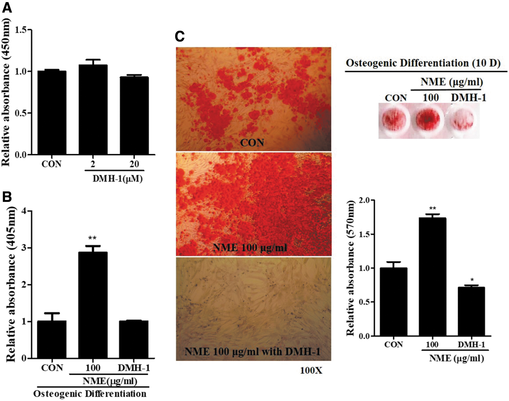
DMH-1 downregulated cell proliferation and osteogenic differentiation in hBMSCs.
To evaluate osteogenic differentiation with DMH-1, we performed the ALP analysis and Alizarin Red S staining, which indicated that DMH-1 reduced NME-treated osteogenic differentiation in hBMSCs (Fig. 3B, C).
Effects of DMH-1 on NME-induced osteogenic differentiation marker gene expression and signaling pathways in hBMSCs
Because of the high reliability of NME effects, we conducted a loss-of-function study using DMH-1. To confirm the osteogenic differentiation promoted by SMAD1/5 cell signaling, DMH-1 was used as a pretreatment before the NME treatment. We used quantitative real-time PCR (RT-PCR) and Western blot to evaluate the osteogenic differentiation marker gene expression (Fig. 4A–C). The analysis of expression of osteogenic marker genes, OC, OPN, and BMP-2, indicated that DMH-1 inhibited osteogenic differentiation, which was accelerated by NME, and SMAD1/5 phosphorylation was decreased in the presence of DMH-1 (Fig. 4D, E). These results indicate that NME plays the role of osteogenic differentiation accelerator in hBMSCs and it is mediated by the SMAD signaling pathway.
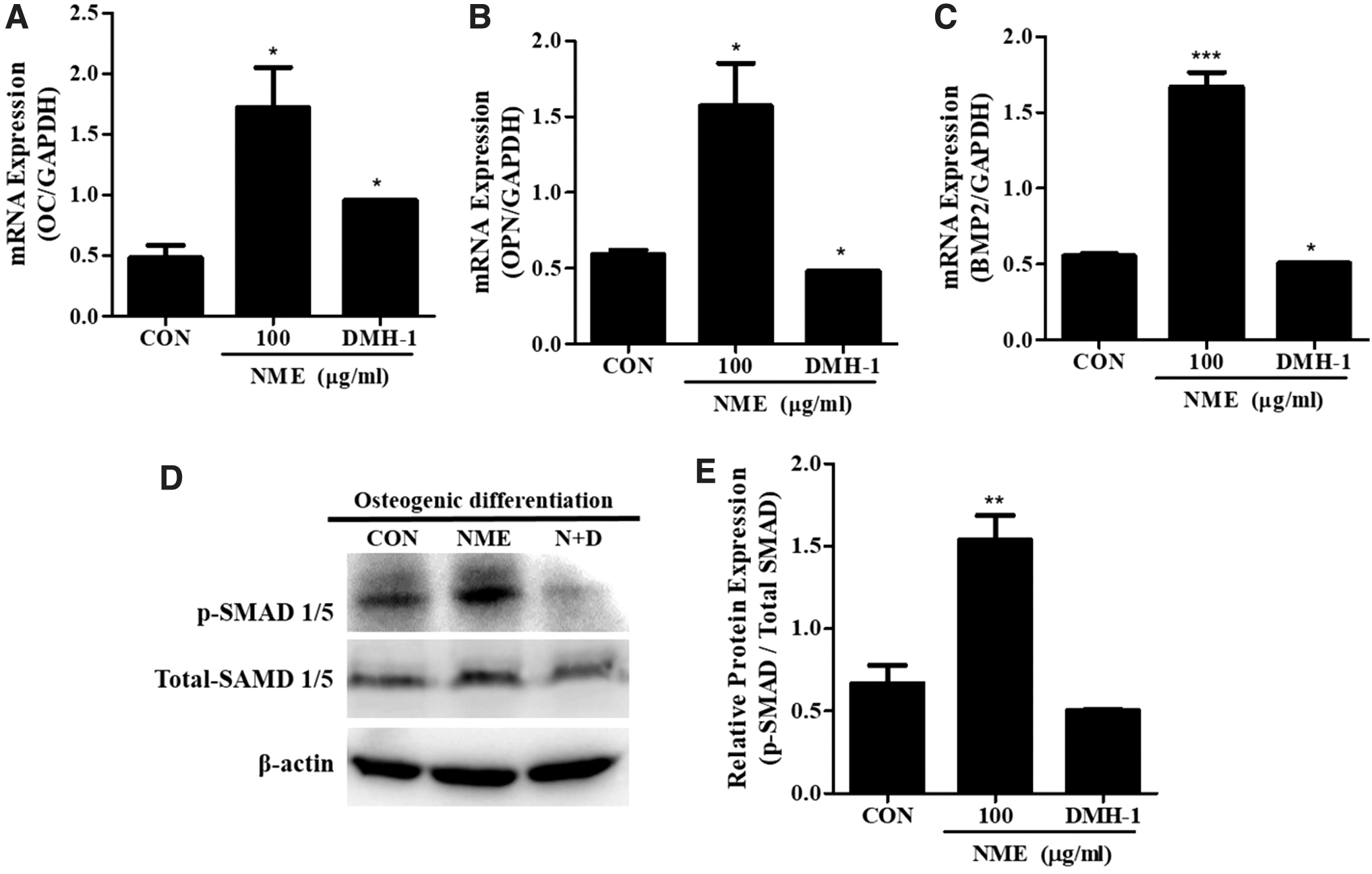
Expression of osteogenic differentiation marker genes decreased. To evaluate osteogenic differentiation, we performed quantitative real-time PCR. We measured the gene expression of osteogenic differentiation markers such as OC, osteopontin, and BMP-2
Discussion
Traditional therapies using natural products have been used to treat a variety of diseases for thousands of years. There are many studies using natural products in the treatment of osteoporosis. 21 Natural products commonly used to treat osteoporosis are Achyranthes, safflower seed, and Acanthopanax. In this study, we used NME mainly comprising safflower seed, hyssop, and rowan. The extract was obtained after two enzyme treatments to increase the extraction yield.
Previous studies on osteoporosis treatment, which have focused on promoting osteoblast differentiation and suppressing osteoclasts, 22 reported that icariin is an effective compound for treating cartilage defects by promoting actin stress fiber formation in fibroblasts through the MAPK signaling pathway and TGF-β and BMP signaling, 23 enhancing the ability of bone marrow-derived mesenchymal cells to migrate. In addition, there is little research on the effects of herbal extracts on differentiation of bone marrow-derived MSCs into bone tissue.
As a result of analyzing the effect of NME on the cell growth rate and ALP activity of hBMSCs, the enzyme-treated extract tended to increase overall activity compared with the extract without enzyme treatment (data not shown). Therefore, in this experiment, the enzyme-treated extract was used for the treatment; the extract was dissolved in water and used during differentiation of hBMSCs into bone cells.
The MTT assay was used to evaluate the toxicity of NME in bone marrow-derived MSCs. At concentrations of 1, 10, and 100 μg/mL, no toxicity was observed and cells proliferated with the passage of time. The lowest concentration of 1 μg/mL was not significantly different from the control group, but 10 μg/mL showed a significant increase from day 2. At the highest concentration of 100 μg/mL, cells were significantly proliferated from day 1 (111%) and increased by 125% on day 3 of culture, promoting cell proliferation.
Bone calcification and bone formation are important markers for bone differentiation. First, the treatment of hBMSCs with NME was confirmed by the ALP assay and ARS staining, which showed differentiation into bone cells. Osteogenic differentiation was significantly induced compared with the control. ALP is an enzyme that hydrolyzes organic phosphate esters to increase the concentration of phosphate ions locally at the site of calcification and is known to perform the function of inducing calcification by depositing calcium phosphate onto the extracellular matrix. 24 –26 De Bemard 26 reported that ALP locally converts proteins to phosphoproteins by increasing phosphate concentrations, which act as nuclei of calcification, with calcium binding properties. In this study, ALP staining for NME was performed 4 days after the beginning of bone differentiation, and the results of ALP synthesis showed that hBMSCs increased significantly at all concentrations.
Expression of the bone differentiation marker gene was confirmed by RT-PCR. In the case of ALP, it is a marker gene for early bone differentiation, and the expression levels of OC and OPN are markers for mid bone differentiation. The expression of bone differentiation marker genes identified at the molecular level also showed higher levels in the experimental group treated with NME than the control group and showed a significant difference.
To verify the cell signaling pathway, we evaluated protein expression of SMAD 1/5 phosphorylation, and NME upregulated SMAD1/5 phosphorylation depending on NME concentration. The SMAD1/5 loss-of-function study, which used pretreated DMH-1, indicated that DMH-1 could reduce osteogenic differentiation. From these results, it was concluded that NME expedites osteogenic differentiation in hBMSCs through SMAD1/5 phosphorylation.
Taken together, the analysis of NME, based on Achyranthes, safflower seed, and Acanthopanax, etc., found that hBMSCs promoted osteogenic differentiation. Therefore, NME can be developed as a product that can induce bone cell differentiation and function. Moreover, it may be useful for developing a therapeutic agent for treating bone diseases. Liquid chromatography was performed to identify the bioactive compound and the result indicated that anserine was the major component among the variety of amino acids in NME (Supplementary Data S1). Anserine is a dipeptide of β-alanine and 1-methylhistidine. It is abundantly present in the muscles of animals. It is not only an antioxidant but it also reduces fatigue and is involved in muscle contraction. There are no articles suggesting that anserine induces osteogenic differentiation until now. Therefore, the effects and mechanisms of action of anserine need to be evaluated in future research; in our opinion, this is the first study to suggest that it induces osteogenic differentiation. For this reason, it should be the subject of further studies for development of food products and medicines for alleviating bone-related diseases.
Footnotes
Authors' Contributions
H.-O.J. and D.-S.K. were involved in conceptualization. S.-K.B. was involved in methodology. H.-O.J. and D.-S.K. were involved in validation. D.-S.K. was involved in formal analysis. K.-S.K., T.-Y.K., and D.-S.K. were involved in investigation. Y.-J.K. was involved in resources. H.-J.K. was involved in data curation. K.-S.K. and D.-S.K. were involved in writing—original draft. S.-K.B. and H.-O.J. were involved in writing—review and editing. H.-O.J. was involved in supervision.
Author Disclosure Statement
No competing financial interests exist.
Funding Information
This work was supported by the National Research Foundation of Korea (NRF-2019R1A6A3A01095696).
Supplementary Material
Supplementary Data S1