




Abstract
The immunomodulatory effects of Lactobacillus rhamnosus HDB1258 were evaluated in mice with colitis induced by Klebsiella oxytoca (KO). L. rhamnosus HDB1258 was cultured in the lava seawater (LS) to improve its probiotic properties. It increased adhesive ability to mucin with mRNA expression levels of chaperone proteins (such as GroEL/ES, DnaKJ, and HtrA). In the in vivo experiments, administration of KO caused an inflammation on the colon with gut dysbiosis. LH group (oral gavage of HDB1258 1.0 × 10⁹ colony forming units/day) showed that inflammatory biomarkers, including IL-1β, TNF-α, IL-6, and PGE2, were significantly decreased to less than half of the KO group, and Th1 cells were decreased in the spleen, but Treg cells were not affected. In contrast, the expression levels of secretory IgA and IL-10 were significantly increased, and the composition of gut microbiota in the LH group tended to recover similar to normal mice without any effect on the α-diversity. In conclusion, L. rhamnosus HDB1258 cultured in the LS could regulate competitively pathogenic bacteria in imbalanced flora with its improved mucin adhesive ability and was an effective immunomodulatory adjuvant for treating colitis by its regulatory function on intestinal inflammation.
Introduction
Probiotics are live microorganisms that provide health benefits to the host. 1 Many studies have been conducted to test the functionality of probiotics on health and diseases related to immune enhancement and modulation, since live microorganisms can directly interact with the gut flora and stimulate intestinal immune cells. 2,3 Recently, lactic acid bacteria (LAB) have attracted much attention as immunomodulatory adjuvants for treating gut diseases like inflammatory bowel disease (IBD).
IBD is an inflammatory condition of the colon and small intestine, which arises from interaction of genetic and environmental factors responsible for immune response and inflammation. 4,5 The immune system collapsed by pathogenic factors induces excessive inflammation, and the reabsorption function of the colon decreases, resulting in diarrhea and gut dysbiosis. Causative factors of IBD are known as dietary patterns, gut microbial dysbiosis, and disrupted intestinal barrier like mucosal layer, as well as inherited genetic factors. 5 For treatment of IBD, anti-inflammatory drugs and antibiotics have been prescribed to attenuate inflammation of the inner wall of the colon and to remove pathogenic bacteria that cause colitis. However, those could be accompanied with a side effect like gut dysbiosis. Since the imbalance of gut microbiota induced by chemical drugs like antibiotics can lead to reoccurrence of the colitis, it is crucial to reduce this side effect to maintain a well-being. 6
Many studies have reported the immunomodulatory effects of probiotics in the colitis model. Lactobacillus reuteri and Lactobacillus johnsonii alleviate intestinal inflammation and gut dysbiosis in mice with colitis induced by chemical drugs like 2,4,6-trinitrobenzenesulfonic acid (TNBS) or antibiotics. 7 –9 Lactobacillus rhamnosus and Lactobacillus casei show regulatory effects on pro-inflammatory cytokines and gut microbiota in pathogenic microbe-infected inflammatory model. 10 These results suggest that probiotics can help alleviate inflammatory symptoms with gut microbiota alteration in the colitis model.
In our previous report, L. rhamnosus HDB1258, which has immune-enhancement functions, had increased intestinal viabilities and adhesive ability to mucin when cultured with Jeju-lava seawater. 11 These properties can be advantageous for survival in stressful environments, like the inflammatory colon. In this study, the lava seawater (LS) culture method was applied for the improvement of mucin adhesive ability of L. rhamnosus HDB1258. The chaperone protein expression levels of L. rhamnosus HDB1258 were investigated as stress response proteins, and the modulatory functions of L. rhamnosus HDB1258 were evaluated for efficacy at controlling excessive inflammatory colon and gut microbiota dysbiosis using the Klebsiella oxytoca (KO)-induced colitis mice model.
Materials and Methods
Materials
Sodium thioglycolate, RPMI 1640 was purchased from Sigma (St. Louis, MO, USA). Antibodies were purchased from Cell Signaling Technology (Beverly, MA, USA). Enzyme-linked Immunosorbent Assay (ELISA) Kits for TNF-α, IL-1β, IL-6, IL-10, and IgA were purchased from eBioscience (San Diego, CA, USA). CD4 T and Natural Killer (NK) Cell Isolation Kits were purchased from Miltenyi Biotec (Teterow, Germany). A Vybrant CFDA SE Cell Tracer Kit was purchased from Invitrogen (Grand Island, NY, USA). A QIAamp DNA Stool Mini Kit was purchased from Qiagen (Hilden, Germany).
Preparation of HDB1258 probiotics
Culture in the LS
L. rhamnosus HDB1258 (named SKB1258 in the previous report 11 ) was inoculated into the lava-seawater LAB media containing 8% glucose, 2% yeast extract, 0.5% soy peptone, 0.5% sodium acetate, 0.1% Tween 80, 0.01% MgSO4, 0.005% MnSO4, 0.2% potassium diphosphate, and 0.2% ammonium sulfate in 30% (v/v) lava-seawater (pH 6.5), incubated at 37℃ for 20 h. The cells were then harvested by centrifugation and mixed with cryoprotectants and freeze-dried. For the in vivo experiments, it was suspended in saline.
Mucin adhesion and expression of chaperone proteins of HDB1258 probiotics
Intestinal adhesive ability was accessed by evaluation of mucin adhesion rate as indicated by Kim et al. 11 Adhesion rate (%) = Log colony forming units (CFU) after incubation for 2 h with mucin/Log CFU at 0 h × 100.
Total RNA of HDB1258 probiotics was extracted using an RNeasy Extraction Kit (Qiagen) according to the manufacturer's instructions. The quantitative real-time PCR (qRT-PCR) was performed using SsoAdvanced Universal SYBR Green Supermix (Bio-Rad) with CFX Connect Real-Time system (Bio-Rad). The conditions were as follows: 3 min at 95°C followed by 40 cycles consisting of 10 sec at 95°C and 30 sec at 55°C. The expression levels of the assayed genes (htrA, dnaK, dnaJ, groEL, groES, and gyrA gene: Supplementary Table S1) were normalized with respect to gyrA.
Evaluation of immunomodulatory function of HDB1258
Animals
C57BL/6 mice (male, 5 weeks old, 19–21 g) were supplied from Orient Bio (Seongnam-si, Korea) and acclimatized for 7 days before the initiation of experiments. All animals were maintained in the plastic cage with the 5 cm raised wire floor under standard conditions (temperature, 20°C ± 2°C; humidity, 50% ± 10%; and lighting, 12 h/day). All mice were fed standard laboratory chow and tap water ad libitum. Animal experiments were conducted according to the NIH and University Guide for Laboratory Animal Care and Usage. All animal experimental procedures were approved by the Institutional Animal Care and Use Committee of the University (IACUC No. KUSASP-20018).
Treatment with HDB1258 in mice with KO-induced colitis
To examine the immunomodulating effect of HDB1258, it was orally gavaged in mice with KO-induced colitis, which was prepared according to the method of Jang et al. 12 Test groups were pretreated with KO 1 × 10⁹ CFU/mouse/day for 5 days and then divided into four groups (n = 6): KO group (vehicle); SSZ group (sulfasalazine 50 mg/kg/day); LL group (HDB1258 1 × 10⁸ CFU/mouse/day); and LH group (HDB1258 1 × 10⁹ CFU/mouse/day). The samples were orally gavaged in KO-treated mice once a day for 14 days. Normal control (NC) mice were orally gavaged with vehicle (saline) instead of HDB1258 for 14 days without pretreatment of KO. Mice were sacrificed 20 h after the final treatment by CO2 inhalation.
Assay of myeloperoxidase activity
Colons were homogenized with cold radio immuno precipitation assay (RIPA) lysis buffer and centrifuged at 10,000 g for 10 min. Myeloperoxidase (MPO) activity was assayed according to the method of Jang et al. 12 One unit was defined as the quantity degrading 1 μmol/mL of peroxide.
Flow cytometry of Th1 and Treg cells in the spleen
For the flow cytometric analysis, the spleens were crushed, lysed with Tris-buffered ammonium chloride, suspended in RPMI 1640 medium, and then filtered. The CD4 T cells were isolated using a Pan T cell Isolation Kit II 13 and fixed. Th1 and Treg cells were stained with anti-IFNγ or anti-Foxp3 antibodies, respectively, and then analyzed by flow cytometry.
Quantitative real-time PCR
Total RNA was isolated from the spleen tissues, and then qRT-PCR was performed, utilizing a Takara thermal cycler (Takara Biology, Inc.), which used SYBR premix agents. The conditions were as follows: activation of DNA polymerase at 95°C for 5 min and 45 cycles of amplification at 95°C for 10 sec and at 60°C for 30 sec. 9 The normalized expression of the assayed genes (TNF-α, IL-1β, IL-10, Foxp3, T-bet, and β-actin: Supplementary Table S1), with respect to β-actin, was computed for all samples using the Microsoft Excel data spreadsheet.
Immunoblotting and ELISA
The spleen and colon tissues were removed and lysed with ice-cold lysis RIPA buffer. 14 The supernatants were electrophoresed by sodium dodecyl sulfate–polyacrylamide gel electrophoresis and transferred to a nylon membrane, probed with the corresponding antibodies (p65, p-p65, and β-actin), and detected with horseradish peroxidase-conjugated secondary antibodies. For the assay of cytokines (IL-1β, IL-6, TNF-α, and IL-10) and secretory IgA (sIgA), the colon tissues and feces homogenate supernatants were transferred in 96-well plate and assayed using ELISA Kits (eBioscience, San Diego, CA, USA). 14
Pyrosequencing
The bacterial genomic DNA was extracted from the feces of mice using a QIAamp DNA Stool Mini Kit. 14 The bacterial 16S rRNA V4 region was amplified and sequenced using Illumina iSeq 100 (San Diego, CA, USA). Functional genes were predicted using the phylogenetic investigation of communities by reconstruction of unobserved states (PICRUSt). 14,15 Linear discriminant analysis (LDA) and cladograms were pictured using the LDA effect size (LefSe) on Galaxy platform Huttenhower lab Galaxy server. 16 Pyrosequencing reads were deposited in the short read archive of NCBI under accession number PRJNA728842.
Statistical analyses
All data are expressed as the mean ± standard deviation, and statistical analyses were conducted using GraphPad Prism 8 (GraphPad Software, Inc., San Diego, CA, USA). The significance was analyzed by Kruskal–Wallis test with Dunn's post hoc test for nonparametric analysis (P < .05).
Results
Improvement of mucin adhesive ability of HDB1258 and its chaperone protein expression
Culturing in the LS increased the adhesion rate to mucin of HDB1258. LS-cultured HDB1258 exhibited adhesion rate to mucin of 38.1%, compared with nonstress treated HDB1258 (20.4%) (Fig. 1A). To examine whether expression of stress response proteins is induced by LS, the transcriptional level by qRT-PCR was measured. In case of LS-cultured HDB1258, the mRNA expression levels of some chaperone proteins (DnaKJ, GroEL/ES, and HtrA) were significantly increased compared to normal cultured HDB1258 (Fig. 1B). Especially, the mRNA level of the HtrA protein was about twofold greater compared with normal cultured HDB1258.

Mucin adhesion rate
Effects of HDB1258 on KO-induced inflammatory responses in the mice
Immunomodulatory effects on inflammation-induced colon
To evaluate the immunomodulatory effect of HDB1258, the colitis mice model was established in KO treated mice. Oral gavage of KO induced severe inflammation, manifested by shortened, thickened, and erythematous colons (Fig. 2 and Supplementary Fig. S1), like previously reported. 12 Especially, MPO activity and inflammatory cytokines, IL-1β and TNF-α, in the colon tissue were significantly increased more than 1.5-fold above those of the NC group, while anti-inflammatory cytokine, IL-10, was decreased. However, HDB1258 and sulfasalazine (SSZ) alleviated expressions of inflammatory biomarkers. Colon length and sIgA expression levels reduced by KO oral gavage were recovered by HDB1258 and SSZ, and MPO activity in colon was significantly decreased compared with KO group (Fig. 2A and Supplementary Fig. S1). Moreover, in the LH (intake of HDB1258 1 × 10⁹ CFU/day) group IL-1β, IL-6, and TNF-α were significantly decreased compared to those of the KO group, but IL-10 was increased (Fig. 2B). HDB1258 inhibited NF-κB activation by regulating phosphorylation of p65 in the colon (Fig. 3). While CD11c+ cells were increased at the end of the villi in the colon of the KO group, they tended to be decreased and diffused in all test groups. These results indicate that HDB1258 can regulate expression of pro-inflammatory cytokines through controlling of NF-κB signaling.
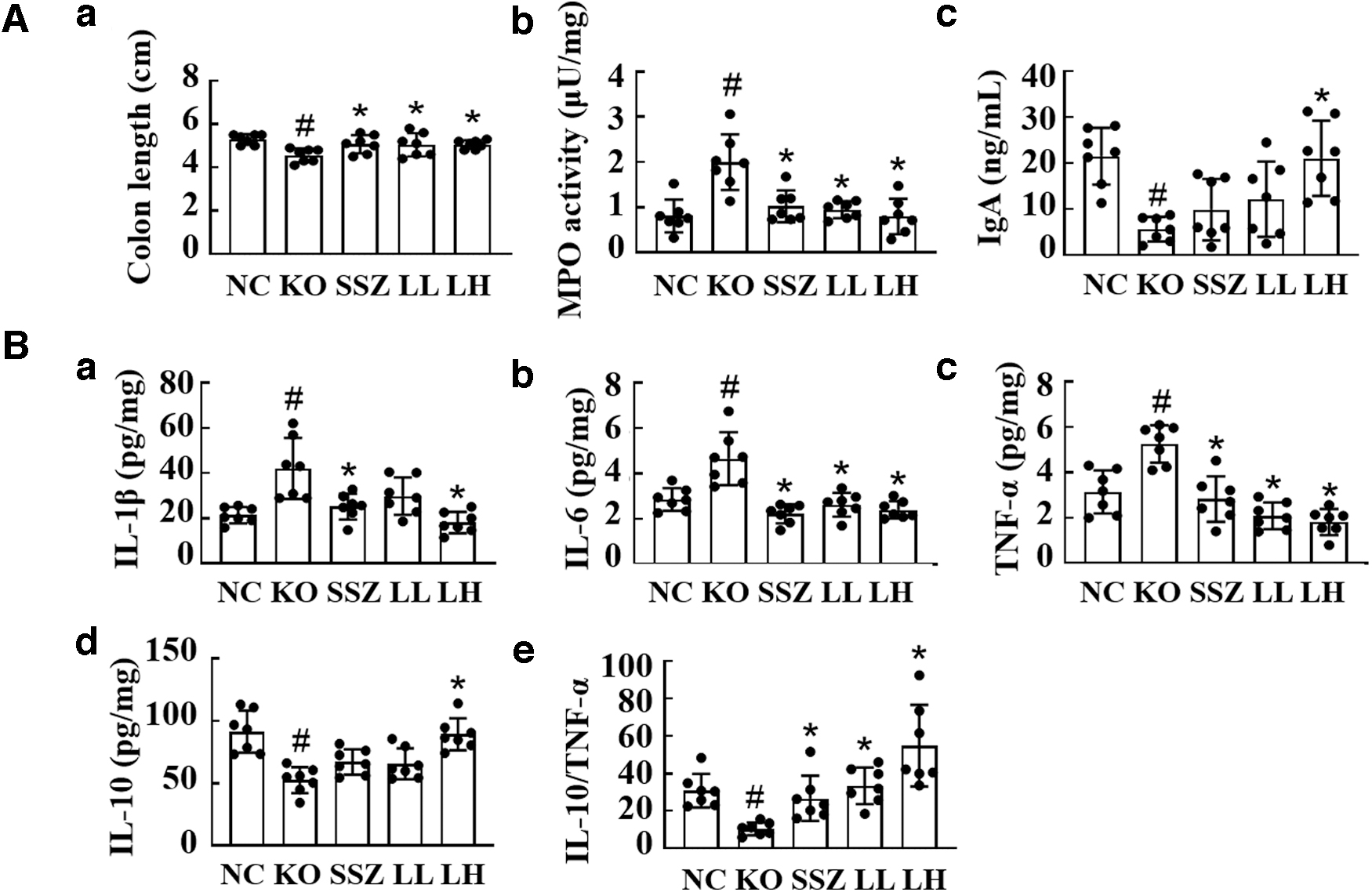
Anti-inflammatory effects of HDB1258 on the colon of KO-induced colitis mice.
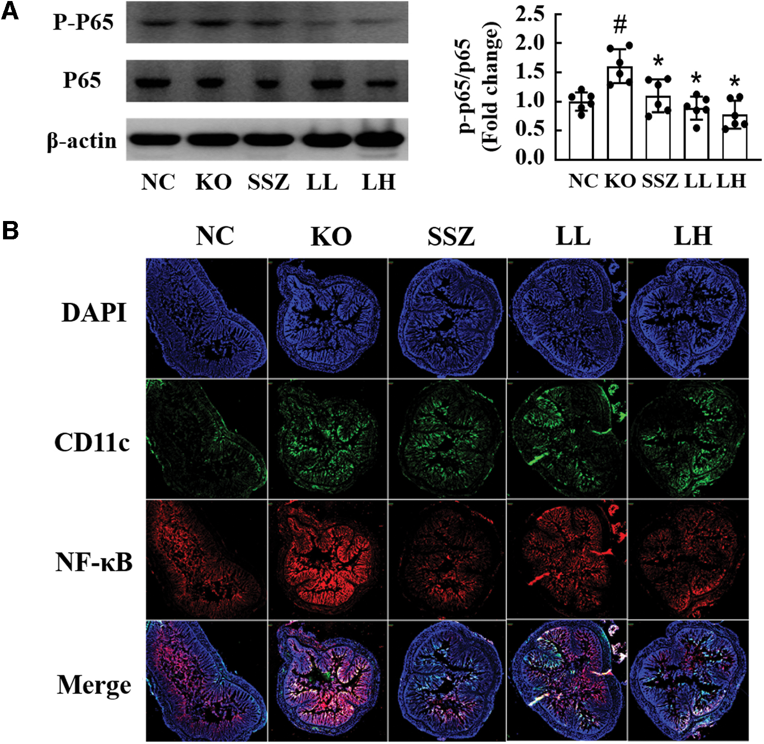
Effects of HDB1258 on NF-κB signaling in the colon of KO-induced colitis mice.
Effects on the spleen in the colitis mice
Oral gavage of KO induced systemic inflammatory response in the mice. The KO group had significantly higher serum PGE2, NK cell cytotoxicity, and phagocytosis of splenic macrophages (Fig. 4A) than those of the normal group. Furthermore, expression level of TNF-α was increased with Th1 cell population, which produces inflammatory cytokines, and its transcription factor, T-bet (Fig. 4B, C, D). In contrast, LH and SSZ group had significantly decreased levels of all these inflammatory markers, while increased IL-10 expression was higher compared with the KO group (Fig. 4). Furthermore, HDB1258 restored the ratios of Th1/Treg cells and Foxp3/T-bet to similar levels of those of the normal group (Fig. 4B, C).
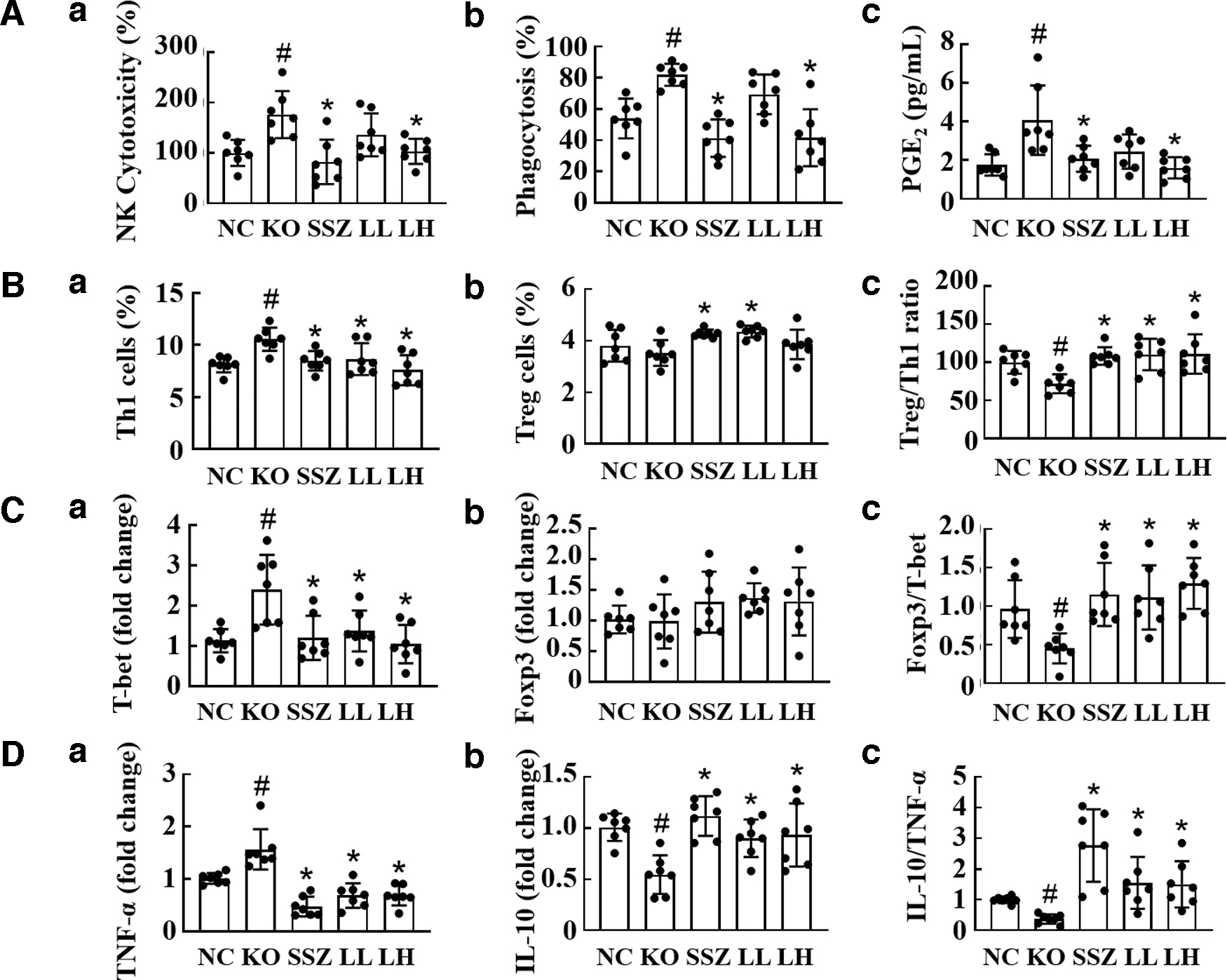
Effects of HDB1258 on the spleen of KO-induced colitis mice.
Regulation of gut microbiota composition in KO-induced colitis mice
Intestinal microflora in mice was affected by KO administration. Treatment of HDB1258 caused gut microbiota alterations: it changed KO-shifted β-diversity, but did not affect α-diversity based on operational taxonomic unit richness (Fig. 5A, B). However, SSZ tended to decrease α-diversity. Oral gavage of KO increased Bacteroidetes and the nondominant phylum Verrucomicrobia, but it reduced Cyanobacteria and Firmicutes (Fig. 5C). In contrast, HDB1258 increased KO-suppressed Firmicutes and Cyanobacteria, and it reduced Verrucomicrobia. At the family and genus levels, some bacteria, which were increased or decreased by administration of KO, were restored to a similar composition as those in the NC group (Supplementary Fig. S2). Among those in the LH group, Erysipelotrichaceae, PAC000198_g, and PAC001066_g were increased with the expression ratio of IL-10 to TNF-α in the colon; Helicobacter was decreased (Fig. 5D). Bacteroidaceae, Faecalibacterium, and PAC001066_g were increased with the ratio of IL-10 to TNF-α in the spleen (Fig. 5E).

Effects of 1258 on gut microbiota of KO-induced colitis mice.
Discussion
LABs have been considered as useful microorganisms to stimulate and develop the immune system, synthesizing and enhancing the bioavailability of nutrients, reducing symptoms of lactose intolerance, and reducing the risk of certain diseases. 17 The important properties for probiotics to function include resistance to gastric acidity, bile acid resistance, adherence to mucus and/or human epithelial cells, and tolerance against toxic metabolites derived from potential pathogens. There are a number of studies that have investigated the molecular bases of the stress response and thereby enhance probiotic properties (see the reviews by Zotta et al. and Derkx et al.). 18,19
In the previous report, the LS successfully improved probiotic properties of L. rhamnosus HDB1258. 11 Therefore, the LS culturing method was applied in this study for improving mucin adhesive ability of HDB1258. As a result, HDB1258 exhibited increased expression levels of chaperone proteins (GroEL/ES, DnaKJ, and HtrA), as well as mucin adhesion. Chaperones might help to disaggregate and renature misfolded proteins, thus protecting the cell from the damage imposed by stressful conditions. Since LS includes minerals, like sodium, calcium, and magnesium, these salts may stimulate the cells as a salt stress factor and then induce the stress response proteins like chaperones. Notably, it has been reported that surface protein HtrA may have an indirect role in regulating expression of cell surface protein. 20 Therefore, the HtrA might influence the expression of several extracellular proteins, including hydrophobic proteins. As a result, those chaperones might help HDB1258 to tolerate, survive, and adhere in the inflammatory bowel, seen as one of the extreme stressful environments.
Many studies have reported that IBD can be induced by opportunistic pathogens in gut microbiota. 21,22 KO is a pathobiont belonging to general gut microbiota and related to gut barrier function and microbiota regulation. 23 In the present study, we found that oral gavage of KO induced excessive inflammation response in the colon. Högenauer et al. reported that KO was found in the feces of patients with antibiotic-associated hemorrhagic colitis and only in the colon of rats that received amoxicillin–clavulanate in addition to being inoculated with KO. 24 Jang et al. reported that KO caused severe colitis in mice. 12 If the mucus barrier is collapsed by chronic inflammation at the colon, sIgA cannot be displayed at the mucus surface and inflammation grows worse by more stimulating epithelial cells and immune cells. 25 Increased sIgA levels of LH group may indicate that the mucus layer destroyed by KO was recovered on the surface of colon tissues, and then cell surface adhesive proteins of HDB1258 induced by stress response proteins, like chaperones, may attach to recovered mucin barrier.
Inflammation is the first step of immune response initiated by IL-1β and TNF-α. 25 When the immune causative factors disappear, the inflammatory response is reduced by the regulatory cytokines, but if the regulatory system is collapsed, it develops into chronic and systemic inflammation and leads to IBD. 5 In the previous study, HDB1258 showed immune-boosting effect by stimulating macrophages and inducing them to produce IL-1β. 11 However, when excessive inflammation occurs, HDB1258 downregulated overexpression of IL-1β and TNF-α and rather upregulated the anti-inflammatory cytokine, IL-10. These results indicate that HDB1258 also has anti-inflammatory functions.
In the spleen of LH group, Th1 cells were significantly reduced compared with KO group. Macrophages initiate the immune response through activation of Th1 cells. 26 As HDB1258 decreased the phagocytosis of splenic macrophages, it is believed that the induction of Th1 cell differentiation was reduced because of the dormant phase of macrophages. Meanwhile, as Treg cells were not affected, it is expected that HDB1258 does not directly affect T cell differentiation. As a result, HDB1258 may regulate excessive inflammatory responses through the control of cytokine expression and innate immune cells only.
Lactobacillus paracasei B21060 and Lactobacillus pentosus var. plantarum C29 attenuated inflammation by regulating cytokines through suppression of NF-κB signaling. 27,28 Berndt et al. reported the role of CD11c+ dendritic cells (DCs) in acute colitis, which are potent antigen-presenting cells regulating innate immunity and initial inflammation. 29 Excessive expression of CD11c in DCs induced acute inflammation, but when they were removed, total colitis index and symptoms were attenuated. 30 HDB1258 significantly inhibited NF-κB activation and decreased NF-κB+/CD11c+ cell population in the colon. The findings indicate that HDB1258 can do therapeutic action to gut inflammation through regulating of NF-κB signaling and differentiation of DCs in colitis model.
In many cases, IBD mice and patients appear to have different gut microbiota than healthy mice and people. 8,14,31 Gut microbiota is a commensal bacteria community that helps to maintain immune homeostasis through interaction with intestinal epithelial cells and immune cells. 32 Gut dysbiosis occurs when opportunistic pathogens in the intestine are unexpectedly increased or are infected from outside. 33 Then, the inflammation is initiated by stimulation of epithelial cells and immune cells, and if the host cannot control the immune response by itself, it progresses to chronic inflammation. General prescription of chemical drugs can relieve symptoms for a short period of time, but it also can re-induce chronic colitis by disrupting the intestinal microbial balance. 5 In this study, HDB1258 successfully alleviated inflammation in the colon while restoring the intestinal flora collapsed by KO similar to that of NC group, while SSZ worsened the imbalance of the flora. HDB1258 regulated the microflora like Helicobacter pylori which may be opportunistic pathogens in the colon. The improved mucin adhesive ability of HDB1258 may be an advantage for the survival and competition against abnormally increased pathogen in the inflammatory condition. As the ratio of pathogens was decreased, which are stimulating factors to immune cells, eventually the inflammatory response in the bowel would have reduced. It could be a beneficial treatment effect different from the side effect of chemical drugs.
In conclusion, L. rhamnosus HDB1258 cultured in the LS exhibited increased mucin adhesive ability and chaperone expression and alleviated the inflammatory response by regulating expression of cytokines in the KO-induced colitis mouse model. With improved adhesive ability, it survived competitively against pathogenic bacteria increased by KO and restored the disturbed gut microflora. Based on these results, L. rhamnosus HDB1258 could be an excellent gut microbiota-mediated immunomodulatory adjuvant for therapy of IBD.
Footnotes
Author Disclosure Statement
No competing financial interests exist.
Funding Information
No funding was received for this article.
Supplementary Material
Supplementary Table S1
Supplementary Figure S1
Supplementary Figure S2