




Abstract
Inflammatory bowel disease is associated with multiple extraintestinal disorders, including hepato-nephrological disruptions. The aim of this study was to evaluate the hepato-nephroprotective effect of Salvia officinalis leaf decoction extract (SLDE) on acetic acid (AA)-induced colitis accompanied with liver and kidney injuries. Wistar albinos rats were pretreated with SLDE (50, 100, and 200 mg kg−1, b.w., p.o.) during 10 days and intoxicated for 24 h by acute rectal administration of AA (3%, v/v, 5 mL kg−1, b.w.). Our results showed that S. officinalis treatment protected against AA-induced liver and kidney injuries by plasma transaminase activities and preservation of the hepatic and renal tissue structures. The level of high-density lipoprotein-cholesterol was also reverted back to near normalcy by treatment. Lipid peroxidation was decreased significantly by officinal sage supplementation. Treatment with SLDE increased enzymatic (superoxide dismutase, catalase, and glutathione peroxidase) and nonenzymatic (–SH groups and reduced glutathione) antioxidants in liver and kidney tissues. Also, SLDE treatment significantly protected against inflammation markers and reversed all intracellular mediator perturbations. This study suggests that the S. officinalis has a beneficial effect in controlling kidney and liver injuries by reducing lipid peroxidation and increasing antioxidant enzyme activities and nonenzymatic contents, which reduce the risk of developing extraintestinal complications.
Introduction
Extraintestinal disorders can involve almost any organ system. The liver, biliary tract, and renal involvement are considered to be the most frequently affected organs. These pathologies have been considered secondary complications of inflammatory bowel diseases (IBD), such as ulcerative colitis (UC) and Crohn's disease. 1 The prevalence of extraintestinal manifestations in IBD can reach 46%. 2 They may arise from the same pathophysiological mechanism of bowel disease, or secondary complications of IBD, or susceptibility to autoimmune diseases. 3
Pathologies range from primary sclerosing cholangitis to hepatic steatosis, considered the most common hepatobiliary complication. It has been suggested that primary biliary cholangitis, hepatic amyloidosis, granulomatous hepatitis, cholelithiasis, portal vein thrombosis, and liver abscess are considered the least common hepatobiliary disorders that are associated with IBD. 4
However, the most common renal involvement in patients with IBD has several aspects ranging from simple urinary sediment abnormality to nephropathy, tubulointerstitial nephritis, nephrolithiasis, and renal failure. 1,3,5,6
To protect and prevent these pathologies, marketed drugs such as mesalazine (5-ASA), infliximab, and azathioprine are usually used. 7 However, prolonged consumption or with relatively high doses can cause unpredictable side effects 8 and the installation of oxidative stress state through reactive oxygen species (ROS) overproduction, which can cause significant damage such as membrane lipoperoxidation, oxidation of enzymatic and structural proteins, as well as degradation of nucleic acids leading to cell death. 9,10
Nowadays, increasing attention has been drawn to finding new antioxidants from natural sources for further use in food processing and medicinal materials to replace synthetic antioxidants. However, a number of medicinal plants, rich in bioactive molecules, have been studied for their possible anti-inflammatory and antioxidant activities and used against IBD and associated hepatic and renal pathologies. 11,12
Salvia officinalis L. is an aromatic and medicinal plant (Lamiaceae family), which prefers hot and calcareous soils. It grows spontaneously in areas in the Mediterranean regions in southern Serbia and Kosovo. 13,14 Due to its richness in essential oils, and antioxidant and anti-inflammatory properties, sage extracts exhibit many beneficial health effects such as antibacterial 15 and antiviral 16 activities. More importantly, this plant has been widely used in the treatment of most gastrointestinal diseases like peptic ulcer and diarrhea. 17,18 Recently, we demonstrated that the flower extract has important hepato-nephroprotective effects. 12
The aim of this study is to show the in vivo hepato-nephroprotective effect of S. officinalis leaf decoction extract against injuries and oxidative stress produced by acute rectal administration of acetic acid (AA) in rats' liver and kidney.
Materials and Methods
Chemicals
AA, β-carotene, bovine catalase, butylated hydroxytoluene (BHT), chloroform, 5,5-dithio bis (2-nitrobenzoic acid), 2,2-diphenyl-1-picrylhydrazyl, methanol, epinephrine, 2-Thio-barbituric acid (TBA), and trichloroacetic acid were from Sigma chemicals Co. (Germany). All other chemicals used were of analytical grade.
Preparation of S. officinalis leaf decoction extract
The sage flowers were cultivated in the region of Tabarka (NW-Tunisia) during April 2020 and identified by Dr. Chokri Hafsi, Associate professor in the University of Jendouba-Tunisia. The plant material was dried at room temperature and stored in a dry place before use. The dried leaves were crushed to a fine powder. The decoction was made with distilled water (10 g/100 mL) at 100°C during 5 min under magnetic agitation and the homogenate was filtered through a Whatman filter paper. The filtrate was lyophilized and the S. officinalis leaf decoction extract (SLDE) was immediately used for experiments.
Free radical-scavenging activity on 2,2′-azino-bis [3-ethylbenzthiazoline-6-sulfonic acid]
The antioxidant capacity of SLDE was evaluated using the 2,2′-azino-bis [3-ethylbenzthiazoline-6-sulfonic acid] (ABTS•+) method. 19 Various SLDE solutions were prepared at different concentrations from 10 to 250 μg/mL. In each tube, 1 mL of the sample solution at all concentration was reacted with 3 mL of 7 mm ABTS radical solution (ABTS•+) and was kept in dark at room temperature for 60 min. The absorbance was measured at 734 nm. Ascorbic acid was used as a reference antioxidant molecule in the same concentration as the test extract.
Antioxidant capacity by the β-carotene bleaching inhibition method
The antioxidant potential was also performed by the method of β-carotene bleaching inhibition according to Nickavar and Esbati, 20 with some modifications. In this respect, 0.2 mg of β-carotene, 20 mg of linoleic acid, and 200 mg of tween 40 are dissolved in 0.5 mL of chloroform. The solvent was then evaporated and the mixture obtained was diluted with 50 mL of water bubbled with oxygen. Four milliliters of the homogenate was expelled into tubes, respectively, containing 0.2 mL of SLDE, 0.2 mL of BHT for the comparative test, and 0.2 mL of the solvent used, which will serve as a negative witness.
The absorbance of the samples is measured at 470 nm at initial time and every 15 min during 120 min. The blank test is an emulsion prepared as above, but without β-carotene. Ascorbic acid was also used as a reference antioxidant molecule in the same concentration as the test extract.
Animals and ethics statement
Adult male Wistar rats, weighing 220–250 g, were purchased from the Central Pharmacy (SIPHAT, SIPHAT, Ben-Arours, Tunisia Tunisia), housed five per cage and acclimatized for 2 weeks with a standard pellet diet (Badr-Utique-TN) and water ad libitum, and maintained under the conditions of a controlled temperature (22°C ± 2), and a daily 12-h photoperiod. They were used following the local ethics committee of Tunis University of the use and care of animals and in accordance with the National Institutes of Health (NIH) recommendation. The protocol was approved by the “Comite d'Ethique Bio-medicale (CEBM)” (JORT472001) of the “Institut Pasteur de Tunis.”
Experimental design
Rats were divided into six groups of 10 animals each. Group 1 and 2 served as a control and received 10 mL kg−1, b.w., p.o. of physiological solution (NaCl, 0.9%, p.o.); groups 3, 4, and 5 were pretreated with various doses of the SLDE (50, 100, and 200 mg kg−1, b.w., p.o.). Preliminary test indicated that those selected doses of SLDE were the lowest concentrations that give a significant protective effect. Finally, group 6 received gallic acid (GA) (50 mg kg−1, b.w., p.o.).
All the animals were kept fasting overnight. UC was induced for each animal, except group 1 by the infusion of AA (3%, v/v, 5 mL kg−1, b.w.) for 30 sec using a polyethylene tube inserted through the rectum into the colon up to a distance of 8 cm. This dose represents the lowest concentration, which induces liver and kidney injuries and no mortality during the entire period of intoxication. Twenty-four hours later, rats were anaesthetized and sacrificed. Blood was collected in heparinized tubes. After centrifugation at 3000 g for 15 min, plasma was processed for biochemical parameter determinations. The liver and kidney were weighted and rapidly excised and homogenized in phosphate-buffered saline. After centrifugation at 10,000 g for 10 min at 4°C, supernatants were used for biochemical analysis.
Histopathological analysis
Small pieces of the liver and kidney were immediately gathered and washed with a solution of NaCl (0.9%). Tissue fragments were then fixed in a 10% neutral buffered formalin solution, embedded in paraffin, and used for histopathological examination. Five micrometer thick sections were cut, deparaffinized, hydrated, and stained with hematoxylin and eosin. Liver and kidney sections thus obtained were examined in a blind manner for all treatments. 21
Assessment of liver function
Plasma aspartate aminotransferase (AST), alanine aminotransferase (ALT), and phosphatase lactate dehydrogenase (LDH) were measured using commercially available diagnostic kits (Biomaghreb, Ariana, TN; ISO 9001 certificate).
Assessment of renal function
Plasma urea, creatinine, and uric acid analyses were measured using commercially available diagnostic kits (Biomaghreb; ISO 9001 certificate).
Metabolic parameters
Plasma high-density lipoprotein-cholesterol (HDL-C) concentration was detected using commercially available diagnostic kits Biomaghreb. Atherogenic index of plasma (AIP) was determined according to the Friedewald
22
equation:
Lipid peroxidation
Lipid peroxidation was detected by the determination of malondialdehyde (MDA) production determined by the method of Draper and Hadley. 23 MDA levels were determined by using an extinction coefficient for MDA-TBA of 1.56 × 10⁵ M−1 cm−1.
Antioxidant enzyme activity assays
Superoxide dismutase (SOD) activity in liver and kidney was determined using modified epinephrine assays. 24 Changes in absorbance were recorded at 480 nm.
The activity of catalase (CAT) was assessed by measuring the initial rate of hydrogen peroxide (H2O2) disappearance at 240 nm. 25 The reaction mixture contained 33 mM H2O2 in 50 mM phosphate buffer, pH 7.0, and the activity of CAT was calculated using the extinction coefficient of 40 mM−1 cm−1 for H2O2.
Nonenzymatic antioxidant levels
The total concentration of thiol groups (–SH) was performed according to Ellman's method. 26 The –SH groups concentration was calculated using a molar extinction coefficient of 13.6 × 10³ M−1 cm−1. The results were expressed as nmol of thiol groups per mg of protein.
Reduced glutathione (GSH) was estimated in kidney and liver tissue by the method of Sedlak and Lindsay. 27 The absorbance was read at 412 nm against blank tube without homogenate.
H2O2 determination
H2O2 levels in liver and kidney were determined according to Dingeon et al. 28 However, the H2O2 reacts with p-hydroxybenzoic acid and 4-aminoantipyrine in the presence of peroxidase leading to the formation of quinoneimine, which has a pink color detected at 505 nm.
Iron measurement
Free iron levels in liver and kidney tissue was measured calorimetrically using the ferrozine method using a commercially available kit from Biomaghreb, Tunisia.
Calcium determination
The calcium levels in liver and kidney were performed using a colorimetric method according to Stren and Lewis. 29 However, at alkaline medium, calcium reacted with cresolphthalein and lead to a colored complex measurable at 570 nm.
Protein determination
Total protein concentrations in plasma, kidney, and liver were performed using commercially available diagnostic kits (Biomaghreb; ISO 9001 certificate).
Statistical analysis
The data were analyzed by one-way analysis of variance (ANOVA) and were expressed as mean ± standard error of the mean. The data are representative of 10 independent experiments. All statistical tests were two tailed and the differences were considered significant at P < .05.
Results
In vitro antioxidant capacity
Concerning the antioxidant capacities, we showed in Table 1 that the inhibition of β-carotene bleaching effect and the radical scavenging activity against ABTS radical of SLDE and ascorbic acid increased significantly in a dose-dependent manner (data not shown). The inhibitory concentration 50 of SLDE (37.18 and 69.54 μg/mL for ABTS radical and β-carotene bleaching, respectively) appears significantly higher than ascorbic acid (IC50 = 29.54 ± 1.17 μg/mL) used as reference antioxidant molecule.
2,2′-Azino-Bis [3-Ethylbenzthiazoline-6-Sulfonic Acid] Radical-Scavenging Activity and β-Carotene Bleaching Inhibition of Salvia officinalis Leaf Decoction Extract and Ascorbic Acid
Data are expressed as mean ± SEM (n = 3).
ABTS•+, 2,2′-azino-bis [3-ethylbenzthiazoline-6-sulfonic acid]; IC50, inhibitory concentration 50; SEM, standard error of the mean; SLDE, Salvia officinalis leaf decoction extract.
Effect of SLDE on organs weight
To test the in vivo effect of S. officinalis leaf decoction extract (SLDE) and GA against anal administration of AA-induced liver and kidney injuries, rats were treated with various doses of SLDE during 10 days. Liver and kidney weights were significantly increased in animals exposed to AA (Fig. 1). This increase was significantly and dose dependently improved by the SLDE (50, 100, and 200 mg kg−1, p.o.) treatment as well as by GA at 50 mg kg−1, p.o. (Fig. 1).
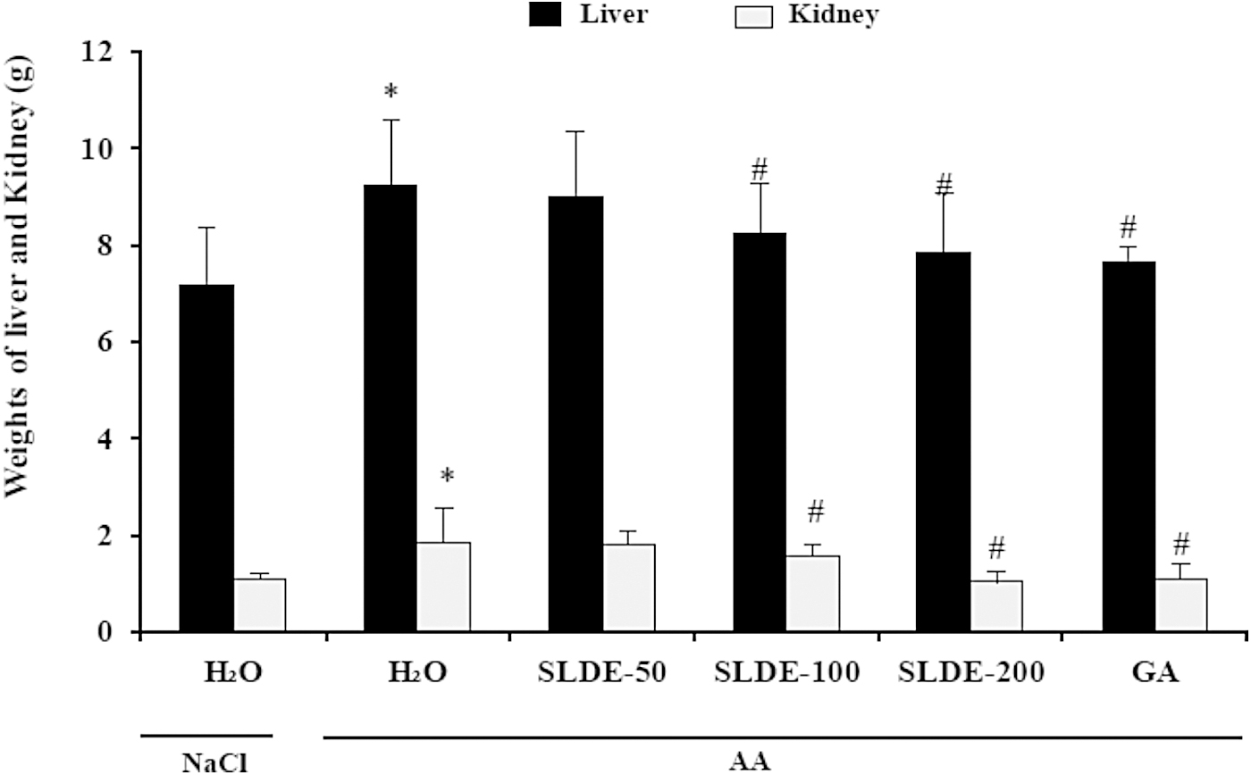
Subacute effect of SLDE on AA-induced deregulations in liver and kidney weights. Animals were pretreated with various doses of SLDE (50, 100, and 200 mg kg−1, b.w., p.o.) and GA (50 mg kg−1, b.w., p.o.) or vehicle (NaCl 0.9%) for 10 days. All animals, except those in the control group, were intoxicated for 24 h by acute anal administration of AA (3%, v/v, 5 mL kg−1, b.w.). *P < .05 compared to control group and # P < .05 compared to AA group. Values are mean ± SEM (n = 10). AA, acetic acid; GA, gallic acid; SEM, standard error of the mean; SLDE, Salvia officinalis leaf decoction extract.
Histological study
The increase of organ weights induced by AA has been associated with modifications in the liver and kidney aspects. In fact, the liver of AA-treated rats was characterized by mononuclear cell infiltration and congestion compared to control (Fig. 2Ab). Similarly, the histological sections of kidneys showed an increase in the glomerular area in rats treated with AA (Fig. 2Bb). However, SLDE pretreatment recorded obvious dose-dependent protection of the liver and kidney. The most important protection was detected in the group receiving the high dose of SLDE. A similar protective effect was also observed in GA-pretreated rats (Fig. 2).

Liver
Effect of SLDE on liver and kidney functions
The effects of AA and SLDE treatment on liver and kidney functions were investigated and the results are presented in Table 2. AA-induced hepatotoxicity was shown by a significant increase of plasma AST, ALT, and LDH activities. Importantly, when animals were treated with SLDE and GA, the levels of these parameters were significantly restored in a dose-dependent manner. Creatinine, uric acid, and urea are considered indicators of toxicity in the kidney. Our study confirmed that the elevation of plasmatic creatinine and urea levels and decrease of those of uric acid observed in the AA-treated group may be explained by kidney damage (Table 2). The treatment with SLDE has shown a restoration of plasmatic creatinine, urea, and uric acid levels reaching normal values compared to AA group (Table 2).
Subacute Effect of Salvia officinalis Leaf Decoction Extract and Gallic Acid on Acetic Acid-Induced Alterations in Liver (Aspartate Aminotransferase, Alanine Aminotransferase, and Lactate Dehydrogenase) and Kidney (Urea, Uric Acid, and Creatinine) Functions in Rats
Animals were pretreated with various doses of SLDE (50, 100, and 200 mg/kg, b.w., p.o.) and GA (50 mg kg−1, b.w., p.o.) or vehicle (NaCl 0.9%) for 10 days. All animals, except those in the control group, were intoxicated for 24 h by acute anal administration of AA (3%, v/v, 5 mL kg−1, b.w.).
Values are mean ± SEM (n = 10).
P < .05 compared to control group.
P < .05 compared to AA group.
AA, acetic acid; ALT, alanine aminotransferase; AST, aspartate aminotransferase; GA, gallic acid; LDH, lactate dehydrogenase.
Effect of SLDE on lipid metabolic parameters
In this research work, we observed a significant increase in plasma levels of HDL-C in the AA group compared to the control group (Table 3). The treatment with SLDE or GA reduced lipid parameters and AIP in treated rats compared to the AA-treated group.
Effects of Salvia officinalis Leaf Decoction Extract and Gallic Acid on Acetic Acid-Induced Alterations of High-Density Lipoprotein-Cholesterol and Atherogenic Index of Plasma in Rats
Animals were pretreated with various doses of SLDE (50, 100, and 200 mg/kg, b.w., p.o.) and GA (50 mg kg−1, b.w., p.o.) or vehicle (NaCl 0.9%) for 10 days. All animals, except those in the control group, were intoxicated for 24 h by acute anal administration of AA (3%, v/v, 5 mL kg−1, b.w.).
Values are mean ± SEM (n = 10).
P < .05 compared to control group.
P < .05 compared to AA group.
AIP, atherogenic index of plasma; HDL-C, high-density lipoprotein-cholesterol; TC, total cholesterol.
Effects of SLDE on hepatic and renal lipid peroxidation increase
The acute anal administration of AA (3%, v/v, 5 mL kg−1, b.w.) to rats recorded oxidative damage to the liver and kidney, as suggested by the magnitude of lipid peroxidation, which was demonstrated by the increase in hepatic and renal MDA levels compared to the control group (Fig. 3). SLDE and GA pretreatment significantly and dose dependently reversed overproduction of MDA level induced by AA intoxication.

Subacute effect of SLDE and GA on AA-induced changes in liver and kidney MDA levels in rats. Animals were pretreated with various doses of SLDE (50, 100, and 200 mg kg−1, b.w., p.o.) and GA (50 mg kg−1, b.w., p.o.) or vehicle (NaCl 0.9%) for 10 days. All animals, except those in the control group, were intoxicated for 24 h by acute anal administration of AA (3%, v/v, 5 mL kg−1, b.w.). *P < .05 compared to control group and # P < .05 compared with AA group. Values are mean ± SEM (n = 10). MDA, malondialdehyde.
Effect of SLDE on liver and kidney antioxidant enzyme activities
The comparison between control and AA groups revealed lower activities in enzymatic antioxidant, including SOD and CAT in the liver and kidney of the AA group. However, the SLDE (50, 100, and 200 mg kg−1, b.w., p.o.) or GA (50 mg kg−1, b.w., p.o.) pretreatment significantly protected against this decrease when compared to the AA group (Fig. 4).
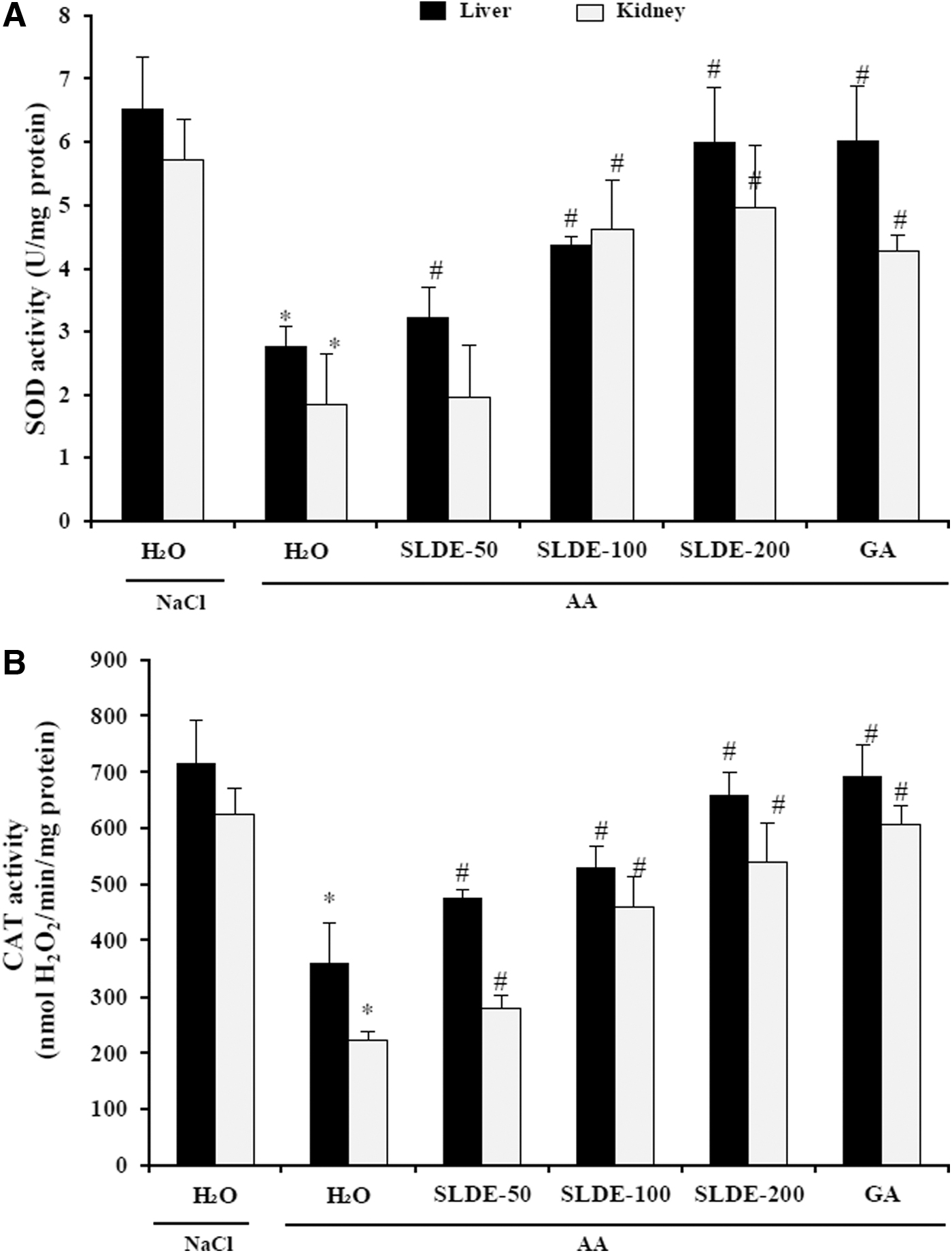
Subacute effect of SLDE and GA on AA-induced deregulations in liver and kidney antioxidant enzymes activities: SOD
Effect of SLDE and AA on liver and kidney nonenzymatic antioxidant levels
The –SH groups and GSH levels in the liver and kidney were also evaluated. Treatment with AA caused a considerable decrease in the sulfhydryl groups and GSH contents, which evidenced the induction of oxidative stress and significant thiol and GSH depletion after anal administration of AA. This effect was significantly and dose dependently corrected by subacute SLDE and GA pretreatment (Fig. 5).
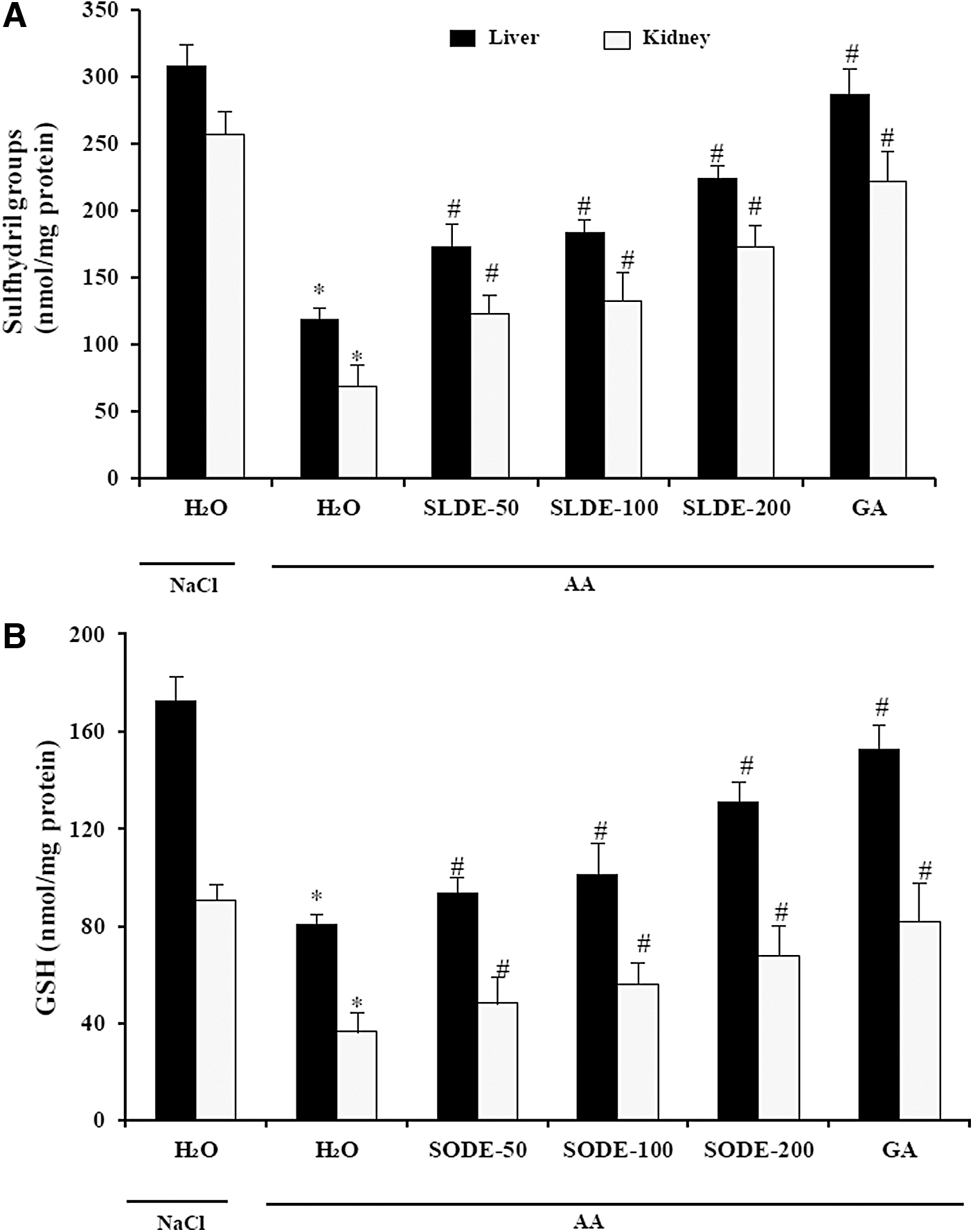
Effects of SLDE and GA on liver and kidney nonenzymatic antioxidants levels: GSH
Effect of SLDE on free iron and calcium levels
We further looked at the effect of AA and the SLDE on some intracellular mediators such as H2O2, free iron, and calcium in kidney and liver. As expected, AA administration increased iron, H2O2, and calcium levels. The SLDE and GA pretreatment significantly protected against AA-induced intracellular mediator disturbances in a dose-dependent manner (Table 4).
Subacute Effect of Salvia officinalis Leaf Decoction Extract and Gallic Acid on Acetic Acid-Induced Changes in Liver and Kidney Hydrogen Peroxide, Free Iron, and Calcium Levels in Rats
Animals were pretreated with various doses of SLDE (50, 100, and 200 mg/kg, b.w., p.o.) and GA (50 mg kg−1, b.w., p.o.) or vehicle (NaCl 0.9%) for 10 days. All animals, except those in the control group, were intoxicated for 24 h by acute anal administration of AA (3%, v/v, 5 mL kg−1, b.w.).
Values are mean ± SEM (n = 10).
P < .05 compared to control group.
P < .05 compared to AA group.
H2O2, hydrogen peroxide.
Discussion
In this study, we investigated the protective effect of S. officinalis on AA-induced colitis-related hepato-nephrological damage in rat.
The phytochemical analysis showed that SLDE is rich in phenolic potential therapeutic candidates. The use of the high-performance liquid chromatography with diode-array detection technique revealed the identification of eight phenolic compounds, distributed in three major groups such as phenolic acids, acylated sugar, and flavan-3-ols. 30 In addition, using the ABTS•+ radical-scavenging activity and β-carotene bleaching inhibition, we found that SLDE exhibits a high scavenging capacity, but lesser than ascorbic acid, which was used as a reference molecule.
The IBD causes a state of oxidative stress, which plays a role in the pathogenesis and progression of several conditions, including damages of the liver and kidney, which are considered two organs of detoxification. 11,30
The first purpose of this study was to induce liver and kidney damage on animal models that would imitate the physiopathology characteristic of human pathology. In this respect, the acute anal administration of AA (3% v/v, 5 mL kg−1, b.w.) clearly induced severe injuries in the kidney and liver tissues. These morphological perturbations are accompanied by an increase in the absolute weights of the kidney and liver, which is considered a reliable and sensitive indicator of the severity and extent of the inflammatory response. 31
The increased absolute weights of liver and kidney were also observed with several cases of intoxication, in rat and mice, such as ethanol, 12,32 dextran sulfate sodium, 11 and malathion. 33 Thus, for liver, hepatomegaly could be explained by the vacuolization and binucleation observed in hepatocytes of AA-treated rat. The ulcerogenic agent induced the expression of CYP 2B apoenzyme associated with hypertrophy of smooth endoplasmic reticulum and an increase in binucleation of hepatocytes. 31
In addition to the morphological liver changes in AA-treated rat, alteration of hepatic function was observed. Indeed, Rtibi et al. 11 have reported that subchronic exposure to dextran sulfate sodium provokes physical alterations of cell membranes objectified by a decreased membrane fluidity, which disturbs the liver function. Thus, enzymes normally in the cytoplasm leak into the blood circulation. According to Selmi et al., 33 elevated activities of plasma transaminases reflect a cellular leakage resulting from the loss of functional integrity of hepatocyte membrane. As observed in our work, activities of AST, ALT, and LDH in the plasma of treated rats were high. Treatment with SLDE caused a decrease in the activities of these enzymes.
On the other hand, anal administration of AA also induced renal dysfunction as assessed by an increase of plasma creatinine and urea levels when uric acid content decreased significantly. The kidney is an organ involved in the metabolism and elimination of toxic products. Indeed, according to Attessahin et al., 34 urea and creatinine are considered major products of protein metabolism, as well as sensitive markers of renal toxicity. More impressively, SLDE treatment significantly and dose dependently protects against AA-induced renal hemodynamic parameter deregulation.
Another important aspect has been studied in this work, which is the alteration of lipid metabolism often associated with AA treatment. This can be due to the change in the permeability of hepatic cells. 32 On the other hand, a significant decrease of HDL-C and an increase in the atherogenic index ratio (TC/HDL-C) following the administration of AA were observed. Our results are in line with the research published by Dreon and Krauss, 35 who showed that the heptotoxicity increases the production and secretion of apolipoprotein, lipoprotein particles, or triglyceride lipases. Moreover, the increase in plasma total cholesterol levels 30 may be due to the blockage of the liver bile ducts, which reduces or stops cholesterol secretion into the duodenum. 36
According to Liu et al., 37 the alteration of membrane fluidity is linked to the induction of oxidative stress and the impairment of enzymatic antioxidant mechanisms. In this context, oxidative stress induced by anal administration of AA enhances ROS overproduction due to the alteration of mitochondrial respiration or through their redox cycles.
In our study, the administration of AA was found to increase the formation of MDA, reflect of lipoperoxidation, and ROS leading to the inactivation of enzymatic antioxidant defense systems, including SOD, CAT, and nonenzymatic antioxidants like GSH and –SH in the liver and kidney. Our results agree well with the previous studies of Rtibi et al., 11 Jedidi et al., 12 Ghorbel Koubaa et al., 31 and Kamoun et al., 32 who suggested that the inhibition of these antioxidants might be caused by the accumulation of H2O2 or by the products of its decomposition. Indeed, SOD converts the reactive superoxide radical to H2O2, which was decreased in hepatic and renal tissues. When this intracellular mediator was not scavenged by CAT, it could lead to lipid peroxidation after generation of hydroxyl radical. 38
More importantly, SLDE treatment protected against AA-induced hepatic and renal oxidative stress. These data fully corroborated all previously reported in vivo and in vitro antioxidant and anti-inflammatory properties of Salvia officinalis. 12,39 These findings are similar with previous study, which has reported that the sage decoction extract contains a high level of total polyphenols, flavonoids, and tannins. Qualitatively, phenolic acids constituted the largest group accounting for 70.63% of the total identified compounds, from which rosmarinic acid and salvianolic acid B were the main compounds. 30
Importantly, we showed an increase of intracellular mediators such as calcium, free iron, and H2O2 in kidney and liver in response to oxidative stress induced by AA administration. However, we can now suggest that SLDE exerts a beneficial effect by chelating free iron and scavenging H2O2 and regulation of the calcium homeostasis. Our results also suppose that pretreatment with SLDE protects against overcharge of cells of the liver and kidney by free iron and H2O2 induced by acute anal administration of AA. Moreover, these later are involved in the generation of hydroxyl radical, which plays the major role in oxidative damage by affecting the molecular structures. 40 It was suggested that living organisms create a complex endogenous and exogenous antioxidant defense system to limit the overproduction of this ROS. 41
Our recent results showed that sage decoction extract caused the reduction of plasma CRP level, used as biological marker of inflammation, 30 and therefore a strong anti-inflammatory capacity of this plant.
In conclusion, this study suggests that treatment with SLDE may be beneficial in the prevention/treatment of extraintestinal disorders, in particular, hepato-nephrological disturbances caused following experimental colitis due to its antioxidant and anti-inflammatory properties, which were observed in reducing biological inflammatory/oxidative/metabolic biomarker levels and total antioxidant capacity.
Footnotes
ACKNOWLEDGMENTS
The financial support of the Tunisian Ministry of Higher Education and Scientific Research and the Institution of Agricultural Research and Higher Education (IRESA) Tunisia is appreciatively acknowledged.
Ethical Approval
All animal procedures on animals of this study were approved with the National Institute of Health recommendations for the use and care of animals. The protocol was approved by the “Comite d'Ethique Bio-medicale (CEBM)” (JORT472001) of the “Institut Pasteur de Tunis.”
Author Disclosure Statement
No competing financial interests exist.
Funding Information
No funding was received for this article.