




Abstract
Fungi are a source of a variety of secondary metabolites of importance in different areas of biotechnology. Several compounds have been characterized with antioxidant, antimicrobial, and anti-inflammatory activity from fungi of the division of the Ascomycota, among which is the species Daldinia eschscholtzii, an endophyte fungus of pantropical distribution. In this study, we evaluated the effect of an ointment made with D. eschscholtzii on the wound healing of BALB/c mice. The species was corroborated using a molecular marker Internal Transcribed Spacer (ITS1 and ITS4). The extracts and dust of the fungus were considered nontoxic as they caused a mortality of <15% in the nematode Panagrellus redivivus, and experimental ointments had no adverse effects on the skin of BALB/c mice. Wounds treated with the D. eschscholtzii ointments had 99.9–100% wound contraction after 17 days, which was similar to commercial healing (positive control). As such, the ointment of D. eschscholtzii is a natural alternative to improve wound healing.
INTRODUCTION
Fungi are known for having a wide range of properties of importance in different areas of biotechnology. This is largely due to molecules that are present in the fruiting body, mycelium, and culture broth, 1 including polysaccharides, terpenes, fatty acids, phenolic compounds, amino acids, and minerals (calcium, potassium, magnesium, iron, and zinc), these compounds have been attributed immunomodulatory, anticarcinogenic, antimicrobial, hypocholesterolemic, and antihyperglycemic effects and have been used in wound healing. 2 The genus Daldinia spp. presents several bioactive metabolites, including derivatives of benzophenones, polyketides (azaphilones, daldinins A-C, cytochalasans, dalmanol A, daeschol A, dalesconol A, B and C), terpene (triterpenoids, concentricols, concentricolide), daldiniapyrone, benzoquinones, steroids, lactone (helicascolide C and helicascolide A). 3 The ascocarps of two Daldinia species are known to be eaten by humans: D. fissa in Guatemala 4 and D. concentrica in Chiapas, Mexico. 5 They have also been reported to have analgesic and antibacterial properties, 6 leading to their use in traditional medicine for wound healing in tribes of diverse regions of Africa. 7
It has been noted that some natural products can accelerate wound healing through a number of different mechanisms, including the proliferation and migration of epithelial cells or fibroblasts, decreased production of reactive oxygen species, and the modulation of inflammatory mediators.
8
With fungi, it was possible to accelerate wound healing by increasing proliferation, migration, and invasion of fibroblasts and keratinocytes and improved collagen synthesis by using an extract rich in polysaccharides of the Auricularia auricula-judae. The extract had antioxidant properties, which favored the equilibrium between pro- and anti-inflammatory mediators in the local environment of the wound.
9
In another example, the polysaccharide β-
MATERIALS AND METHODS
Biological material
We used D. eschscholtzii stroma cultured on a combination of substrates: avocado leaf, tomato, and maize straw (20/20/60%; Dc), and maize straw (100%; Dm).
Molecular identification
The mycelium was grown for 7 days in potato dextrose broth. We extracted deoxyribose nucleic acid (DNA) from the mycelium using the Quick-DNA Fungal/Bacterial (Zymo Research) kit following the manufacturer’s instructions. DNA was amplified by polymerase chain reaction (PCR) using the universal primers ITS1 (5′-TCCGTAGGTGAACCTGCGG) and ITS4 (5′-TCCTCCGTCTATTGATATGC). 11 The reaction mixture volume was 50 μL: 5 μL (5 μM) of each primer, 25 μL of Q5 High-Fidelity 2X Master Mix, 2 μL of DNA (5 ng/μL) and 13 μL of water. The PCR amplification protocol was: 98°C for 3 min for one cycle, 98°C for 30 sec, 55°C for 30 sec, 72°C for 30 sec, and 72°C for 5 min. The reaction was purified with AMPure XP Beads. It was sequenced on an Illumina NextSeq 500 sequencer (at the Unidad Universitaria de Secuenciación Masiva y Bioinformática, UNAM). The sequence was compared to the database using BLASTn for homology (http://www.ncbi.nlm.nih.gov; https://www.ebi.ac.uk/Tools/sss/ncbiblast/) in order to identify the organism. The ITS was compared with the ITS in the GenBank database using BlastN. The phylogenetic analysis of the matrix was done using the neighbor-joining model in the program MEGAX. 12
Preparation of D. eschscholtzii extracts
We used 10 g of dehydrated and pulverized stroma from each culture. For the extract from the culture of the combination substrate (Dc1) and maize straw (Dm2 maize), we added a hydroalcoholic solution (70% ethanol), shook for 24 h (120 rpm/25°C), then centrifuged for 10 min at 3500 rpm (Centurion Scientific Limited), 13 concentrated using a rotavapor (Heidolph modelo Hei-VAP Advantage) and lyophilized (LABCONO FreeZone). The extracts were stored at 4°C until use.
Preparation of D. eschscholtzii ointment
The ointment was prepared with propylene glycol, sodium lauryl sulfate, liquid petroleum jelly, and stearyl alcohol. To this ointment base, we added 75 mg/mL of the D. eschscholtzii extracts (Dc1, Dm2 maize), powder (Dc3, Dm4 maize), or neither ointment base (OB).
Evaluation in vivo
We carried out two tests in vivo to evaluate the treatments (Dc1, Dm2 maize, Dc3, Dm4 maize). First, we tested for acute dermal toxicity, where we observed that there was no irritation or damage to the skin of the experimental animal at the end of the exposure period. Second, we tested wound healing. Here, we added two additional treatments, ointment base (OB; negative control) and ulcoderma (UI; positive control), considering in this test as variables the contraction of the wound and the epithelialization period.
Acute dermal toxicity
The acute dermal toxicity test was carried out following the practices for the experimental use of the animal. 14 A total of 20 six-week-old BALB/c mice (10 females and 10 males) were divided into 5 groups (Dc1, Dm2 maize, Dc3, Dm4 maize, no treatment) of four animals. The animals were kept in a cage and acclimated to the laboratory conditions for 1 week before the test. We shaved the fur from 10% of the dorsal surface of the body 24 h before the test. In each group, we placed the same amount (120 ± 10 mg) of the corresponding ointment and observed no irritation or damage to the skin of the experimental animal. At the end of the exposure period (24 h), we eliminated the residual test substance and observed daily to detect the presence of any adverse cutaneous reaction for the next 14 days. 14,15
Evaluation of wound healing in vivo
Were used 36 six-week-old BALB/C mice (18 females and 18 males) weighing 19.5 ± 2.1 g provided by the National Public Health Institute (INSP). The mice were handled according to national standards NOM-062-ZOO-1999. 14 They were housed individually in polypropylene cages with bedding of sterilized sawdust and covered with wire mesh (Lab Diet 5008) in a room with a temperature of 25°C, 55% humidity, 12 h: 12 h light-dark cycle. They had free access to water (sterile) and food (Lab Diet 5008). The mice were acclimated to laboratory conditions for 1 week before the beginning of the experiment.
The mice were randomly split into 6 groups: (1) Dc1, (2) Dm2 maize, (3) Dc3, (4) Dm4 maize, (5) OB, and (6) UI (positive control) (n = 6). We shaved the dorsal surface and placed naproxen/lidocaine (10 g/2 g) on the surface of the skin just before making the excision. We made a 100 mm2 wound using a disposable biopsy punch (Kai medical, Japan). Treatments (ointments) were applied daily in the morning until the last day of treatment (healing). Treatments were evaluated by progressive changes in the wound area of mice, measured with a digital calibrator (BEC, Taiwan), and photographs were taken as a record. The wound contraction was calculated as a percentage of reduction in the wound area.
16
Measurement of the epithelialization period
We calculated the time in days required for the epithelium to form, i.e., for the remaining dead tissue to fall off with no residual open wound. 17
Toxicity assay with P. redivivus
This nematode is a free-living bacterivore, found in various nutrient-rich habitats (soil, rotten fruits, insects, etc.). It has been used in ecotoxicity tests previously.
18
We analyzed the toxicity of the stroma extracts (EDc1, EDm2) and powder of D. eschscholtzii (PDc3, PDm4) at different concentrations (12.5, 25, 50, 75, and 100 mg/mL). We used M9 as the negative control
19
and ivermectin as the positive control (5 mg/mL). We harvested the nematodes from hydrated oats (200 g/300 mL) left at room temperature (23°C). The nematodes were separated from the oats by washing them through sieves. The assays were carried out in a 96-well plate. In each well, we placed 50 µL (150 juvenile larvae) of P. redivivus and 50 µL of each treatment. The nematode exposure to the treatment lasted for 48 h at room temperature. Then, a transmission stereoscopic microscope quantified each well’s number of live and dead nematodes (Olympus SZH 10). We did six repetitions for each treatment. No standard guide is available for mortality tests in this species, so we chose 20% mortality as a validity criterion.
20
We calculated the percent mortality using the following formula, and the percent survival was obtained by subtracting the percent mortality from 100.
21
Preliminary phytochemical evaluation of D. eschscholtzii
Were added to 5 mg of extracts (EDc1, EDm2) and powder of D. eschscholtzii (PDc3, PDm4) and were read after 5 min. A color change indicated a positive result. The Liebermann–Burchard reaction suggested the presence of triterpenes when the color changed to purple and consisted of adding 1 mL of the acetic anhydride reagent, 100 µL H2SO4, and 10 drops of HCl. The Shinoda reaction suggested the presence of flavonoids when the color changed to red and consisted of adding 1 mL CH3OH, 2–3 Mg chips and 2- drops HCl. Molisch’s test (carbohydrate-violet): 1 mL of water, 2 drops of alphanaphthol (1%) and 7 drops of H2SO4. The reaction to identify coumarins suggested the presence of those substances when the yellow color of the extract disappeared and consisted of adding 1 mL of CH3OH, 1 mL of alcoholic NaOH, and 8 drops of HCl. The presence of saponins was confirmed by foam formation when adding the extract to 1 mL of water. The Wagner reaction indicated the presence of alkaloids when the color changed to brown and consisted of adding one chip of Iodine, KI, and 1 mL of distilled water. Each test was performed in triplicate. 22
Mass spectrometry analysis
Gas-mass chromatography was performed on a Thermo Scientific TRACE GC with an ITQ900 ion trap mass detector (Thermo Electron Corporation, Milan, Italy). The carrier gas was helium with a flow rate of 1 mL/min. The column used was TRACE-5MS (30 m, 0.25 μm film, and 0.25 mm internal diameter). The GC was equipped with a split-splitless injector that was held at 270°C. The oven was programmed as follows: initially 50°C for 1 min, then the temperature was ramped in three steps: from 50°C to 300°C at 7°C/min and finally held at 300°C by 5 min, for a total chromatographic time of 56 min. The mass spectra were acquired by electron impact ionization at 70 eV, and the detector was set in TIC/Scan (Total Ion Current) mode from 50–650 m/z with a scanning rate of 0.2 scan s-1 (Dwell). The transfer line and ion source temperatures were held at 270°C and 200°C, respectively. The raw data were processed using Xcalibur™ software version 4.0 (Thermo Scientific, USA).
Statistical analysis
We report the mean and standard deviation of the results. We did the analyses in the program Statistic 7 using a one-way ANOVA followed by post-hoc tests. We considered statistical significance at P < 0.05.
RESULTS
Identification of the fungal species
It confirmed the identity of the fungus included in the treatments using molecular identification. The sequence was deposited in the GenBank of the National Center for Biotechnology Information (NCBI) with the accession number OP735354. Comparing the sequence with existing entries in two databases confirmed that the fungal species was D. eschscholtzii (Ehrenb.) Rehm [as “eschscholzii”] from the phylum Ascomycota (Hypoxylaceae, Xylariales). 23 The sequence had a 100% match with two strains of D. eschscholtzii (EM_FUN:MF495455 and MF495455), and a 99.8% match with three additional strains (EM_FUN:MG490699; EM_FUN:MZ270647; MG490699.1; MZ270647.1). A phylogenetic analysis showed that the D. eschscholtzii samples were grouped (Fig. 1).
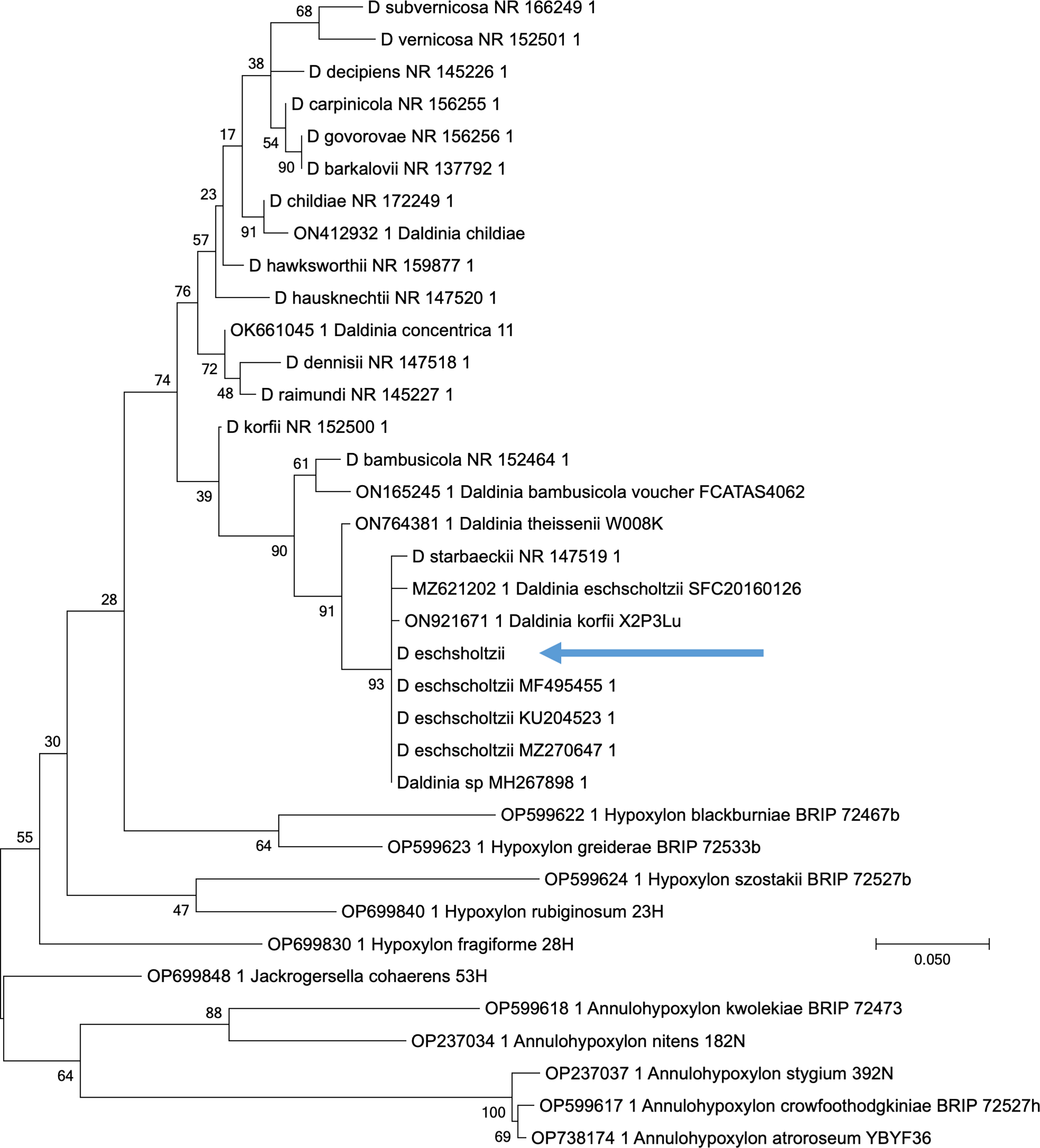
Molecular phylogenetic analysis by the Maximum Likelihood method of the nucleotide sequence of D. eschscholtzii. The tree with the highest log likelihood (−2157.1937) is shown. The percentage of trees in which the associated taxa clustered together is shown next to the branches. Initial tree
Evaluation in vivo
Acute cutaneous toxicity
Application of the experimental ointments containing D. eschscholtzii extracts or powder showed no adverse skin reactions. During the 14 days of observation, the experimental individuals showed no signs of irritation, erythema, inflammation, or toxicity, nor were there changes in the animals’ skin or behavior.
Wound healing in vivo
The ointments D. eschscholtzii favored wound healing in the excision test in live mice, obtaining results similar to a positive control (ulcoderma). The wound had closed completely by 17 days after excision (wound area 0 mm2) for the OB, three of the four treatments: Dc1 (extract from D. eschscholtzii cultured on a combined substrate), Dm2 maize (extract from D. eschscholtzii cultured on maize), and Dm3 (powder of D. eschscholtzii cultured on a combined substrate). For the Dm4 maize treatment (powder of D. eschscholtzii cultured on maize), the wound size at 17 days was 0.04 mm2. When considering daily comparisons of wound size, the treatments generally did not present significant differences by day, except for days 1 and 3. After 72 h, the positive control (UI) wound area was 91 mm2; for the Dc1, Dm2 maize and Dm4 maize, on average, it was 86 mm2, and the largest area was for Dc3 (90.45 mm2). However, in this last treatment experimental individuals showed a 20% reduction in the epithelization period (Table 1).
Wound Area and Time to Epithelialization in Mice Treated with Ointments Containing D. eschscholtzii Extract (Dc1, Dm2) or Powder (Dc3, Dc4)
Ul = Ulcoderma (positive control), ointment of D. eschscholtzii extracts from the culture of the combination substrate (Dc1), ointment of D. eschscholtzii extracts from the culture of the maize straw (Dm2 maize), ointment of D. eschscholtzii powder from the culture of the combination substrate (Dc3), ointment of D. eschscholtzii powder from the culture of the maize straw (Dm4 maize). Data are represented as mean ± SD values of six mice, by Tukey’s multiple range test, different letters in the same column are significantly different (p< 0.005).
In general, the percent contraction was 100% by day 17, except for Dm4 maize (99.9%). On the seventh day, Dm4 maize had the highest percent contraction (48.69%), Dc1 (38.54%) and Dm2 maize (36.05%) had the lowest, but there were no significant differences among the experimental groups. By day 11, the four treatments were already closed by approximately 80% (Fig. 2), except for Dm2 maize (76.6%).

Percentage of wound contraction in the presence of extracts and powder of D. eschscholtzii. Data are represented as mean ± SD. values of six mice.
The fungus D. eschscholtzii could be used for wound closure since it improved the appearance of the wound architecture and did not present any cytotoxic effects in the contraction model; said results were similar to those obtained with UI (Fig. 3).
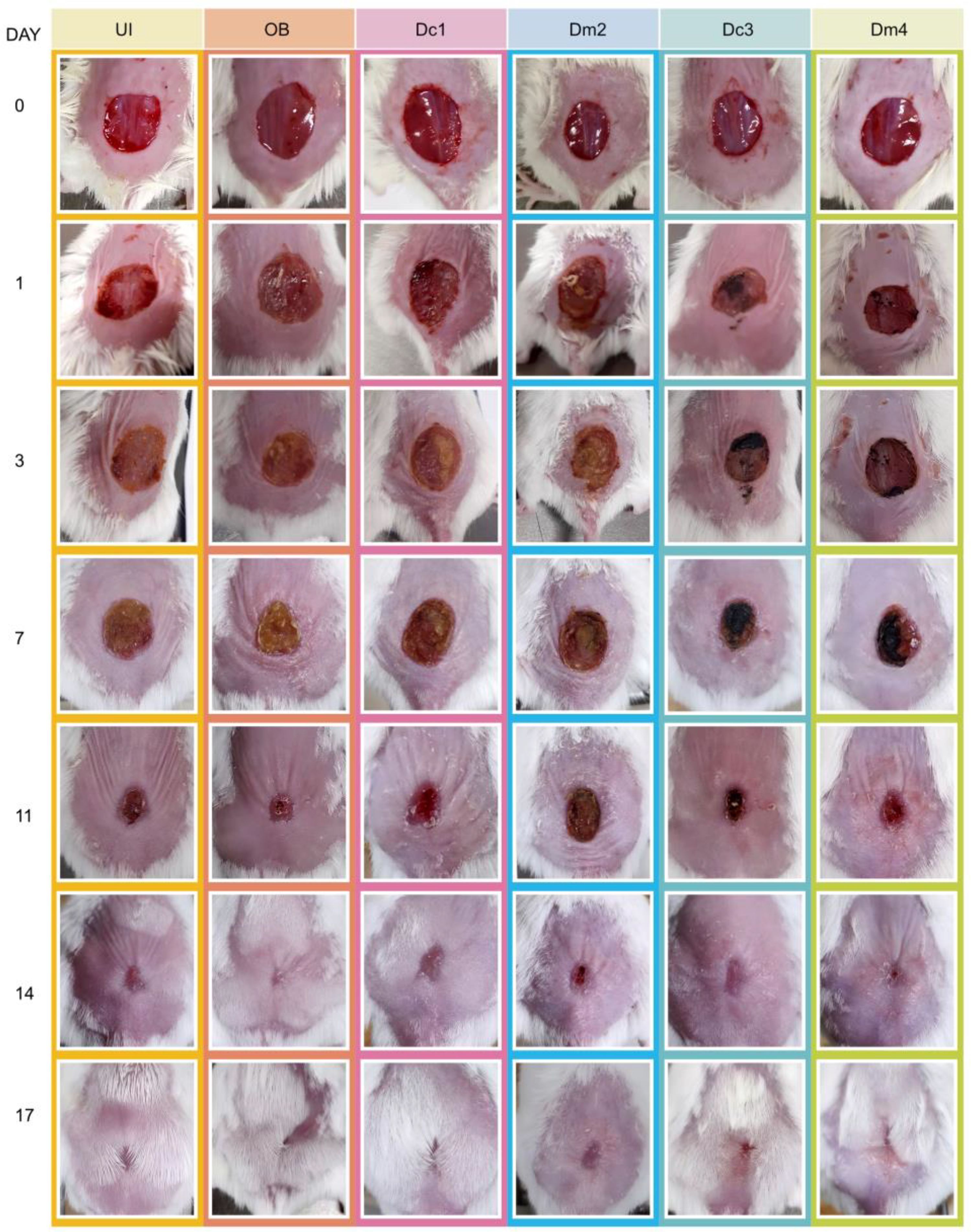
Closing of the excision in BALB/C mice in each of the experimental groups on days 0, 1, 3, 7, 11, 14, and 17.
Toxicity in vitro with P. redivivus
The experimental samples had low toxicity to the nematode P. redivivus. At the highest concentration (100 mg/mL), the D. eschscholtzii treatments all had <15% mortality, compared with 100% mortality with the positive control, ivermectin (5 mg/mL), and 3.2 ± 0.72% mortality with the negative control (M9). This was reflected as 98.56% survival for the stroma powder (PDc3), for PDm4 it was 98.24%, for the stroma extract it was 95.9% (EDc1) and in the case of EDm2 it was 85.5%, which was significantly different. This trend was similar for the 75 mg/mL concentration (Fig. 4). The nematode P. redivivus is an amphimictic species with oviparous females, the larvae mature after hatching into adults in approximately 4 days, they progress through four larval stages to reach adulthood (L1–L4 o J1–J4). 24 Toxicity tests are important during the standardization of the use of new molecules and to maintain reproducibility over time and among different laboratories.
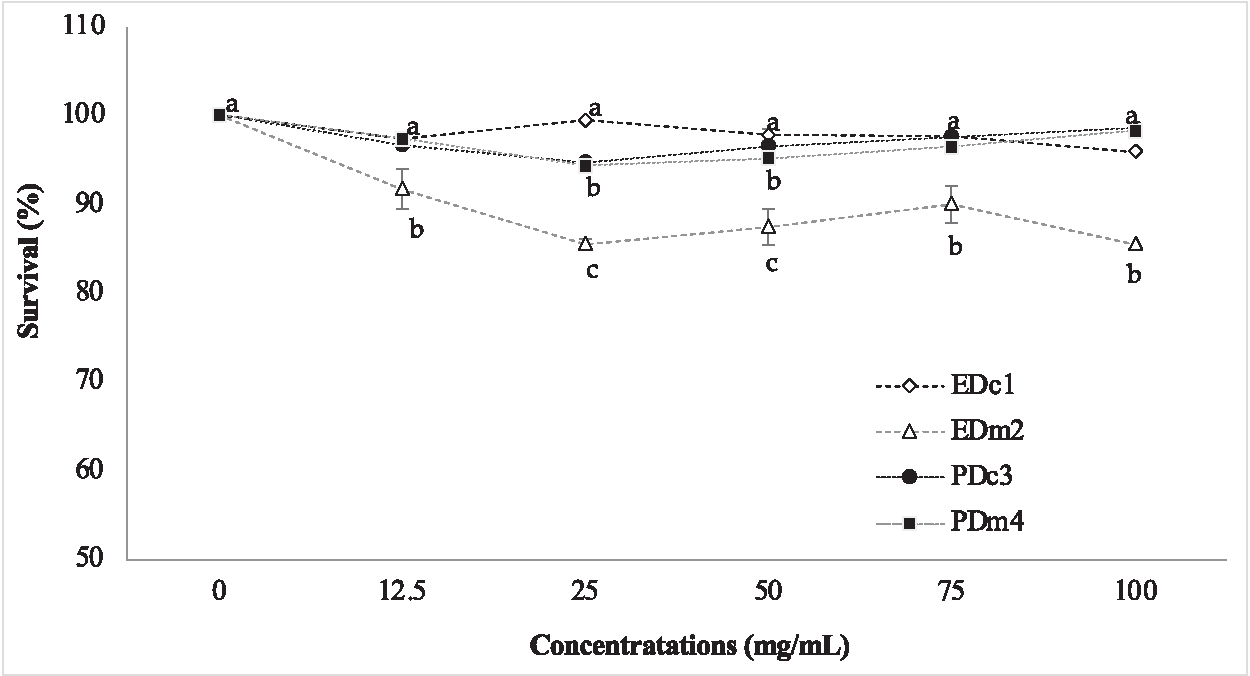
Percentage of survival of P. redivivus in the presence of extracts and powder of D. eschscholtzii. Data are represented as mean ± SD values of six replicates by Tukey’s multiple range test, different letters are significantly different (p< 0.005).
Phytochemical components of the fungal extracts and powder
According to the qualitative phytochemical study, the treatment ointments have flavonoids, saponins, triterpenes. We only detected coumarins in EDm1 and alkaloids in PDc4.
Mass spectrometry of the fungal extracts and powder
The results of the GS-MS chromatographic profile are shown in Table 2. Identified 28 molecules from three samples (EDc1, EDm2, and PDc3). We found naphthalene derivatives. The EDm2 sample contained six compounds, which included anthraquinone derivatives. In PDc3, there were three unknown compounds and five identified compounds, which included azoic compounds. The EDc1 sample had the highest content of compounds. Compound 1 from that sample has been reported to have activity against human prostate carcinoma cells (PC-3). 25 Molecule 6 (EDc1) has a pyrazolo[3,4-b]pyridine core, and these compounds have been attributed to biomedical applications (antitumoral, anti-inflammatory). 26 We also detected a sugar alcohol, arabinitol, which has been shown to eliminate hydroxy radicals and protect endothelial cells under hyperglycemic conditions. 27
Compounds Identified by GS-MS Analysis of Three Samples of D. eschscholtzii
D. eschscholtzii extracts from the culture of the combination substrate (EDc1), D. eschscholtzii extracts from the culture of the maize straw (EDm2), D. eschscholtzii powder from the culture of the combination substrate (PDc3).
DISCUSSION
The strain was identified as D. eschscholtzii, and the fungus is native to Guerrero, Mexico, it is important to note that there’s no previous work with this species regarding wound healing properties in the country, so it is important to know the application of ascomycetes. Barbosa-Reséndiz et al. 28 reported that D. eschscholtzii is characterized by its placentiform to semi-globose stroma, reddish–brown to black, which releases a violet pigment in the presence of KOH, and the spores are 10–14 × 5–6.5 µm. The fungi of the genus Daldinia are endophytic and can remain in the host tissue for long periods without causing any symptoms or evident damage. They grow gregariously on decomposing wood (legumes) in tropical deciduous forests and xerophytic scrub, and the species has a pantropical distribution.
Regarding our analysis of secondary metabolites in D. eschscholtzii, we found 28 compounds, these could be of importance as secondary metabolites obtained from fungi are compounds that usually have a beneficial application for human health (pharmacological or biological activities), so it is essential to analyze these metabolites and their toxicity tests to ensure that the use of said natural products is safe. 29 In our toxicity tests, we found <15%, which is relevant compared with other similar studies that tested the effect of extracts of natural products on P. redivivus in general and which reported toxic effects. In the case of leaf extract of Ficus carica (5 mg/mL), it had 96.2% mortality after 72 h in P. redivivus, and the LC50 of psoralen (isolated compound) was 0.18 mg/mL after 72 h. 30
Concerning toxicity tests with fungi, Liu et al. 31 isolated three compounds from Coprinus xanthothrix (Basidiomycota) and indicated that the LC50 for P. redivivus were 0.25, 1.0, and 0.125 mg/mL for each compound. Li et al. 32 reported that the culture broth of Stereum sp. (Basidiomycota) presented mortality of P. redivivus, the nematocidic activity of two aromatic compounds, the compound 3,5-dihydroxy-4-(3-methylbut-2-enyl)benzene-1,2-dicarbaldehyde (2) presented 50% (100 ppm) mortality and butyl 2,4-dihydroxy-6-methylbenzoate (4) had 90% (200 ppm) mortality after 24 h, compound 2 presented a CHO group, compound 4 and ester group, and these groups seem to be responsible for the activity. Has been reported nematicides activity for three compounds of Daldinia; in the case of 1-methoxy-8-hydroxynaphthalene it presented a median lethal concentration (LC50) of 10 μg/mL (1), for 1,8-dimethoxynaphthalene it was 25 μg/mL (2) and for 5-hydroxy-2-methylchromanone and more than 100 μg/mL (3) was required in Caenorhabditis elegans, but when using Meloidogyne incognita in all cases the LC50 was above 100 μg/mL. 33 In this study, we used higher concentrations (100 mg/mL) and found only 14.5% mortality for EDm2, the ascocarps of D. eschscholtzii do not present toxic effects in the model used and can be considered a safe species for use in natural products for therapeutic purposes.
In relation to the secondary metabolites reported for genus Daldinia, it has been indicated one of the main compounds concentricol is within the family of cytochalasin molecules (cytochalasin I; cytochalasin II; cytochalasin III; cytochalasin IV), which may be derived from the acetate-mevalonate pathway, but other species of Daldinia contain terpenoids as the main stroma metabolites. In other species, the main metabolites are 1,1′-binaphthalene-4,4′−5,5′-tetrol; daldinal A; daldinal B; daldinin C, which is probably of polyketide origin, and the main metabolites: 1,1′-binaphthalene-4,4′−5,5′-tetrol and daldinol, which is considered to be metabolites that are generated through a lateral pathway of melanin biosynthesis mediated by polyketide synthase, which arises from 1,8-dihidroxinaftaleno, which is a melanin precursor in ascomycetes. 34,35 The presence of 1,8-dihidroxinaphthalene in the secondary metabolism of Daldinia could suggest the presence of naphthalene derivatives in the majority of the species of the genus. 36,37
The mycelia of D. concentrica contain aromatic metabolites: chromanone (V) and mono- and di-methyl ester of 1,8-dihidroxinaphthalene, derived from benzene. 35 In two Japanese strains of Daldinia spp., over 20 metabolites were characterized. In D. concentrica, four aromatic compounds, three azophilones, and 16 cytochalasins were determined, which had cytotoxic activity in a KB cell line, and in D. vernicosa, a nitrosamine was found. 36 It has been shown that the stroma of D. childiae contains benzo-phenones (daldinal A; daldinal B) and azaphilones (daldinin C), but in some stroma of D. eschscholzii cytochalasins have been found (terpene, alkaloids), but in other stroma of the same species the cytochalasins were not the main metabolites. The compounds present were similar to D. concentrica, which presents concentricol and 1,1′-Binaphthalene-4,4′-5,5′-tetrol, 34 which suggests the existence of subpopulations of D. eschscholzii, which probably arose due to host specificity and geographic isolation. 37 In this work, derivatives of naphthalene and anthraquinones were found, which is why it is important to characterize the compounds present in species of the country and to know their possible use in the pharmaceutical industry.
The four ointments (Dc1, Dm2 maize, Dc3, and Dm4 maize) presented healing activity and on most days, the effect was without significant difference (P < 0.05) between them. The ointments promoted the stages of healing (hemostasis, inflammation, proliferation, and remodeling). However, more experiments must be carried out to define exactly the process in which each of the ointments is involved. Considering the results, it is suggested that the stroma powder of D. eschscholtzii grown in the substrate mixture (Dc3) would be the best option for the preparation of healing ointments from an economic and methodological point of view. The healing process observed in this work was better than that reported by Rajeshwaran et al. 13 who used 5% and 10% methanolic extracts of wild specimens of D. concentrica, the authors reported that the greatest healing activity in vivo was in the 10% extract (75–87% contraction), while with Neosporin (commercial ointment) they observed 92% contraction at 21 days. It is important to mention that the cultivation of D. eschscholtzii, as in this study, allows us to have biological material to avoid the collection of wild species, contributing to the conservation of natural resources.
This fungus is widely used in traditional medicine; for example, in Spain, the D. vernicosa fungus is used for muscle cramps as an analgesic and sedative. 38 In Mexico, the Tzeltal Indians used Daldinia sp. in warts, pimples, or ringworm of the scalp. 39 In Nagaland, India, D. concentrica is used to treat skin allergies and heal wounds, 40 and certain tribes in regions of Africa have used it to heal wounds. 7 Additionally, it has been reported to have analgesic and antibacterial properties. 6
Gupta et al. 41 reported a lyophilized aqueous extract of Ganoderma lucidum that had high polyphenol and flavonoid contents had better healing activity than povidone-iodine cream; the fungal extract increased wound contraction, reduced inflammation, improved reepithelialization, increased angiogenesis, and collagen (hydroxyproline) and hexosamine accumulation. Krupodorova et al. 42 reported the healing activity of aqueous extract of mycelium (100 mg/mL) from two fungi Basidiomycota; Crinipellis schevczenkovi and G. lucidum. C. schevczenkovi presented wound healing on the third day of topical application in albino mice, while G. lucidum showed healing on the fifth day, but by the sixth day, the results were similar between the two extracts. In another example, an extract of Antrodia camphorata (Basidiomycota) significantly improved wound closure in rats by increasing the proliferation of fibroblasts; there was a reduction in the number of inflammatory cells, increased collagen production, and proliferation of capillaries (angiogenesis) compared with the vehicle. 43 The ointment of D. eschscholtzii did not have toxic effects, it improved wound closure and prevented the formation of hypertrophic and keloid scars. We, therefore, suggest that it is a natural alternative for wound healing. Sharifi-Rad et al. 44 reported that mushroom extracts might contribute to certain mechanisms, such as stimulation of immune, epithelial cells, extracellular matrix, cytokines, growth factors, reactive oxygen species, and various inflammatory intermediates. Therefore, it is important to continue searching for natural alternatives to improve the wound healing process.
CONCLUSION
The ointments with D. eschscholtzii did not have adverse toxic effects on the model used. They improved wound closure and prevented the formation of hypertrophic and keloid scars, making them a natural alternative for use in wound healing.
Footnotes
ACKNOWLEDGMENTS
This study was part of the master’s degree thesis of Reyna I. Cueva Clavijo (No. 793587) at master’s degree in Natural Resource Management, from the Biological Research Center of the Autonomous University of the State of Morelos, Mexico. The authors thank Tigram Contreras for his help in editing the picture.
AUTHORS’ CONTRIBUTIONS
M.T.T.: Conceptualization. R.I.C.C., G.S.C.R., L.F.M.P., M.T.T., E.H.N.: methodology. R.I.C.C., L.A.M., A.W.V., and E.H.N.: Software. M.T.T., L.A.M., and A.W.V.: Validation. M.T.T. and L.A.M.: Formal analysis. R.I.C.C.; M.T.T.: Investigation. M.T.T. and M.L.A.U.: Resources. R.I.C.C. and M.T.T.: Writing—original draft preparation. M.T.T. and L.A.M.: Writing—review and editing. M.T.T. and R.I.C.C.: Visualization. M.T.T: Supervision. All authors have read and agreed to the published version of the article.
INSTITUTIONAL REVIEW BOARD STATEMENT
The management of experimental animals was carried out under the standard NOM-062-ZOO-1999, following the technical specifications for the production, care, and use of laboratory animals. These guidelines specify that all procedures performed in studies involving animals must follow Federal Law and Official Rules strictly in accordance with the ethical standards of INSP. Furthermore, animal welfare and unnecessary animal suffering are Good Management Practices and Policies well established at the Institution. Thus, this is to certify that the animals used in the procedures performed for the generation of results included in the article were handled and treated in accordance with the ethical standards and by strictly following the Federal Law and Official Rule above cited.
AUTHOR DISCLOSURE STATEMENT
The authors declare no conflict of interest.
FUNDING INFORMATION
This research received no external funding.