




Abstract
The aim of this study is to investigate the efficacy and the underlying mechanism of Veronica incana in osteoarthritis (OA) induced by intraarticular injection of monosodium iodoacetate (MIA). The selected major four compounds (A–D) of V. incana were found from fractions 3 and 4. Its structure elucidation was determined by liquid chromatography-electrospray ionization-mass spectrometry (LC-ESI-MS) data analysis and nuclear magnetic resonance (NMR) data comparison with literature. MIA (50 μL with 80 mg/mL) for the animal experiment was injected into the right knee joint. The V. incana was administered orally every day to rats for 14 days from 7 days after MIA treatment. Finally, we confirmed the four compounds: (A) verproside; (B) catalposide; (C) 6-vanilloylcatapol; and (D) 6-isovanilloylcatapol. When we evaluated the effect of V. incana on the MIA injection-induced knee OA model, there were a noticeable initial decreased in hind paw weight-bearing distribution compared to the Normal group (P < .001), but V. incana supplementation resulted in a significant increase in the weight-bearing distribution to the treated knee (P < .001). Moreover, the V. incana treatment led to a decrease in the levels of liver function enzymes and tissue malondialdehyde (P < .05 and .01). The V. incana significantly suppressed the inflammatory factors through the nuclear factor-kappa B signaling pathway and downregulated the expression of matrix metalloproteinases, which are involved in the degradation of the extracellular matrix (P < .01 and .001). In addition, we confirmed the alleviation of cartilage degeneration through tissue stains. In conclusion, this study confirmed the major four compounds of V. incana and suggested that V. incana could serve as an anti-inflammatory candidate agent for patients with OA.
Introduction
Osteoarthritis (OA) is one of the most common types of arthritis, and especially the knee is powerfully affected by OA. Furthermore, the incidence of OA is well recognized to dramatically increase with obesity and age. 1,2 OA is characterized by cartilage inflammation and degradation and OA is typically accompanied by pain. 1 Consequently, OA leads to disability, physical limitations, mental stress, and socioeconomic burdens. 3 Therefore, OA symptoms cause a much lower quality of life. The direct etiology of OA is not well known; however, it is widely accepted that it is closely related to inflammation. 4 Thus, anti-inflammatory drugs, including nonsteroidal anti-inflammatory drugs (NSAIDs), can alleviate OA symptoms. 5 However, the use of these drugs is associated with cardiovascular, gastrointestinal, and renal adverse events. 6,7 Accordingly, new treatment strategies for safe and effective agents for OA are urgently needed.
Veronica incana, commonly known as silver speedwell, is a plant commonly found in natural habitats in Mongolia, Eastern Europe, and Russia. 8 V. incana has been drunk as a tea for the prevention and treatment of cardiovascular and inflammatory diseases, and has also been used to prevent colds, as an analgesic, and to heal wounds. 9 Veronica is known to have anti-inflammatory, anticancer, angiogenic, and hepatoprotective effects through modern pharmacological evaluation. 10 –13
In the study of Song et al., veronica suppressed nuclear factor-kappa B (NF-κB) activation, and the expression of inflammatory cytokines was reduced. 10 It has also been reported to suppress the expression of proinflammatory proteins such as cyclooxygenase-2 (COX-2) mediated by NF-κB. 14 As components included in V. incana, such as tannins, flavonoids, coumarins, and iridoids were identified, and 37 carboxylic acids were additionally identified in a recent study. 15 Other than that, further details about the chemical profiles and efficacy of V. incana are not well known.
Therefore, in this study, the main compounds of V. incana were isolated and confirmed. In addition, we tried to determine the effect of V. incana on articular cartilage. The hypothesis of this study was that V. incana supplementation could have an anti-inflammatory effect on the articular cartilage. For that reason, such efficacy and the underlying mechanism were evaluated using the monosodium iodoacetate (MIA)-induced OA model.
Materials and Methods
Materials
MIA, phenyl methyl sulfonyl fluoride, dithiothreitol, and diethylenetriaminepenta-acetic acid were supplied by Sigma Aldrich Co., Ltd (St. Louis, MO, USA). The 0.3 mL-insulin syringes were obtained from BD Medical-Diabetes Care (Holdrege, USA). The ethylenediaminetetraacetic acid and protease inhibitor mixture solution were obtained from Wako Pure Chemical Industries, Ltd. (Osaka, Japan). The bicinchoninic acid (BCA) protein assay kit was supplied by Thermo Fisher Scientific (Waltham, MA, USA). Also, the pure nitrocellulose (NC) membranes were supplied from GE Healthcare (Piscataway, NJ, USA).
Primary antibodies against inhibitor of nuclear factor-kappa B alpha (IκBα), phospho-IκBα (p-IκBα), nuclear factor-kappa B p65 (NF-κBp65), tumor necrosis factor-alpha (TNF-α), interleukin (IL) −6, matrix metalloproteinases (MMP) −2, −3, and −13, histone, and β-actin were supplied by Santa Cruz Biotechnology, Inc. (Santa Cruz, CA, USA). Secondary antibodies of Goat anti-rabbit and goat anti-mouse immunoglobulin G (IgG) horseradish peroxidase (HRP)-conjugated were purchased from GeneTex, Inc., (Irvine, LA, USA). Enhanced chemiluminescence (ECL) Western Blotting Detection Reagent was obtained from Cyanagen Srl (Bologna, Italy).
Isolation of major compounds from the extracts of V. incana
The crude extract of V. incana (2.0 mg/mL, 10 μL) was injected to reverse phased analytical high performance liquid chromatography (HPLC) under the following conditions: Shimadzu HPLC system; Agilent C18 column, 4.6 × 250 mm, 5 μm; eluting with a gradient solvent system [H2O:ACN = 10:90 (0 min) →20:80 (2 min) →20:80 (12 min) →0:100 (26 min) →0:100 (34 min) →90:10 (34.01 min) →90:10 (40 min); 1.0 mL/min]; and evaporative light scattering detector detection. The peaks eluted at 9.6, 12.3, 13.2, and 14.1 min were selected as major compounds of the crude extract of V. incana, as shown in Figure 1a.
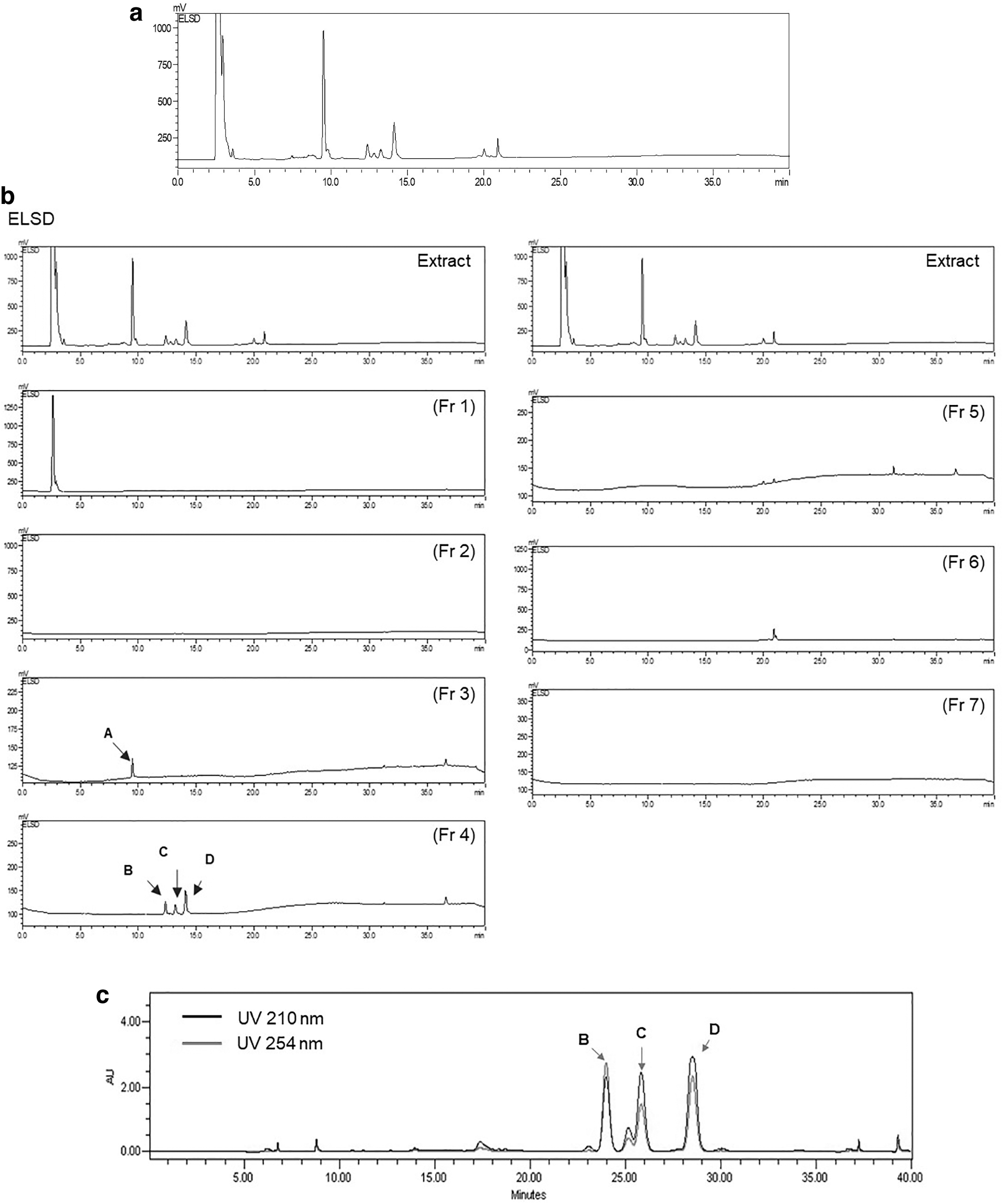
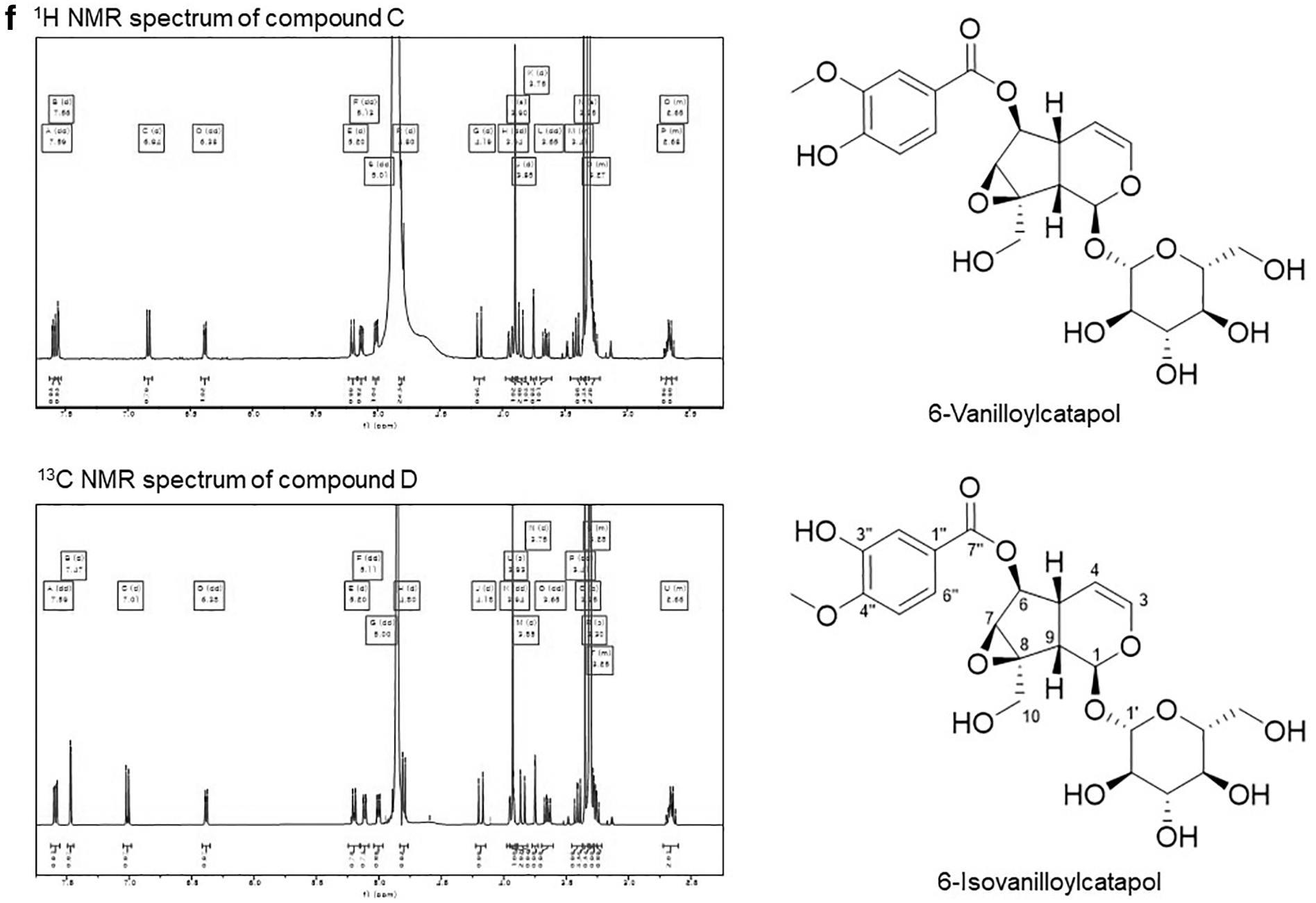
ELSD chromatogram and NMR spectroscopic data.

The extract was also assayed using an LC-HRESIMS system equipped with a Thermo Scientific Q Exactive Hybrid Quadrupole-Orbitrap mass spectrometer coupled to a Thermo Scientific Vanquish UHPLC System (Waltham, MA, USA), equipped with a Phenomenex Kintex 2.6 μm C18 column (100 Å, 100 × 2.1 mm, 0.3 mL/min) at Research Center for Natural Products and Medicinal Material (RCNM) for further confirming the selection of major components (data not shown).
Cosmosil 140C18-OPN stationary phase was used for open column chromatography. HPLC performed on a semiprep Waters HPLC series system (996 PDA, pump) with a YMC-pack ODS column (10 × 250 mm) was applied to isolate compounds. Low-resolution ESI-MS was confirmed with an Agilent 6120 single-quadrupole mass spectrometer with a reversed-phase C18 column (Phenomenex Luna 3 μ C18(2) 100 Å, 4.6 × 150 mm). NMR spectra were recorded in CD3OD on a Bruker (Billerica, USA) AVANCE III HD 400 ( 1 H, 400 MHz, 13 C, 100 MHz) spectrometer.
Development of OA with MIA injections and V. incana administration
This animal experiment was approved by the Animal Care and Use Committee of Daegu Haany University (Approval No. DHU2022-067), and was conducted according to the protocol. Seven-week-old male Sprague-Dawley rats weighing 200–250 g at the start of the experiment were purchased from DBL Co. (Eumseong, South Korea). The animals were maintained with controlled lighting (12-h light/12-h dark cycle), temperature conditions (23 ± 2°C), and humidity (about 55 ± 5%) with access to free food and water. After adaptation (1 week), rats are randomly arranged in descending order of weight and assigned into four groups of equal number (n = 8) without statistically significant differences among the groups:
(i) Normal, normal group; (ii) Control, MIA-induced OA control rats; (iii) Indo, the indomethacin 2 mg/kg-administered and MIA-induced OA rats; and (iv) VI200, the V. incana 200 mg/kg-administered and MIA-induced OA rats.
OA was induced in SD rats employing the method of Wang with minor modification. 16 After anesthetization with an injection of zoletil mixture (Vibrac, France) 0.75 mg/kg intraperitoneally, rats were injected with MIA 80 mg/kg in a 50 μL volume (31G needle 0.3 mL-insulin syringe) inserted through the intra-articular space of the right knee. The rats in all groups were sacrificed after the end of the experimental period. The rats were anesthetized using a mixture of zoletil and xylazine and euthanized by isoflurane overdose. The right knee cartilage was separated for histology and Western blot analysis. The muscle and excess soft tissues were carefully removed and the articular tissues were rinsed with saline. The blood samples were centrifuged (at 3000 rpm for 10 min at 4°C; Hanil Scientific Inc., Gimpo., Korea). Also, the serum was collected and stored at −80°C until analysis.
Measurements of hindpaw weight-bearing distribution
Hindpaw weight-bearing distribution was assessed using incapacitance tester (Linton Instrumentation, Norfolk, United Kingdom).
17
It measures the weight-bearing distribution of each hind limb. A significant shift in weight from the arthritis to the contralateral limb was considered to be an index of pain. The weight distribution ratio was calculated using the following equation:
Measurement of glutamic oxaloacetic transaminase and glutamic pyruvic transaminase levels in serum and malondialdehyde levels in tissue
Hepatic functional parameters such as glutamic oxaloacetic transaminase (GOT) and glutamic pyruvic transaminase (GPT) assay were measured using a commercial kit (Asan Pharm Co., Ltd., Seoul, Korea). Malondialdehyde (MDA) levels were measured as follows (standard sample, 1,1,3,3,-tetra methoxy propane) 18 : the sample and 1% phosphoric acid were mixed. After that, 0.67% thiobarbituric acid was added and boiled for 45 min. After that, butanol was added. It was centrifuged (3000 rpm, 10 min; Hanil Scientific Inc., Gimpo., Korea) to use the supernatant. The absorbance was measured at 540 nm using a spectrophotometer (Infinite M200 Pro, Tecan, Männedorf, Switzerland).
Western blotting
Protein (cytoplasmic and nuclear) extraction was performed by referring to the method of Komatsu. 19 The protein concentration was determined using BCA Protein Assays. The 12 μg proteins of each nuclear and cytosol fraction were electrophoresed through 8–10% sodium dodecyl sulfate-polyacrylamide gel (SDS-PAGE). Separated proteins were transferred to an NC membrane, and then incubated with primary antibodies (1:1000, overnight at 4°C), respectively. After the blots were washed, they were incubated with anti-rabbit and anti-mouse IgG HRP-conjugated secondary antibody (1:3000, for 2 h at room temperature).
Each antigen–antibody complex was visualized using ECL Western blotting detection reagent. And it was detected with Sensi-Q 2000 Chemidoc (Lugen Sci Co., Ltd., Gyeonggi-do, Korea). The densities of the detected band were measured using ATTO Densitograph Software (ATTO Corporation, Tokyo, Japan). They were also quantified as the ratios to β-actin and histone. The protein levels of the groups are expressed relative to those of the normal rat (represented as 1). 20
Histological examination
Histological examinations were performed on the separated knee joints. The knee joints were fixed by using a 10% neutral-buffered formalin and embedded in paraffin and cut into 3 μm sections. The sections were stained using hematoxylin & eosin (H&E) and Safranin-O. The stained slices were then observed under an optical microscope.
Statistical analysis
Data are expressed as mean ± standard error of the mean. Statistical comparisons were assessed by one-way analysis of variance followed by least significant differences tests (SPSS 26.0 for Windows, SPSS Inc., NY, USA). Values of P < .05 were considered significant.
Results
The major four compounds from the extracts of V. incana
The crude extract of V. incana (300 mg) was fractionated into seven fractions through RP open column chromatography (H2O:MeOH = 7:3 to 4:6). The crude extract and the seven fractions were analyzed by reversed-phase analytical HPLC and the four major compounds were identified in fractions 3 and 4, as shown in Figure 1b.
Fraction 3 was analyzed to be the major compound A (33 mg). The fraction 4 containing compounds B–D (Fig. 1c) was subjected to reverse phased semipreparative HPLC with the following conditions: Waters HPLC system; YMC-pack ODS column (10 × 250 mm), 5 μm; eluting with an isocratic solvent system (H2O:ACN = 80:20; 2.0 mL/min); and detection under UV 210 and 254 nm to give the three compounds B–D (0.5, 0.8, and 0.6 mg, respectively) (Fig. 1c).
The 1 H and 13 C NMR spectra of compound A in CD3OD were recorded (Fig. 1d). It was also confirmed by LC-ESI-MS (Fig. 2). The structure of compound A was confirmed to be verproside by NMR data comparison with the previously reported data. 21 The structures of compounds B–D were also determined by LC-ESI-MS data analysis (Fig. 1e, f) and NMR data comparison with literature as shown in Figure 2. 21 –23
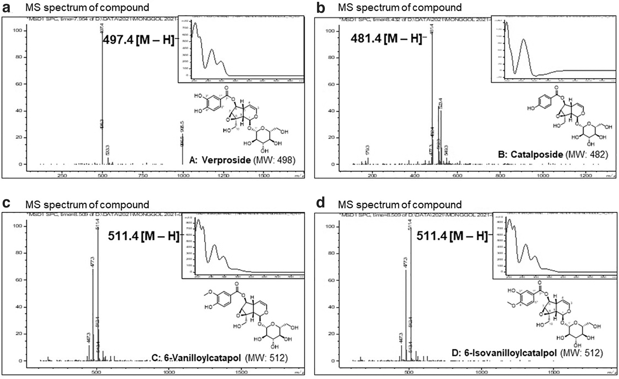
LC–MS data of compounds A–D. Insets show the MS spectrum of compound A–D.
Body weight change and food efficiency ratio
Initially, we measured the body weight (BW) of rats during the experimental period (Table 1). Intra-articular injection of MIA or oral administration of V. incana did not meaningfully affect BW change. The mild increase in food efficacy ratio in the Control group and VI200 group led to the increase in BW. However, it did not show a significant difference.
Body Weight Change and Food Efficiency Ratio
Data are the mean ± SEM. (n = 8).
Control, MIA-induced OA control rats; Indo, indomethacin 2 mg/kg-treated and MIA-induced OA rats; MIA, monosodium iodoacetate; Normal, the normal rats; OA, osteoarthritis; SEM, standard error of the mean; VI200, V. incana 200 mg/kg-treated and MIA-induced OA rats.
The effect of V. incana on serum liver function enzymes and tissue MDA level
OA can cause liver dysfunction due to stress and pain or medications taken to treat it. Therefore, we conducted a blood biochemical analysis to determine whether V. incana administration caused liver dysfunction in vivo (Table 2). Intra-articular injection of MIA dramatically increased GOT and GPT levels in the Control group (P < .01 and .001). However, both GOT and GPT were significantly decreased with the administration of V. incana (P < .05 and .01). Moreover, the tissue levels of MDA were also significantly decreased in Indo and VI200 groups compared with the Control group (P < .05).
Levels of Serum Liver Function Enzymes and Tissue Malondialdehyde
Data are the mean ± SEM. (n = 8). Significance: # p < 0.05, ## p < 0.01, ### p < 0.001 versus the normal rat values and * p < 0.05, ** p < 0.01 versus the control rat values.
MDA, malondialdehyde; GOT, glutamic oxaloacetic transaminase; GPT, glutamic pyruvic transaminase.
Change in hindpaw weight distribution
The pain in knee joints leads to a decrease in Hindpaw Weight Distribution (HWD), which has been proved through the previous study. 24 The HWD on 0, 7, and 14 days is measured in Figure 3. On day 0, the MIA-treated OA control group showed lower HWD compared with the normal group (P < .001). After 14 days, the decreased HWD was significantly increased in Indo and VI200 groups (P < .001).
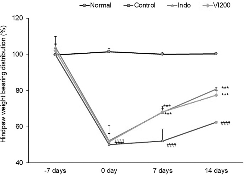
Change in hindpaw weight distribution. Data are the mean ± SEM. (n = 8). Significance: ###P < .001 versus the normal rat values and ***P < .001 versus the control rat values. Control, MIA-induced OA control rats; Indo, indomethacin 2 mg/kg-treated and MIA-induced OA rats; MIA, monosodium iodoacetate; Normal, the normal rats; OA, osteoarthritis; SEM, standard error of the mean; VI200, V. incana 200 mg/kg-treated and MIA-induced OA rats.
The expression of inflammation-related proteins in knee cartilage
The previous study reported that the inhibition of IκBα phosphorylation attenuates the transcriptional activity of the proinflammatory transcription factor, NF-κB. 25 Activated NF-κB regulates the expression of proinflammatory cytokines. 26 The phosphorylation of IκBα affected NF-κBp65 activation (Fig. 4a). The injection of MIA resulted in a significant increase of p-IκBα (P < .01) compared with the Normal group. In contrast, all drug treatments significantly reduced the expression of p-IκBα. Furthermore, the NF-κBp65 expression in the Control group remained noticeably elevated, whereas it was effectively reduced by V. incana administration (P < .01). Figure 4b shows the effect of V. incana on the expression of inflammation-related cytokines, including TNF-α and IL-6. The MIA treatment significantly increased these protein expressions. However, V. incana treatment significantly decreased all inflammatory factors. Especially, the anti-inflammatory effects between Indo and V. incana groups are similar (IL-6; P < .01, TNF-α; P < .01 and P < .001, respectively).
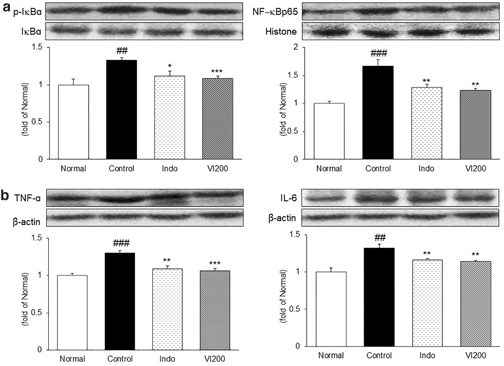
The expression of inflammation-related proteins in knee cartilage. Data are the mean ± SEM (n = 8). Significance: ##P < .01, ###P < .001 versus the normal rat values and *P < .05, **P < .01, ***P < .001 versus the control rat values.
The expression of MMP-2, MMP-3, and MMP-13 in knee cartilage
The MMPs, which are proteolytic enzymes, degrade the extracellular matrix (ECM). 27 The levels of MMP-2, MMP-3, and MMP-13 were notably upregulated in MIA-induced OA control rats (Fig. 5). However, the levels of MMP-3 and MMP-13 were significantly suppressed only in the V. incana group (MMP-3, P < .01; MMP-13, P < .001).
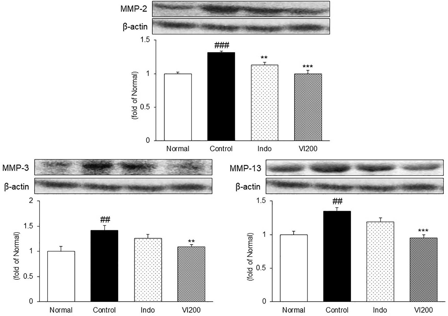
The expression of MMP-2, MMP-3, and MMP-13 in knee cartilage. Data are the mean ± SEM (n = 8). Significance: ##P < .01, ###P < 0.001 versus the normal rat values and **P < .01, ***P < .001 versus the control rat values.
The histological alteration in knee cartilage
We stained the knee cartilage with H&E and Safranin-O to determine whether the administration of V. incana alleviated the histopathological alteration of OA in the knee joint. H&E staining revealed well-preserved knee cartilage in the Normal group. However, the Control group exhibited severely damaged articular cartilage, as evidenced by surface delamination (Fig. 6a), but V. incana treatment alleviated such changes. Moreover, Safranin-O staining results revealed less stained areas in the Control group, which means degraded proteoglycan in ECM. The Indo and VI200 groups showed higher proteoglycan than the Control group (Fig. 6b). These results indicate that V. incana administration reversed the histopathological alterations induced by MIA injection.
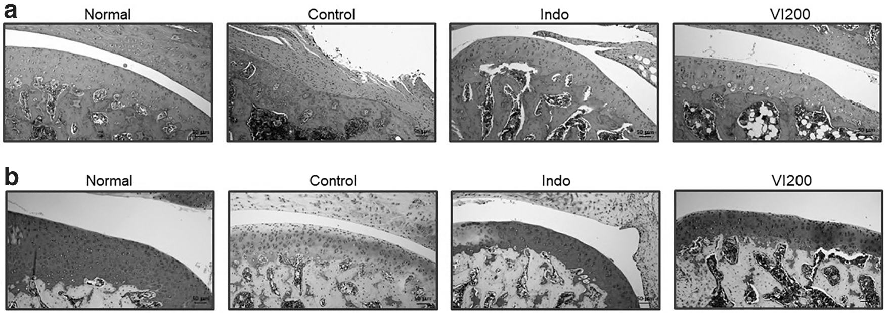
Effect of V. incana on histological changes in knee cartilage. MIA-induced OA rats were administered either indomethacin (2 mg/kg BW/day) or VI (200 mg/kg BW/day) for 2 weeks. Knee cartilage was stained with
Discussion
OA is the most common joint disease in the world, and OA may be painless, but in most cases, it is accompanied by pain. Its prevalence is increasing as life expectancy increases and as the prevalence increases, pain, disability, and socioeconomic burden are increasing. 28 –30 Therefore, a lot of research is currently underway to find new therapeutic agents that can reduce the risk factors of the disease and alleviate the symptoms. Therefore, we also tried to observe the effect of V. incana on articular cartilage to find an effective therapeutic agent for OA.
First, we isolated the main component of V. incana, and finally identified the following four compounds: (A) verproside; (B) catalposide; (C) 6-vanilloylcatapol; and (D) 6-isovanilloylcatapol. These components isolated from V. incana are known to have antioxidant and anti-inflammatory effects. 31 –34 In particular, verproside is known to inhibit the TNF-α/NF-κB pathway, and catalposide has been found to inhibit NF-κB activation and the production of inflammatory cytokines such as TNF-α, IL-6, and IL-1β. 31,34 These study results support that V. incana can play an important role in improving OA, and they confirmed anti-inflammatory effect of V. incana, which was the objective of this study.
We induced OA in the legs of animals using MIA to evaluate the anti-inflammatory and articular cartilage protective effects of V. incana. After that, indomethacin and V. incana were orally administered for 2 weeks. Indomethacin is one of the NSAID drugs used to treat OA. 35 It exerts an anti-inflammatory effect by inhibiting the proinflammatory protein COX. 36 It may also affect inflammatory cytokine expression. Indomethacin has been used as a positive control drug in various studies related to OA. 37 –39 Therefore, in this study, indomethacin was used as a positive control group, and the effect of V. incana on OA was investigated.
In this study, we identified liver function biomarkers and MDA levels. Liver dysfunction is common in patients with arthritis. These markers can be increased by stress and pain caused by OA or medications taken to treat it. 40,41 In our study, the levels of GOT and GPT, which are liver function biomarkers, increased in the OA-induced control group, whereas V. incana treatment significantly reduced the increased GOT and GPT. Also, the level of MDA in the joint tissue was confirmed. Various stimuli such as stress and inflammation overproduce reactive oxygen species in the body, which increases MDA, a lipid peroxide. Increased MDA is involved in inflammation and apoptosis, and transforms key proteins in the body. 42 In our study, V. incana significantly reduced the level of MDA in joint tissue. These results are related to inflammation of the articular cartilage and are judged to be the result of the relief of inflammation.
The weight-bearing distribution is a commonly used index when evaluating the degree of pain in OA, and the degree of OA improvement in the experimental group can be confirmed by numerical values of the weight-bearing distribution. This method has been widely used in previously published studies. 37,43 As in previous studies, weight-bearing distribution was evaluated for 14 days. Before administration, it was confirmed that weight-bearing distribution significantly decreased in all groups, except the Normal group, and V. incana significantly increased the decreased weight-bearing distribution. Accordingly, the pain in knee joints induced by MIA treatment could be alleviated through V. incana supplementation.
We confirmed the expression of inflammatory proteins and proteolytic factors, MMPs, through Western blotting to confirm changes in protein expression in joint tissues caused by OA. In OA-affected joints, NF-κB is activated, and inflammatory cytokines such as TNF-α and IL-6 are overproduced by synovial cells. In addition, NF-κB-derived MMPs are upregulated, resulting in changes such as cartilage erosion, ECM degradation, and synovial inflammation. 44 NF-κB is known as a representative transcription factor of the inflammatory pathway. NF-κB, translocated to the nucleus by various stimuli, activates the expression of proinflammatory cytokines and promotes the inflammatory response. 45 In addition, MMPs produced by NF-κB and inflammatory cytokines degrade all components of the ECM and can greatly exacerbate OA. Among various MMPs, MMP-2 and MMP-3 are increased in OA to degrade noncollagen components of joints.
In addition, MMP-13, in particular, is a product of chondrocytes in cartilage and has a dual role in matrix destruction by degrading aggrecan, a proteoglycan molecule, so current research focuses on the development of MMP-13 inhibitors. 46 –48 In our study, as in several previously published studies, it was confirmed that inflammatory proteins and MMPs were increased through OA induction. Here, V. incana inhibited the activity of NF-κB and reduced the production of inflammatory cytokines. Also, it significantly reduced the expression of MMPs to the level of the Normal group. These results suggest that V. incana protects knee cartilage by inhibiting the production of inflammatory cytokines and MMPs by blocking NF-κB activity in MIA-induced OA.
We confirmed that the four major compounds (A–D) of V. incana are as follows: (A) verproside; (B) catalposide; (C) 6-vanilloylcatapol; and (D) 6-isovanilloylcatapol. Moreover, we demonstrated the anti-inflammatory effects of V. incana in knee cartilage. These results suggest that it is possible to protect knee cartilage by inhibiting proinflammatory cytokines through the blocking of NF-κB activation. Moreover, we further confirmed that V. incana can decrease the degradation of the cartilage matrix in MIA-induced OA rats. Therefore, V. incana treatment may be a potential therapeutic candidate for treating patients with OA.
Footnotes
Authors' Contributions
J.A.L.: writing-original draft. T.H.N.: formal analysis. M.-R.S.: writing-review and editing. J.W.C.: formal analysis. H.C.: methodology and conceptualization. J.-W.N.: writing-review and editing. S.-S.R.: funding acquisition and supervision.
Author Disclosure Statement
No competing financial interests exist.
Funding Information
This study was supported by “Cooperative Research Program for Agriculture Science and Technology Development (No. PJ015272012022) in Rural Development Administration Republic of Korea” and “National Research Foundation of Korea (NRF) grant funded by the Korea government (MSIT) (No. 2018R1A5A2025272).”