
Abstract
Metabolic diseases, including obesity, diabetes, and fatty liver disease, are dramatically increasing around the world. Seaweed is low in calories and rich in many active ingredients that are necessary for maintaining good health, and is expected to be effective for preventing metabolic diseases. The purpose of this study was to examine the effects of a traditional Japanese edible seaweed Hypnea asiatica (H. asiatica) on obesity, using a mouse model. H. asiatica was dried and powdered, mixed with a high-fat diet, and fed to male C57BL/6J mice for 13 weeks. On the last day of the experiment, blood samples were collected under anesthesia and biochemical parameters such as lipids and adipokines were measured. Liver and adipose tissue were excised, weighed, and oxidant/antioxidant parameters were measured. Some mice were perfused with a fixative solution containing formalin, and tissue specimens were prepared. A glucose tolerance test was used to assess insulin resistance. The inhibition of lipase activity was evaluated in vitro. Thirteen-week supplementation with H. asiatica suppressed body weight gain, body fat accumulation, and blood glucose levels. H. asiatica also improved fatty liver and hypercholesterolemia, and reduced the oxidant and inflammatory parameters of serum and liver. H. asiatica increased fecal triglyceride excretion and polyphenol-rich ethanol extract of H. asiatica inhibited lipase activity in vitro. These results suggest that polysaccharides and polyphenols in H. asiatica may ameliorate obesity and diabetes by inhibiting intestinal fat absorption and reducing oxidative stress and inflammation. H. asiatica may be useful in preventing metabolic diseases such as obesity, diabetes, and fatty liver.
INTRODUCTION
The number of patients with metabolic diseases such as obesity and diabetes has dramatically increased worldwide in recent years due to dietary changes and sedentary lifestyles, and has become a major social problem. 1,2 Obesity and diabetes also affect the healthy life expectancy and cause increased medical costs. Obese patients show increased blood lipid levels, fatty liver, and elevated blood pressure, along with increased fat accumulation in the body. In fact, obesity has been proven to increase the risk of numerous chronic noncommunicable diseases, including diabetes, dyslipidemia, myocardial infarction, non-alcoholic fatty liver disease, cerebral infarction, kidney disease, sleep apnea, and certain types of cancer. 3,4 Therefore, the prevention of obesity is critical to maintaining good health.
To prevent obesity, it is important to have a regular diet, moderate calorie intake, and eat more vegetables and other foods. Seaweed is low in calories and is rich in minerals, vitamins, soluble fibers, and amino acids necessary for maintaining good health. 5 People in East Asia, including Japan and Korea, have long consumed seaweed in their daily diets. Seaweed consumption may have affected the health and longevity of people in these regions. A large epidemiological study that followed >80,000 Japanese for 20 years revealed that humans who consumed seaweed more frequently had a lower incidence of cardiovascular disease than those who consumed seaweed less frequently. 6
Hypnea asiatica (H. asiatica), formerly known as Hypnea charoides, is a red seaweed widely distributed on the coastlines from the Pacific Ocean to the Indian Ocean. It has been eaten as a traditional dish in Japan, especially in the Ryukyu region of Japan, for over 200 years. A component analysis showed that H. asiatica contains high levels of the sulfated polysaccharide κ-carrageenan. 7 Carrageenan is extensively used as a thickening agent and stabilizer in food processing due to its gelling properties. Although there are few reports on the health benefits of H. asiatica, limited functional studies revealed cholesterol-lowering and anticoagulation activities in rats. 8
We have previously reported that several types of seaweeds can retard the onset and progression of obesity and diabetes in mice fed high-fat diets (HFs). 9 –11 Polysaccharides and polyphenols contained in seaweeds were found to play important roles in the prevention of these diseases. Given that H. asiatica is rich in polysaccharides and polyphenols, it is suggested that this seaweed may be useful against metabolic diseases. In this study, we examined the effects of H. asiatica, a traditional Japanese food ingredient, on obesity and related metabolic disorders using diet-induced obese mice.
MATERIALS AND METHODS
Polysaccharide purification and molecular weight determination
The red seaweed H. asiatica was collected from the coast of Noto Peninsula, Ishikawa, Japan, washed with water, and allowed to dry naturally. The dried seaweed was then powdered using a food grinder. Seaweed powder was dissolved in distilled water after removing lipids with ethanol, and finally lyophilized. The extraction of polysaccharides was performed by a previously described procedure. 12 The molecular weight of extracted polysaccharides was determined by high-performance size exclusion chromatography with Shodex OHpak SB-807G (Guard), SB-807 HQ, and SB-806 M HQ (Showa Denko KK, Tokyo, Japan) and estimated using dextran standards of four different molecular weights (Sigma-Aldrich Corp., American Polymer Standards Corp., Mentor, OH, USA). Samples (injected volume: 20 μL) were eluted using 0.3 M NaNO3 at a flow rate of 1 mL/min and was detected using a refractive index detector RID10 (Shimadzu Corp., Kyoto, Japan).
Animals and experimental design
Six-week-old male C57BL/6J mice (CLEA Japan, Tokyo, Japan) were purchased and acclimatized in a controlled environment of 22°C, 50% humidity, and a 12-h light/12-h dark cycle. After 1 week, the animals were randomly divided into the following 4 groups, with 12–13 animals per group: normal diet (Normal), HF, HF supplemented with low-dose (2%) H. asiatica (HF+HaL), and HF supplemented with high-dose (6%) H. asiatica (HF+HaH). The compositions of experimental diets were adjusted by considering the nutritional components of H asiatica. 9 The normal diet provided 354 kcal/100 g of energy (14.4% calories from protein, 11.1% calories from fat, and 74.5% calories from carbohydrate), while the HF provided 493 kcal/100 g of energy (17.9% calories from protein, 60.7% calories from fat, and 21.4% calories from carbohydrate).
All experimental diets were based on the AIN-76 diet (Oriental Yeast Co. Ltd., Tokyo, Japan) and animals had ad libitum access to chow and drinking water. Body weights and feed intakes were measured once a week. Thirteen weeks after the start of the experiment, the animals were fasted overnight and blood samples were withdrawn under mixed anesthetic agent (0.3 mg/kg of medetomidine, 4.0 mg/kg of midazolam, and 5.0 mg/kg of butorphanol; Fujifilm Wako Pure Chemical Co., Osaka, Japan). The liver and epididymal, peritoneal, and mesenteric white adipose tissues were excised, weighed, and stored at −80°C. Some of the mice were used for histopathological analysis. All experimental protocols were approved by the Institutional Animal Care and Use Committee of Fukui Prefectural University (Approval No. 19-14).
Serum analyses
Serum was separated by centrifugation at 1500 g for 15 min, 4°C, and levels of total cholesterol, high-density lipoprotein (HDL)-cholesterol, triglyceride, alanine aminotransferase (ALT), and aspartate aminotransferase (AST) were determined using a Hitachi 7060 Automatic Analyzer (Hitachi, Tokyo, Japan) with commercial kits (Fujifilm Wako Pure Chemical Co.). The non-HDL cholesterol levels were calculated by subtracting the HDL-cholesterol from the total cholesterol level. The serum levels of insulin (Morinaga Institute of Biological Science, Yokohama, Japan), adiponectin (Otsuka Pharmaceutical Co. Ltd. Tokyo, Japan), and tumor necrosis factor-α (TNF-α; Fujifilm Wako Pure Chemical Co.) were also measured using a commercial ELISA kit. Serum malondialdehyde (MDA) levels were measured using a commercial kit (Japan Institute for the Control of Aging Co. Ltd., Shizuoka, Japan).
Liver analyses
Fifty milligrams of frozen liver tissue was homogenized in five volumes of isopropanol to extract lipids. 13 The homogenates were left at room temperature for 2 days and then centrifuged at 1000 g for 10 min. The supernatants were analyzed for triglyceride content using a commercial kit (Fujifilm Wako Pure Chemical Co.). For biochemical analysis, liver samples were homogenized in five volumes of cold 20 mM Tris-HCl buffer (pH 7.4), and centrifuged at 12,000 g for 15 min, at 4°C. The lipid peroxidation was assessed by measuring MDA using a commercial kit (Japan Institute for the Control of Aging Co. Ltd.). Liver glutathione (GSH) levels and superoxide dismutase (SOD) activities were determined using commercial assay kits (Fujifilm Wako Pure Chemical Co.).
Histological analyses
At the end of the experiment, the mice were anesthetized by injecting a mixed anesthetic (0.3 mg/kg of medetomidine, 4.0 mg/kg of midazolam, and 5.0 mg/kg of butorphanol; Fujifilm Wako Pure Chemical Co.), and transcardially perfused with a fixative solution containing 4% paraformaldehyde and 1.5% glutaraldehyde in phosphate-buffered saline (PBS). Then, the livers and white adipose tissues were excised and allowed to stand in the same fixative solution for 1 day. The tissues were washed several times with PBS and embedded in paraffin. Tissues were cut into 5-μm-thick sections and stained with hematoxylin and eosin (HE). Adipocyte diameters were measured in HE-stained epididymal white adipocytes.
Glucose tolerance test
To assess insulin resistance, a glucose tolerance test was performed before the end of the study. Mice were fasted overnight, and glucose solution was administered intraperitoneally (2 g/kg body weight). Blood was collected from the tail vein before and 15, 30, 60, 90, and 120 min after glucose injection, and blood glucose concentrations were measured using a blood glucometer (Nipro, Osaka, Japan).
Fecal analyses
Mice were housed individually, and fecal samples were collected for a 24-h period from each mouse. Fecal samples were powdered and 300 μL of distilled water was added to 50 mg of feces to extract polysaccharides. After centrifugation at 16,000g, for 30 min, at 4°C, ethanol was added to the supernatant to a final concentration of 85%. The resulting residue was washed with 85% ethanol and then resuspended in distilled water and centrifuged at 16,000 g, for 10 min, at 4°C. The amount of polysaccharides in the supernatant was determined using a phenol sulfuric acid method, with galactose as the standard. 14 To extract lipids, isopropanol (10 times by weight) was added to the powdered fecal sample. Samples were then dried and redissolved in isopropanol. The amount of triglyceride was determined using a commercial kit (Fujifilm Wako Pure Chemical Co.).
Lipase assay
The effects of H. asiatica extracts on lipase activity were examined according to previously described methods. 15 Commercially available lipase (type II, from porcine pancreas, 400 U/mg protein; Sigma-Aldrich, St. Louis, MO) was dissolved in distilled water at 5 mg/mL and centrifuged at 1000 g, for 5 min, and the supernatant was used as the enzyme source. 4-Nitrophenyl butyrate (4-NPB; Sigma-Aldrich) was dissolved in dimethylsulfoxide and used as a substrate for the enzyme reaction.
The reaction mixture contained 100 μL of enzyme solution and 100 μL of H. asiatica extract in 4 mL of 20 mM Tris-HCl buffer, pH 8.5. After preincubating the enzyme reaction solution for 10 min, 100 μL of 5 mM 4-NPB solution was added and incubated at 37°C for 30 min, and the absorbance of the reaction solution was measured at 400 nm. To prepare the seaweed extract, 70% ethanol or water was added to the dried H. asiatica powder, stirred, left overnight at room temperature, and then centrifuged. The resulting supernatant was used as ethanol and water extracts for the assay.
Statistical analysis
The results are expressed as the mean ± the standard error of the means. Data were analyzed by a one-way analysis of variance followed by Turkey's multiple range tests. P values of P < .05 were considered to indicate statistical significance.
RESULTS
Isolation of polysaccharides
The polysaccharide fraction of H. asiatica showed a symmetric peak on high-performance size exclusion chromatography, with an estimated molecular weight of 2.2 × 10⁴ Da.
Food intake and body weight
The addition of H. asiatica to the HF did not affect feed intake. The average daily feed intake of each group was as follows: 3.0 g (Normal), 2.5 g (HF), 2.4 g (HF+HaL), and 2.4 g (HF+HaH). The average caloric intakes in Normal, HF, HF+HaL, and HF+HaH groups were 10.6, 12.3, 11.8, and 11.8 kcal, respectively, per day per mouse, with no significant difference among the groups. This result indicated that the lower weight gain of H. asiatica-treated mice was not due to lower caloric intake. Mice fed an HF showed a more pronounced weight gain in comparison to mice fed the normal diet, with significant differences beginning at week 5 (Fig. 1A). The body weight gain in the HF group was twice that in the Normal mice (Fig. 1B). Mice fed an HF with high-dose H. asiatica (HF+HaH) showed significantly suppressed weight gain (Fig. 1A, B).
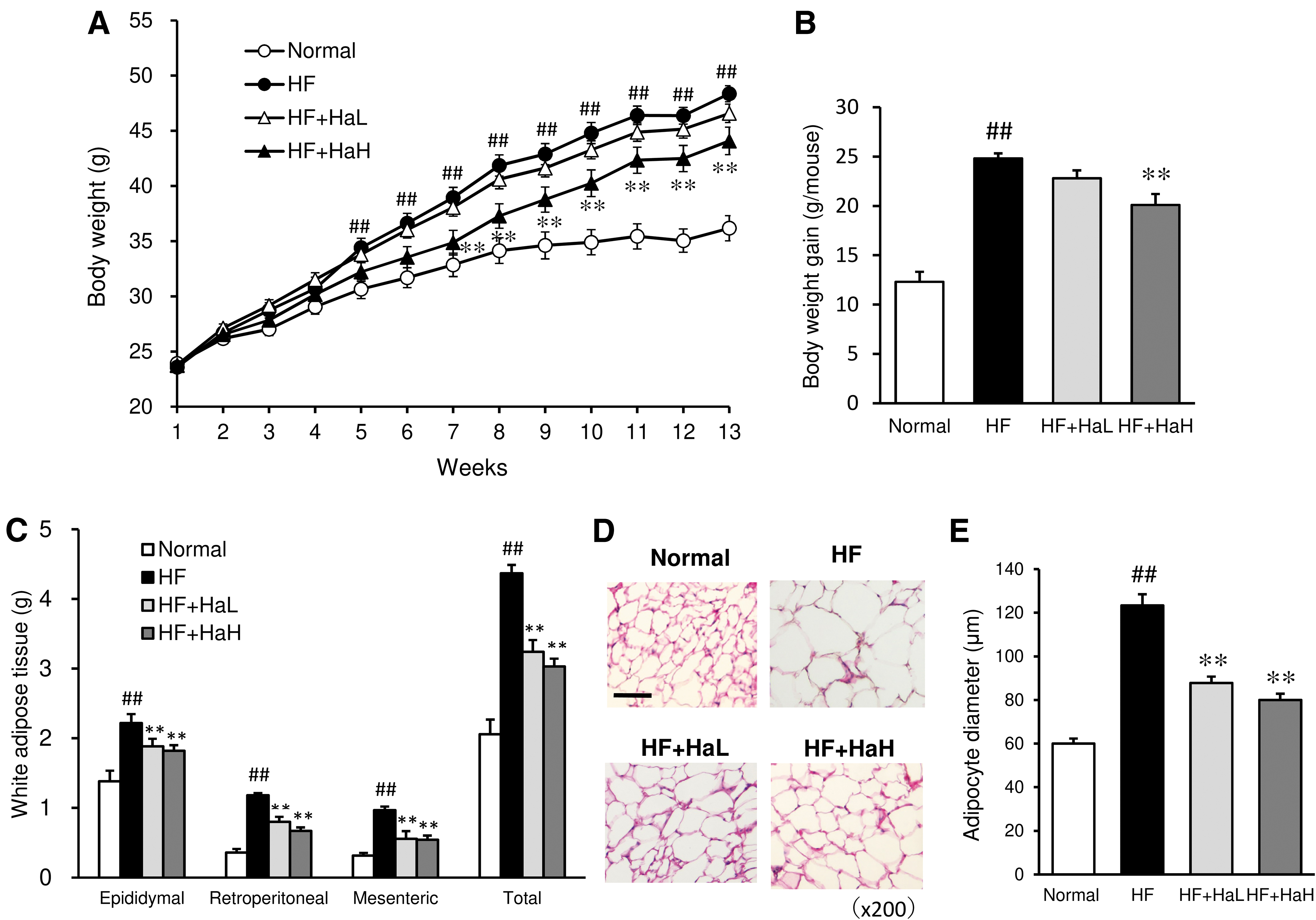
Effects of H. asiatica on weekly body weight
White adipose tissue mass and morphology
The weights of epididymal, retroperitoneal, and mesenteric adipose tissue in the HF group were significantly higher than those in the Normal group, as was the total adipose tissue mass (Fig. 1C). In the H. asiatica-treated group, total adipose tissue weight and the weight of each adipose tissue were significantly lower than those in the HF group (Fig. 1C). Cell hypertrophy was observed in the adipose tissue of the HF group, and the diameter of adipocytes was significantly increased compared to that of the normal group (Fig. 1D, E). H. asiatica treatment visibly reduced the diameter of adipocytes (Fig. 1D, E).
Serum glucose and insulin levels, and insulin resistance
Serum glucose and insulin levels were significantly higher in the HF group compared to the Normal group (Fig. 2A, B). Thirteen weeks of treatment with H. asiatica prevented HF-induced increases in glucose and insulin levels. A glucose tolerance test was used to evaluate the effect of H. asiatica on insulin resistance. Blood glucose levels were higher in the HF group than in the Normal group at any time after intraperitoneal glucose injection. On the other hand, the H. asiatica-treated group exhibited a reduced increase in blood glucose, and blood glucose levels at 30 and 60 min after glucose injection were significantly lower than those in the HF group (Fig. 2C). The increase in the area under the curve induced by HF was also significantly suppressed by H. asiatica treatment (Fig. 2D).
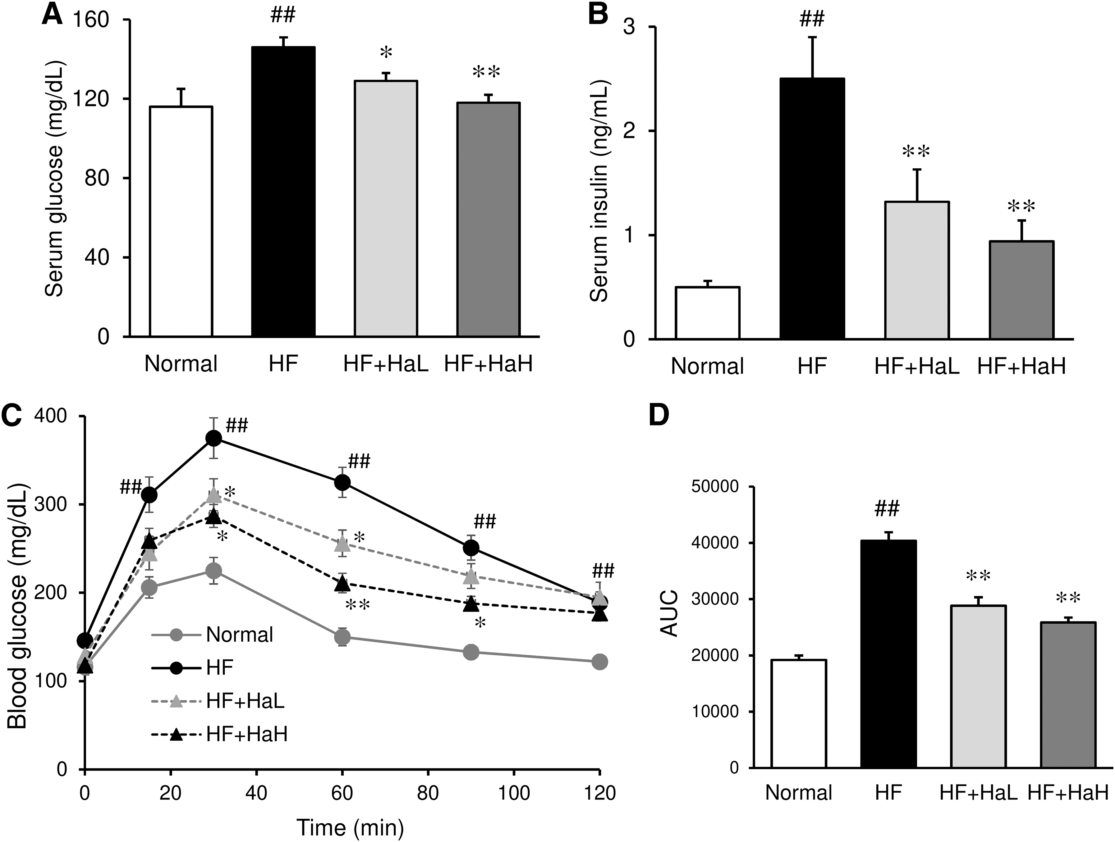
Effects of H. asiatica on serum glucose
Hepatic steatosis and liver damage markers
The liver weight and triglyceride content of the HF group were significantly higher than those of the Normal group (Fig. 3A, B); these increases were accompanied by an increase in serum liver function parameters, including ALT and AST (Fig. 3C, D). Macroscopically, the livers of the HF group were enlarged and whitish in color compared to the Normal group (Fig. 3E). Histological examination revealed that the livers of the HF group presented hepatic steatosis, as evidenced by the occurrence of vacuoles, fat droplets, and cellular infiltrates in the liver tissue (Fig. 3F). Treatment of the HF group with H. asiatica reduced liver weight and liver triglyceride content (Fig. 3A, B), and ameliorated hepatic steatosis both macroscopically and histologically (Fig. 3E, F). Elevated serum liver function markers, AST and ALT levels, were also suppressed by treatment with H. asiatica (Fig. 3C, D).
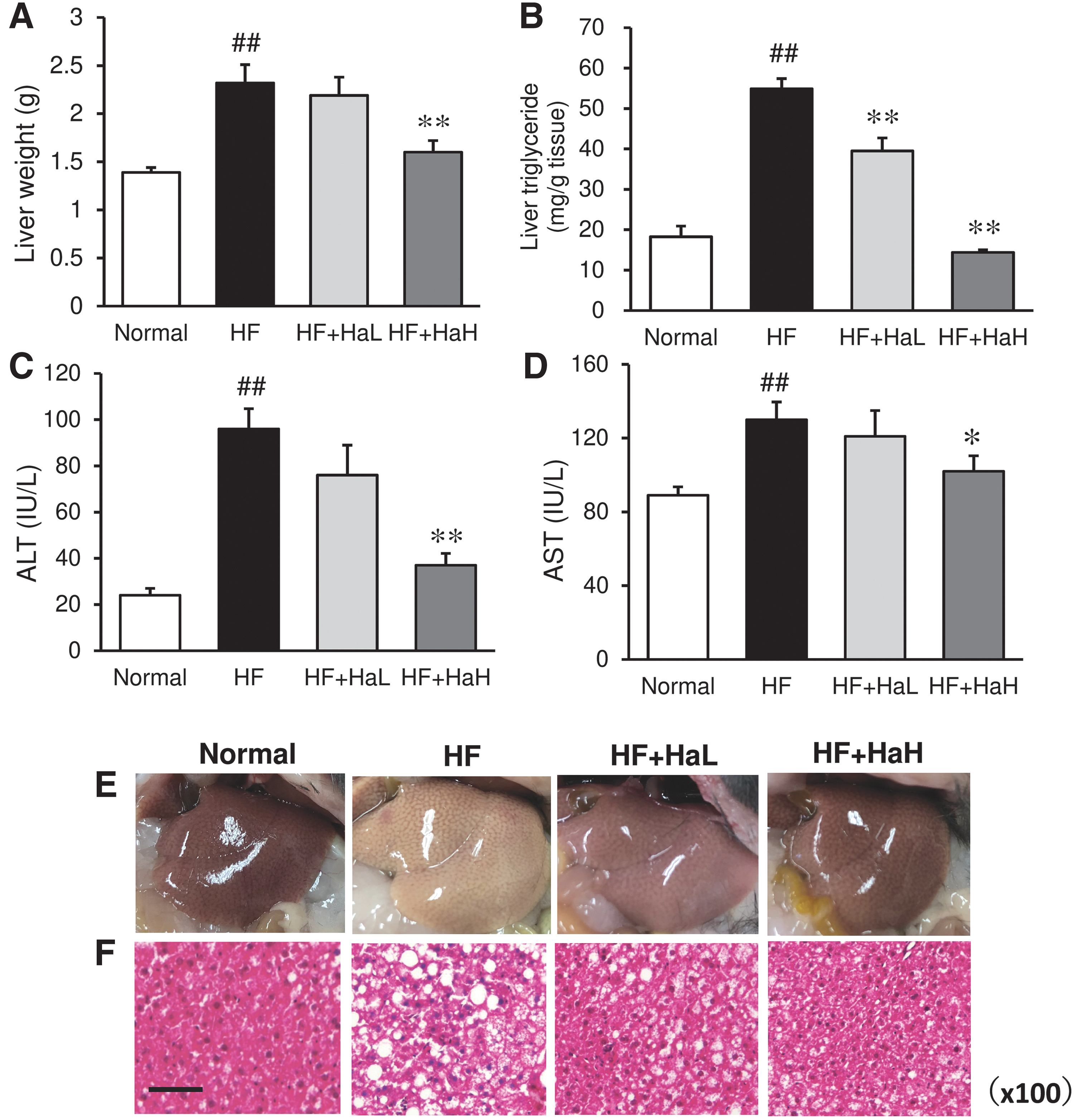
Effects of H. asiatica on the liver weight
Serum lipid levels
Serum total cholesterol and non-HDL cholesterol levels were significantly elevated by consumption of an HF, which was suppressed by treatment with H. asiatica (Fig. 4A, B). The HF did not significantly affect blood HDL and triglyceride levels, nor did H. asiatica administration significantly alter these lipid levels (Fig. 4C, D).

Effects of H. asiatica on serum lipid levels in C57BL/6J mice fed a high-fat diet. Serum levels of total cholesterol
Lipid and polysaccharide content in feces
The amount of triglycerides in the feces was significantly increased in the HF group compared to the Normal diet group (Fig. 5A). Administration of H. asiatica to HF mice further increased the amount of triglycerides in the feces, suggesting that H. asiatica promotes fecal excretion of triglycerides (Fig. 5A). The fecal polysaccharide content in the H. asiatica-treated group was markedly increased in comparison to the HF and Normal groups (Fig. 5B).
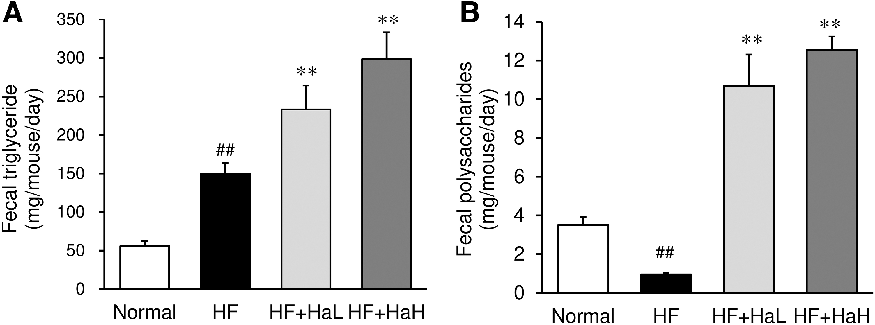
Effects of H. asiatica on fecal triglyceride
Adipokines and oxidative stress marker levels in serum and liver
Serum and liver levels of MDA, a marker of lipid peroxidation, were increased by the HF (Fig. 6A, D). H. asiatica administration dose-dependently suppressed serum and liver MDA levels increased by the HF. Serum levels of anti-inflammatory adiponectin were significantly decreased by the HF (Fig. 6B), while the level of the proinflammatory adipokine TNF-α was increased by the HF (Fig. 6C). These changes in anti-inflammatory and inflammatory markers induced by the HF were improved in the H. asiatica-treated group (Fig. 6B, C). GSH content and SOD activity, which form the antioxidant defense system of the liver, were decreased by the HF, but recovered with H. asiatica treatment (Fig. 6E, F).
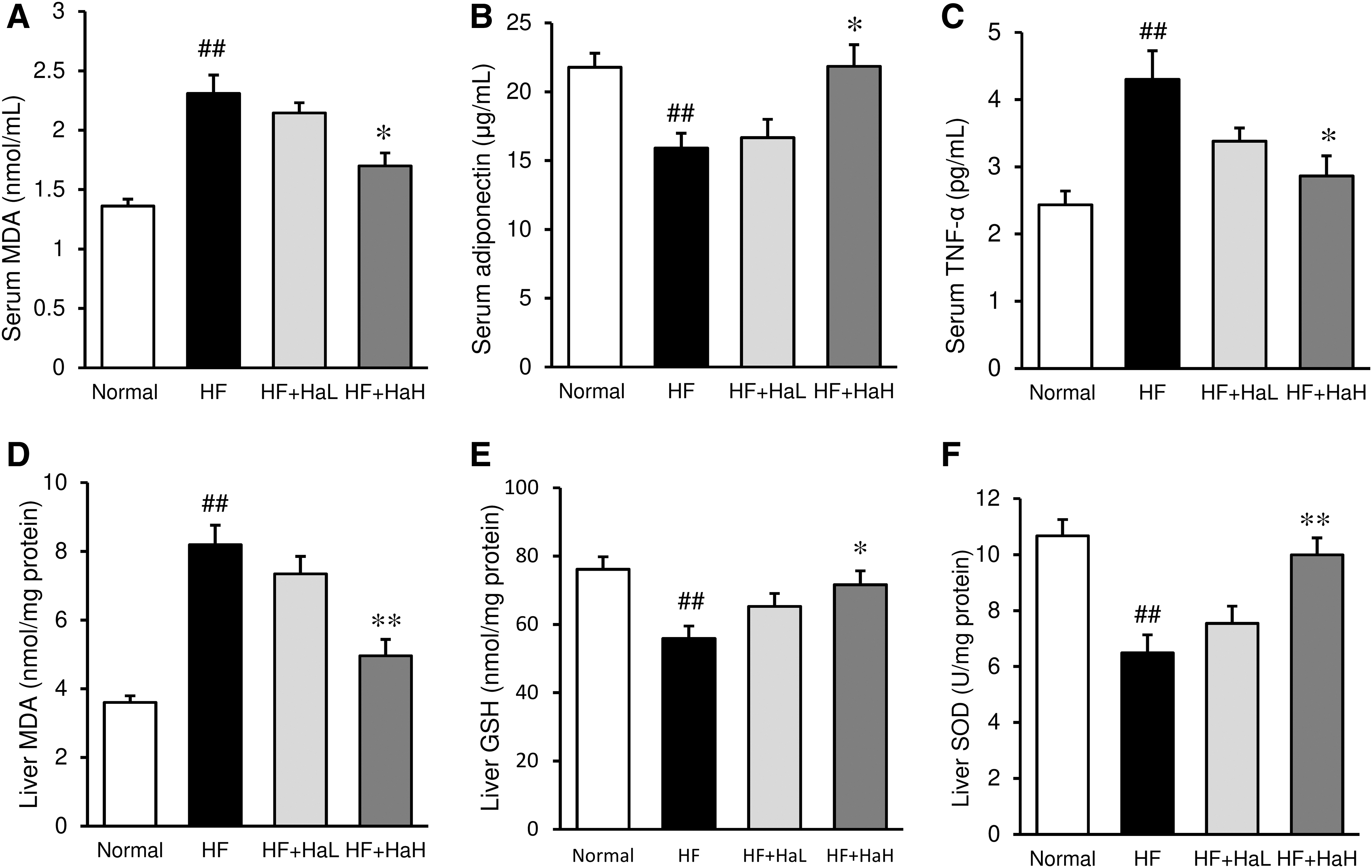
Effects of H. asiatica on adipokines and oxidative/antioxidant markers of the serum and liver in C57BL/6J mice fed a high-fat diet. Serum levels of MDA
Inhibition of lipase activity
The inhibitory effect of H. asiatica on the activity of pancreatic lipase, a triglyceride-degrading enzyme, was evaluated in vitro. Ethanol extract of H. asiatica inhibited the enzyme activity in a dose-dependent manner, while polysaccharide isolated from H. asiatica had little or no effect (Fig. 7).
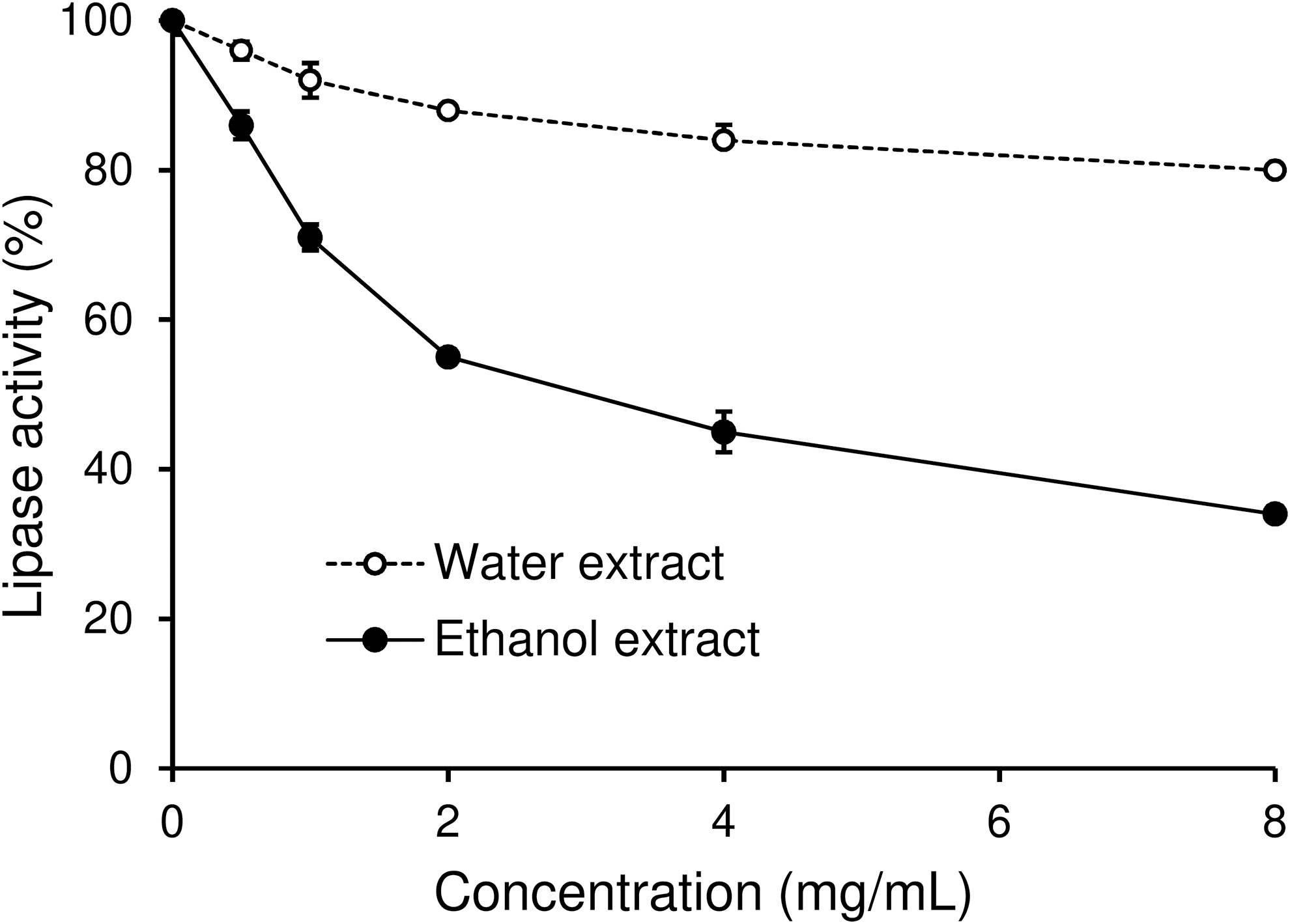
Effects of polysaccharides and ethanol extracts from H. asiatica on the pancreatic lipase activity. Each point represents the mean ± SEM of triplicate experiments.
DISCUSSION
This study indicates that H. asiatica suppresses the progression of metabolic disorders, including obesity, diabetes, fatty liver, and hypercholesterolemia, in mice fed HFs. Results of serum, liver, and fecal analyses suggest that the antiobesity effects of H. asiatica may be related to the inhibition of intestinal fat absorption. In fact, polyphenol-rich ethanol extract of H. asiatica inhibited the activity of the lipolytic enzyme lipase. In addition, the amelioration of oxidative stress in the body, which is increased in mice fed the HF, may also be involved in antiobesity effects of H. asiatica.
Pancreatic lipase is a key enzyme involved in the digestion of triglycerides, hydrolyzing dietary lipids to monoglycerides and free fatty acids in the intestine. 16 Orlistat and cetilistat, which are approved as prescription antiobesity drugs, inhibit pancreatic lipase in the intestinal tract, thereby blocking fat absorption and ultimately leading to weight loss. 17,18 Thus, pancreatic lipase is a promising target for antiobesity drugs, and many plant-derived compounds, including polyphenols and saponins, which target lipase inhibition, are being explored. 19 In in vitro experiments, polysaccharides of H. asiatica had little effect on lipase activity, while ethanol extract of H. asiatica inhibited lipase activity in a concentration-dependent manner.
Since ethanol extract contains polyphenols such as flavonoids, it is likely that the polyphenols and other hydrophobic compounds in H. asiatica inhibited lipase activity. It has already been reported that polyphenols prevent metabolic diseases such as obesity by inhibiting lipase activity. 19 Polyphenols from seaweed have also been shown to be effective against obesity and other metabolic diseases. 20 In a previous study, we reported that an ethanol extract of some seaweeds inhibits lipase activity, which is associated with inhibition of fat absorption in the intestine and a subsequent reduction in body fat accumulation in a mouse model of dietary obesity. 9
H. asiatica has been reported to contain κ-carrageenan as a major polysaccharide. 7 In this study, carrageenan purified by high-performance size exclusion chromatography had a molecular weight of 2.2 × 10⁴ Da. Carrageenan is a negatively charged polysaccharide found in the intercellular spaces of red seaweed and is widely applied in foods as a thickening and stabilizing agent because of its property of forming stable gels. 21 Carrageenan has been reported to show various pharmacological and biological activities, including antiviral and anticancer effects. 22,23 Carrageenans obtained from the red seaweed Sarconema filiforme have been shown to attenuate diet-induced metabolic syndrome in rat models. 24
Although polysaccharide carrageenan isolated from H. asiatica did not show direct lipase inhibition in vitro, 25 carrageenan may inhibit fat absorption in the intestinal tract due to its physicochemical properties. Some soluble dietary fibers are highly viscous, and many studies have been conducted to determine the relationship between their physiological activities and viscosity. 26 Several studies have shown that polysaccharides with high viscosity have a strong inhibitory effect on postprandial hyperglycemia. 27 This has been attributed to the physicochemical actions of polysaccharides that prolong the residence time of food in the stomach and reduce the rate of sugar diffusion in the small intestine.
Similarly, it is possible that high viscosity H. asiatica polysaccharides may delay fat absorption by affecting gastrointestinal motility and the interaction of fat with gastrointestinal enzymes. Therefore, it is speculated that multiple mechanisms other than lipase inhibition are involved in the suppressive effect of H. asiatica on fat absorption in vivo. In fact, the administration of H. asiatica increased fecal triglyceride excretion, which correlated with the amount of polysaccharides in the feces. This suggests that polysaccharides are also involved in the fecal excretion of triglycerides, in addition to the inhibition of lipase activity by polyphenols.
It is well established that oxidative stress is intimately involved in the onset and progression of a wide variety of diseases. Oxidative stress is also highly implicated in metabolic diseases such as obesity, diabetes, and fatty liver. 28 It is known that HFs increase reactive oxygen species productions and reduce the expression of antioxidant enzymes in diet-induced obese mice. Obesity and diabetes cause cellular dysfunction, which is associated with redox imbalance and an environment of oxidative stress. Livers and adipocytes of obese animals and humans exhibit increased oxidative stress and impaired antioxidant defenses. 29
In this study, MDA, a lipid oxidation marker, was increased in the liver and blood of obese mice in comparison to the Normal group. In addition, antioxidant defense systems, including GSH and SOD, were decreased in the liver of obese mice in comparison to the Normal group. The administration of H. asiatica to obese mice restored the redox balance. Adipose tissue produces and releases a wide variety of proinflammatory and anti-inflammatory active substances, including the adipokines, adiponectin, and leptin, as well as cytokines such as TNF-α and IL-6. The increased adipose mass associated with obesity has been linked to a low-grade and chronic inflammatory response, which is characterized by altered production of adipokines and increased inflammatory markers. 30
Adiponectin has been shown to exhibit a wide range of physiological properties, including inhibition of inflammation and oxidative stress, regulation of glucose and lipid metabolism, and improved insulin sensitivity 31 In contrast, proinflammatory cytokines produced by adipose tissue are closely associated with the development of insulin resistance and increased risk of cardiovascular disease in obese individuals. 32 In this study, the serum levels of anti-inflammatory adiponectin were decreased in mice fed the HF. In contrast, the serum levels of proinflammatory TNF-α were markedly elevated. The administration of H. asiatica normalized the serum adipokine balance, which paralleled the inhibition of adipocyte hypertrophy induced by the HF. Thus, it is thought that antioxidants such as polyphenols, which are contained in H. asiatica, inhibit oxidative stress in tissues and suppress adipocyte hypertrophy, which may improve adipokine secretion and insulin resistance in the adipose tissue and liver, leading to the suppression of obesity and diabetes.
In summary, this study indicates that H. asiatica retarded the progression of obesity and diabetes in a mouse model of diet-induced obesity through the stimulation of fecal fat excretion and antioxidant and anti-inflammatory actions. The polysaccharide κ-carrageenan and polyphenols are thought to be the main components involved in these effects. Although further clinical evaluation is needed, the results suggest that the consumption of this seaweed, which has long been used as an ingredient in Japanese local cuisine, may be effective for preventing metabolic diseases.
Footnotes
AUTHORs' CONTRIBUTIONS
S.M. conceived and designed the study. S.M., C.H., T.M., T.O., R.Y., N.M., T.Ik.,
ETHICAL APPROVAL
All experiments were conducted in accordance with relevant guidelines and regulations. The animal study was approved by the Institutional Animal Care and Use Committee of Fukui Prefectural University (Approval No. 19-14). The animal care and experimental procedures were carried out in compliance with the ARRIVE guidelines.
AUTHOR DISCLOSURE STATEMENT
No competing financial interests exist.
FUNDING INFORMATION
This work was supported by a grant for scientific research from Fukui Prefectural University.