
Abstract
The discovery of limocitrin in Sedum sarmentosum Bunge, a compound known for its potent antitumor activity, has sparked interest in understanding its molecular mechanisms and bioactive effects. Breast cancer, particularly triple-negative breast cancer (TNBC), presents a challenging prognosis with a higher likelihood of recurrence, metastasis, and lower survival rates compared to most other cancer types. This study aimed to explore the anticancer potential of limocitrin on two different human breast cancer cell lines. The results of the study revealed that limocitrin effectively reduced the viability of breast cancer cells, with IC50 values of 29.33 ± 0.010 and 28.70 ± 0.030 μM for MDA-MB-231 and MCF-7 cells, respectively. Further investigations demonstrated that limocitrin induced apoptotic cell death, characterized by an increase in the population of apoptotic cells and the formation of apoptotic bodies. Limocitrin induced the upregulation of apoptosis-related protein expressions such as apoptosis-inducing factor, Bax, endonuclease G, and cleaved-poly ADP-ribose polymerase, while downregulating the expression of proteins associated with cell survival, including Akt, Bcl-2, Bid, mTOR, PI3K, procaspases, and p70 S6 kinase. Notably, the response to limocitrin treatment varied between the two types of breast cancer cells, indicating a differential effect of limocitrin on the intracellular signaling pathways related to cell survival in breast cancer. These findings open up avenues for further research and exploration of limocitrin as a potential therapeutic agent for breast cancer treatment, especially for challenging subtypes like TNBC.
Introduction
Breast cancer is a prevalent form of cancer affecting women worldwide, occurring both before and after menopause, and it is the most common cancer overall. 1 Hormones play a significant role in breast cancer development. 2 Around 80% of breast cancers are classified as estrogen receptor (ER) or progesterone receptor (PR)-dependent, whereas the remaining 20% comprise triple-negative and/or human epidermal growth factor receptor 2 (HER2)-positive breast cancers. Triple-negative breast cancer (TNBC) does not express ER, PR, and HER2 receptors. 3
Treatment strategies for breast cancer typically involve surgical resection and/or hormonal chemotherapy. For patients with TNBC, chemotherapy is the primary therapeutic approach since this subtype does not respond to hormone-based therapies. TNBCs that respond well to chemotherapy exhibit similar cure rates as other types of breast cancer. 4 However, TNBC with a poor initial response tends to recur within three years, leading to lower survival rates compared to other breast cancer types.
The MDA-MB-231 cell line is widely used as a representative model for studying TNBC due to its characteristics. 5,6 In this study, we focused on investigating the anticancer effects of limocitrin specifically in MDA-MB-231 cells. To facilitate a comparative analysis, the MCF-7 cell line, which represents ER-positive breast cancer, was also utilized.
This study aims to shed light on the potential therapeutic role of limocitrin in TNBC, and by comparing its effects with ER-positive breast cancer cells, the researchers could gain valuable insights into its differential impact on different breast cancer subtypes. Understanding the specific effects of limocitrin in these breast cancer cell lines may offer new possibilities for targeted therapies and contribute to the development of improved treatment strategies for breast cancer patients.
Limocitrin, a compound with demonstrated antitumor activity, can be found in both Sedum sarmentosum Bunge (also known as stonecrop) and orange peel. Traditional medicine has long utilized S. sarmentosum Bunge for treating hepatitis and inflammatory conditions, making it a well-known medicinal plant with multiple health benefits. 7 –9 Previous research has indicated that S. sarmentosum Bunge exhibits various pharmacological effects, including antifibrotic, anticancer, and estrogenic properties. 8,10 –12
The antifibrotic activity of S. sarmentosum Bunge suggests its potential for preventing or reducing fibrotic tissue formation, which is a common complication in chronic liver diseases and other conditions. In addition, studies have indicated that this plant possesses anticancer properties, which could have significant implications in cancer treatment and management. Furthermore, the estrogenic effects of S. sarmentosum Bunge may have relevance in hormone-related health conditions.
With limocitrin as one of its bioactive components, S. sarmentosum Bunge holds promise as a valuable natural resource in pharmaceutical and medical research. Understanding the molecular mechanisms and bioactive effects of limocitrin could provide valuable insights into its potential therapeutic applications, including its role in cancer treatment and inflammation-related diseases. Further exploration of these effects could lead to the development of novel drugs or therapies for various health conditions.
Recent reports have highlighted the antioxidant and antithrombotic effects of limocitrin, which is found in various sources such as S. sarmentosum Bunge, Malus halliana, and orange peel. 13,14 These properties suggest that limocitrin may have potential health benefits in terms of reducing oxidative stress and preventing thrombotic events.
Despite these promising findings, the molecular mechanisms and specific bioactive effects of limocitrin are still not fully understood. Further research is required to elucidate the precise cellular pathways and targets through which limocitrin exerts its antioxidant and antithrombotic effects.
Furthermore, the potential anticancer effects of limocitrin warrant thorough investigation. Given its presence in traditional medicinal plants and its known antitumor activity, exploring limocitrin's role in cancer prevention and treatment could have significant implications for cancer therapy. Understanding the mechanisms underlying its anticancer effects can potentially pave the way for the development of novel cancer drugs or complementary treatments. In conclusion, while limocitrin has shown promising antioxidant and antithrombotic properties, its specific bioactive effects and potential as an anticancer agent remain areas of interest for further research. Investigating the molecular mechanisms of limocitrin can contribute to expanding our understanding of its health benefits.
Programmed cell death, commonly known as apoptosis, plays a vital role in maintaining intracellular homeostasis and suppressing tumor growth. These processes are characterized by distinct morphological changes. 15 Previous research has demonstrated that apoptosis plays a crucial role in cancer inhibition and can be modulated by various natural resources and their constituents. 16 –23
Numerous studies have shown that natural resources and components derived from them exhibit cancer-inhibiting effects by activating the apoptotic signaling pathway in different cancer cells. In this particular study, we investigated the impact of limocitrin in promoting apoptotic mechanisms in human breast cancer cells.
In the present study, we investigated the effects of limocitrin on inducing apoptosis in human breast cancer cells, examining both caspase-dependent and -independent apoptotic pathways. This study is the first documented evidence of the specific apoptotic mechanisms triggered by limocitrin in human breast cancer cells.
By exploring these mechanisms, we aim to contribute to a deeper understanding of limocitrin's potential as a therapeutic agent for breast cancer treatment. The elucidation of limocitrin's role in apoptosis induction could offer valuable insights for the development of novel strategies for cancer therapy. The findings from this study may pave the way for further research and clinical investigations to harness the apoptotic effects of limocitrin in combating breast cancer and other malignancies.
Materials and Methods
Chemicals and reagents
The structural formula of limocitrin is shown in Figure 1A and was obtained from ChemFaces, located in Wuhan, Hubei, China. The cell lines used in the study were purchased from ATCC (MD, USA). Antibiotic-antimycotic, fetal bovine serum (FBS), Eagle's Minimum Essential Medium (EMEM), Roswell Park Memorial Institute (RPMI), and trypsin-ethlyenediaminetetraacetic acid solution were acquired from either ATCC (Rockville, MD, USA) or GIBCO BRL Co. (Gaithersburg, MD, USA). The caspase inhibitor, z-VAD-fmk, was purchased from R&D Systems (MN, USA). Antibodies used in the experiments were purchased from Cell Signaling (MA, USA) or Santa Cruz (CA, USA). The Western Blot Detection Kit was sourced from iNtRON Bio (Seongnam, Korea). In addition, Hoechst 33258 and insulin were purchased from Sigma-Aldrich (MO, USA). These reagents and materials were instrumental in conducting various experimental procedures and analyses as described in the study.

Effect of limocitrin on the proliferation of breast cancer cells.
Cell culture and tetrazolium salt assay
MDA-MB-231 and MCF-7 cell lines were cultured in EMEM or RPMI medium supplemented with 10% FBS and 1% antibiotic–antimycotic solution. The cell viability was analyzed using the tetrazolium salt (WST-1) assay following the manufacturer's protocols. The WST-1 assay provides a colorimetric readout that is proportional to the number of viable cells. By analyzing the absorbance at 450 nm, we could quantify the effect of limocitrin on cell viability at different concentrations and time intervals for both cell lines. This assay is a commonly used method for assessing cell viability and is essential for evaluating the cytotoxicity or growth-inhibitory effects of compounds like limocitrin on cancer cells in vitro.
Annexin V staining assay
The cells were processed following the protocols provided by the Annexin V manufacturer. Annexin V is a fluorescent probe that specifically binds to phosphatidylserine, a cell membrane phospholipid that becomes externalized on the surface of apoptotic cells. Therefore, Annexin V staining allows the detection of apoptotic cells. The stained cells were then analyzed using a Muse cell analyzer from Merck KGaA (Darmstadt, Germany). Using the Annexin V staining assay and the cell analyzer, we were able to accurately quantify and analyze the percentage of apoptotic cells induced by limocitrin treatment in the breast cancer cell population.
Morphological changes
The cells were processed following the protocols provided by Won et al. The stained cell suspension was then measured using a fluorescence microscope from Olympus (Tokyo, Japan). The fluorescence microscopy facilitated the detection and observation of the morphological changes associated with apoptosis in response to limocitrin treatment in the breast cancer cells.
Caspase inhibitor on cell viability
In this experiment, a caspase inhibitor was used to assess its impact on cell viability. The WST-1 assay was used to measure cell viability. The use of the caspase inhibitor allowed us to investigate whether the observed effects on cell viability induced by limocitrin were mediated through the caspase-dependent apoptotic pathway. By assessing cell viability using the WST-1 assay, the impact of caspase inhibition on the limocitrin-induced responses in the breast cancer cells could be evaluated.
Western blot analysis
The cells were processed following the protocols provided by Won et al. Protein detection was accomplished using a Western Blot Detection Kit. This methodology allowed for the precise examination of protein expression levels and enabled the assessment of the effects of limocitrin on specific proteins involved in the signaling pathways under investigation.
Statistical analysis
The data were analyzed using Prism software from GraphPad (CA, USA). For statistical analysis, one-way analysis of variance was performed to determine the significant differences between the experimental group and the control group. The significances are reported as **P < .01 and ***P < .001. The data values are presented as the means ± standard deviations of measurements obtained in triplicate. This rigorous statistical analysis provides confidence in the observed results and their significance in the context of the experimental study.
RESULTS
Limocitrin's impact on breast cancer cell growth and survival
To assess the impact of limocitrin on the growth and survival of breast cancer cells, cells were exposed to different concentrations (15, 30, and 60 μM) of limocitrin for varying time intervals (24, 48, and 72 h). Cell viability was assessed through WST-1 assay, and morphological changes were examined (Fig. 1B–E). Limocitrin demonstrated significant inhibition of breast cancer cell growth. The IC50 values were 29.33 ± 0.010 and 28.70 ± 0.030 μM, respectively. In addition, limocitrin induced noticeable morphological alterations in both cell types, resulting in irregular cell shapes and reduced cell numbers. Notably, limocitrin selectively inhibited breast cancer cells and had no significant effect on the epithelial keratinocyte cells (HaCaT) (Supplementary Fig. S1). Importantly, limocitrin exhibited inhibitory effects on both ER (+) and ER (−) breast cancer cells.
Limocitrin on the apoptosis signaling of breast cancer cells
The effects of limocitrin on apoptosis signaling in ER-positive and TNBC cells were investigated in this study. To evaluate the induction of apoptotic cell death, various biological indicators of apoptosis were measured using Annexin V staining and Hoechst 33258 staining techniques (Fig. 2).
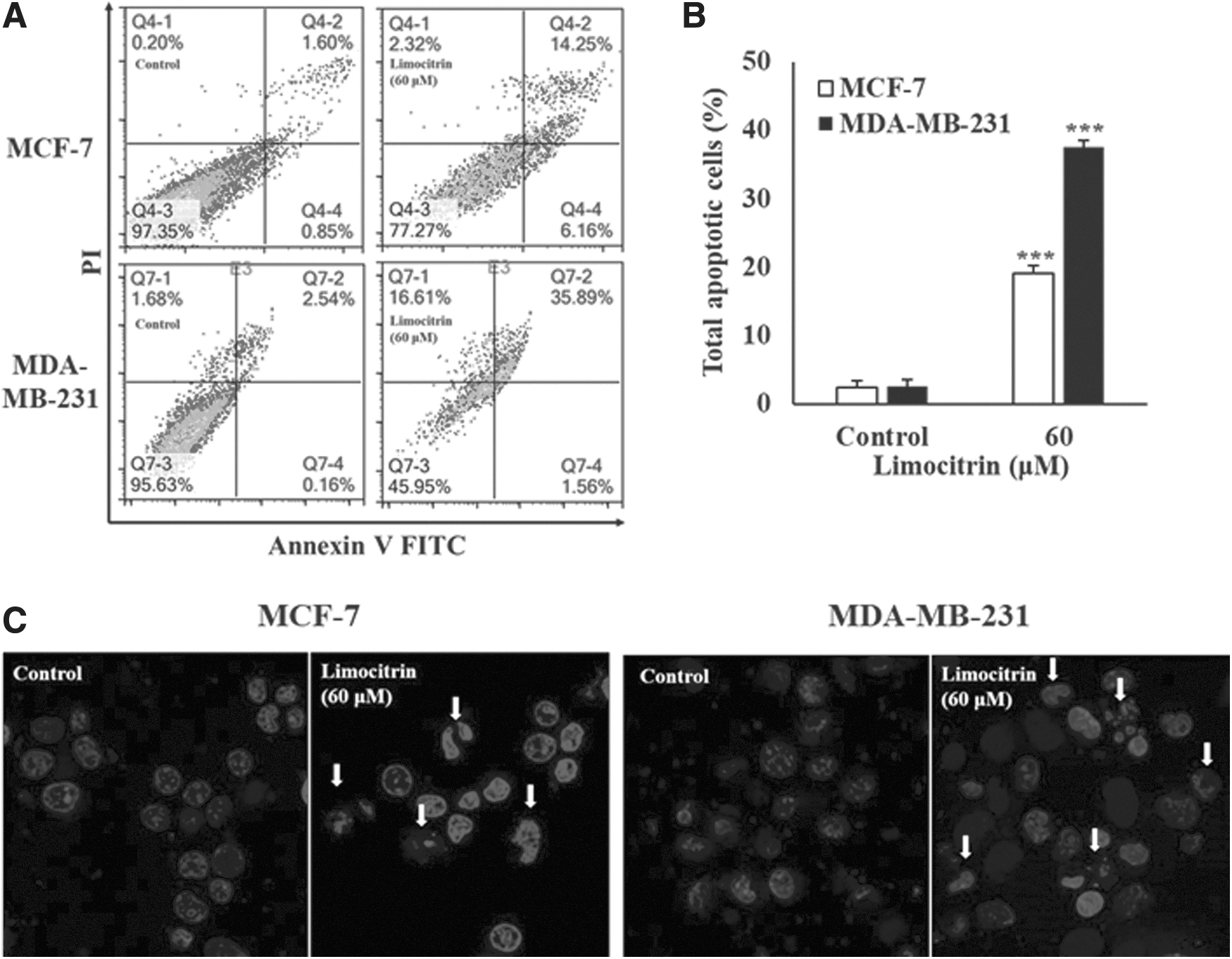
Effect of limocitrin on the apoptosis rate of human breast cancer cells. Cells were cultured and treated with 60 μM limocitrin for 48 h.
After treating the cells with 60 μM limocitrin for 48 h, a noticeable increase in the percentage of apoptotic cells was observed (Fig. 2A, B). In addition, limocitrin treatment led to the appearance of characteristic biological markers of apoptosis, including apoptotic bodies and condensed chromatin (Fig. 2C). These findings suggest that limocitrin effectively enhances apoptotic processes in both ER positive and TNBC.
Particularly noteworthy is the high apoptosis rate induced by limocitrin in TNBC cells, indicating its potential as a therapeutic agent against this aggressive breast cancer subtype. Furthermore, the study highlights that limocitrin can induce apoptosis not only in ER-positive breast cancer cells but also in ER-negative breast cancer cells, suggesting its broad applicability in targeting different breast cancer subtypes. Overall, these results provide valuable insights into the apoptotic effects of limocitrin on breast cancer cells and underscore its potential as a promising candidate for further investigation in breast cancer treatment.
Limocitrin on MDA-MB-231 and MCF-7 cell apoptosis mediated through caspase activation
In this study, we explored the effect of limocitrin on apoptosis in breast cancer cells, focusing on the involvement of the caspase activation pathway. To investigate whether limocitrin induces apoptosis through a caspase cascade-dependent mechanism, the cells were pretreated with a universal caspase inhibitor, z-VAD-fmk, for 3 h, followed by incubation with 60 μM limocitrin for 48 h (Fig. 3A, C). The results showed that pretreatment with the caspase inhibitor inhibited the cell death triggered by limocitrin, indicating the involvement of caspase activation in limocitrin-induced apoptosis in breast cancer cells. Furthermore, we assessed the levels of various proteins associated with apoptosis through western blot analysis (Fig. 3B, D). Limocitrin treatment led to a downregulation of the proapoptotic protein Bid, the antiapoptotic protein Bcl-2, and procaspases (caspase-3, -8, and -9) in both cell types. Conversely, the expression of the proapoptotic protein Bax and the cleaved form of Poly ADP-ribose polymerase (PARP) was elevated by limocitrin treatment.

Effect of limocitrin on apoptosis in MDA-MB-231 and MCF-7 cells mediated through caspase activation.
The data obtained from triplicate experiments were statistically analyzed (Fig. 3E), confirming that limocitrin effectively induces apoptosis through the mitochondrial apoptosis signaling pathway in both triple-negative and ER-positive breast cancer cells. Overall, these findings provide valuable insights into the molecular mechanisms underlying limocitrin-induced apoptosis and suggest its potential as a promising agent for targeting breast cancer cells, regardless of their ER status.
Limocitrin on the rate of MDA-MB-231 and MCF-7 cell apoptosis mediated through apoptosis-inducing factor activation
In this study, we explored the impact of limocitrin on the apoptosis in both cell lines, with a focus on the activation of caspase-independent signaling, specifically Apoptosis-Inducing Factor (AIF) and Endonuclease G (Endo G). Caspase-independent signaling pathways involving AIF and Endo G are associated with the level of PARP, leading to the destruction of cancer cells. The activation of AIF and Endo G occurs through pathways that do not involve caspases. To investigate the mechanism of caspase-independent apoptosis induced by limocitrin, the breast cancer cells were cultured with 60 μM limocitrin for 48 h. The results showed that the levels of AIF and Endo G gradually increased following limocitrin treatment in both cell lines (Fig. 4A, C). These findings were quantified and expressed as triplicate measurements (Fig. 4B, D).

Effect of limocitrin on apoptosis in MDA-MB-231 and MCF-7 cells mediated through the activation of AIF.
Based on these results, we concluded that limocitrin induces an anticancer effect on breast cancer cells through both caspase-dependent and caspase-independent apoptosis mechanisms. The activation of caspase-independent pathways, particularly involving AIF and Endo G, contributes to the apoptotic effects of limocitrin on breast cancer cells. This study provides insights into the diverse apoptotic pathways triggered by limocitrin, suggesting its potential as a multifaceted therapeutic agent in targeting breast cancer cells.
Limocitrin on the inhibition of MDA-MB-231 and MCF-7 cell proliferation as mediated through the suppression of PI3K/Akt/mTOR/S6K signaling pathway activation
In this study, we investigated the effect of limocitrin on the inhibition of proliferation in both cell types, with a focus on its impact on the PI3K/Akt/mTOR/S6K signaling pathway. The PI3K/Akt/mTOR/S6K signaling pathway is known to play a crucial role in various cellular processes, including cell metastasis, growth, survival, and proliferation. 24 To assess whether limocitrin can inhibit this signaling pathway in breast cancer cells, the expression levels of key proteins involved in the pathway were measured (Fig. 5). Following limocitrin treatment, the levels of PI3K, p-PI3K, Akt, p-Akt, mTOR, p-mTOR, p70 S6 kinase, and p-p70 S6 kinase were found to decrease in both cell types (Fig. 5). These findings suggest that limocitrin treatment leads to the suppression of the PI3K/Akt/mTOR/S6K signaling pathway in breast cancer cells.

Effect of limocitrin on the inhibition of MDA-MB-231 and MCF-7 cell proliferation as mediated through the suppression of PI3K/Akt/mTOR signaling pathway activation.
The data, obtained from triplicate measurements, indicate that limocitrin exerts an anticancer effect on breast cancer cells, and this effect can be attributed to its ability to suppress the PI3K/Akt/mTOR/S6K signaling pathway. By inhibiting this pathway, limocitrin may interfere with critical cellular processes that contribute to the growth and proliferation of breast cancer cells. Overall, these results provide important insights into the molecular mechanism underlying the anticancer effects of limocitrin and suggest its potential as a therapeutic agent for targeting breast cancer cells by modulating the PI3K/Akt/mTOR/S6K signaling pathway.
Discussion
S. sarmentosum Bunge, known for its medicinal and culinary uses in treating inflammatory diseases and cancer, has been extensively studied. 7 –9 Limocitrin, categorized as a flavone compound, is present in S. sarmentosum Bunge, M. halliana, and orange peel. 13,14,25 Notably, S. sarmentosum Bunge extract has shown promising effects in mitigating acute liver and kidney injuries by modulating the intracellular cell signaling pathway and M1 macrophage polarization, respectively. 9,26 In addition, it has demonstrated potential in alleviating liver-related conditions, such as hepatitis, liver fibrosis, and fatty liver, through its anti-inflammatory properties. 7,10,27 Moreover, the extract has exhibited significant apoptotic and antiproliferative effects on hepatocellular carcinoma and pancreatic cancer cells. 11,28 Despite these encouraging findings, further research on the biological effects of S. sarmentosum Bunge and limocitrin is warranted.
Most current anticancer approaches primarily focus on targeting hormone receptors or gene protein receptors. For patients with triple-negative tumors, the treatment typically involves the same chemotherapy regimens as those used for other types of cancer, including hormone-related breast cancers. 4 However, despite similar cure rates among different breast cancer types, TNBC poses a unique challenge due to its propensity for recurrence, especially when the initial response to treatment is inadequate. Consequently, the survival rate for TNBC tends to be lower than other forms of breast cancer.
In research settings, MCF-7 and MDA-MB-231 cells are commonly used as representative models of breast cancer. 20,29 –31 As part of this study, we aimed to investigate the apoptotic effect of limocitrin specifically on TNBC cell lines. Furthermore, we conducted a comparative experimental study involving the TNBC cell line and an ER-positive breast cancer cell line to gain insights into the potential differential effects of limocitrin on these distinct breast cancer subtypes.
The findings from this study revealed a significant inhibition of human breast cancer cell proliferation by limocitrin (Fig. 1). Importantly, limocitrin exhibited its effects on both cell types. To the best of our understanding, this is the first documented evidence demonstrating the anticancer and apoptotic properties of limocitrin in breast cancer cells.
Apoptosis, a programmed cell death process, is distinguished by distinct morphological features such as chromatin condensation and apoptotic body formation. This process is governed by both mitochondria-dependent and death receptor-mediated signaling pathways. 15,32 In this study, exposure to limocitrin led to the activation of apoptotic cell death in two breast cancer cell types (Fig. 2). These findings demonstrate that treatment with limocitrin increased the evidence of apoptosis, including an elevated apoptosis rate and the presence of apoptotic bodies, in human breast cancer cells.
Apoptotic cell death mechanisms involve initiator caspases (such as caspase-2, -8, -9, and -10) and effector caspases (including caspase-3, -6, and -7), which play pivotal roles in apoptosis. 33 Recent reports have highlighted that natural products exhibit anticancer activity predominantly through a caspase-dependent pathway. 17,23 Prior research has also indicated that apoptotic death in all breast cancer cell types is attributed to caspase-dependent apoptosis. 19,20,34 In the present study, the expressions of procaspase-3, -8, -9, Bid, and Bcl-2 were downregulated, while the expression of Bax and cleaved PARP was upregulated in both breast cancer cells following treatment with limocitrin (Fig. 3). These results suggest that limocitrin induces apoptosis in breast cancer cells through caspase-dependent mechanisms, thereby regulating the expression of key apoptotic proteins.
AIF and Endo G proteins are known to play crucial roles in inducing apoptotic death in various cancer cells. 35 –39 The permeabilization of the mitochondrial outer membrane leads to the release of AIF and Endo G, signifying the loss of mitochondrial function and increased DNA degradation during apoptotic cell death. 40–41 Several natural products, including celastrol, α-mangostin, lupiwighteone, sanggenol L, and isoquercitrin, have been reported to inhibit the growth of breast, skin, and prostate cancer cells by activating AIF. 19,42 In our study, treatment with limocitrin induced the expression of AIF and Endo G in both cell lines (Fig. 4). These results indicate that limocitrin has potential as a natural product with anticancer properties.
The PI3K/Akt/mTOR signaling pathway is a major intracellular pathway involved in the regulation of cell survival and proliferation. 43 Targeting this pathway has been considered to be a strategy for suppressing cancer growth. Several studies have reported that natural products inhibit the PI3K/Akt/mTOR/S6K signaling pathway in various cancer cells. 17,42,44 –46 In our study, we found that limocitrin effectively inhibited PI3K-Akt-mTOR-S6K signaling expression in breast cancer cells (Fig. 5). These results indicate that limocitrin may have potential as a natural product for suppressing cancer growth by targeting this crucial signaling pathway in breast cancer cells.
This study successfully demonstrated that limocitrin effectively inhibited cell growth in both MCF-7 and MDA-MB-231 breast cancer cells. These findings suggest that the anticancer effects of limocitrin in these two different types of breast cancer cell lines can be attributed to its regulation of the PI3K/Akt/mTOR pathway and apoptotic cell death signaling. Notably, the study revealed that limocitrin exerted distinct regulatory effects on the PI3K/Akt/mTOR pathway in breast cancer cells. These results are consistent with other previous studies involving substances such as Tamoxifen or curcumin, which have also been shown to modulate the PI3K/Akt/mTOR pathway which is differentially regulated in ER(+) and ER(−) breast cancer cells. 47,48 In conclusion, the current research supports the potential of limocitrin as a promising agent for breast cancer treatment, with its ability to target the PI3K/Akt/mTOR pathway and induce apoptotic cell death in breast cancer cells, particularly in both ER(+) and ER(−) subtypes.
Indeed, the current study provides groundbreaking evidence that limocitrin effectively suppresses human breast cancer cell growth through the activation of the mitochondrial apoptotic cell death signaling pathway and concurrent inhibition of the PI3K/Akt/mTOR signaling pathway. These findings are novel and highlight the potential of limocitrin as a promising therapeutic agent for breast cancer treatment.
By promoting apoptotic cell death in breast cancer cells, limocitrin exhibits a key mechanism that could be leveraged in cancer therapy. The ability of limocitrin to induce apoptosis in breast cancer cells offers a valuable strategy for targeting cancer cells and potentially overcoming resistance to traditional treatments.
In summary, this study offers critical insights into the multifaceted anticancer effects of limocitrin and underscores its potential as a valuable candidate for further investigation and development in breast cancer therapy. The promotion of apoptosis in breast cancer cells represents a significant step forward in the quest for effective and targeted treatments for breast cancer patients.
Footnotes
Authors' Contributions
H.-J.L. and J.B.: Writing—original draft, conceptualization, and formal analysis. E.H.P., S.-G.B., and N.C.: Investigation. H.T.: Conceptualization. S.-I.P.: Review and editing. Y.-S.W.: Conceptualization, investigation, data curation, formal analysis, writing—review and editing, project administration. S.-J.L.: Conceptualization and writing—review and editing.
Author Disclosure Statement
No competing financial interests exist.
Funding Information
This work was supported by a National Research Foundation of Korea (NRF) grant funded by the Korean government (MSIT) (No. 2019R1G1A1094747) and the KRIBB Research Initiative Program (KGM5242322).
Supplementary Material
Supplementary Figure S1