




Abstract
Baicalin has been acknowledged for its anti-inflammatory properties. However, its potential impact on osteoarthritis (OA) has not yet been explored. Therefore, our study aimed to examine the effects of Baicalin on OA, both in laboratory and animal models. To evaluate its efficacy, human chondrocytes affected by OA were treated with interleukin-1β and/or Baicalin. The effects were then assessed through viability tests using the cell counting kit-8 (CCK-8) method and flow cytometry. In addition, we analyzed the expressions of various factors such as FOXO1, autophagy, apoptosis, and cartilage synthesis and breakdown to corroborate the effects of Baicalin. We also assessed the severity of OA through analysis of tissue samples. Our findings demonstrate that Baicalin effectively suppresses inflammatory cytokines and MMP-13 levels caused by collagenase-induced osteoarthritis, while simultaneously preserving the levels of Aggrecan and Col2. Furthermore, Baicalin has been shown to enhance autophagy. Through the use of FOXO1 inhibitors, lentivirus-mediated knockdown, and chromatin immunoprecipitation, we verified that Baicalin exerts its protective effects by activating FOXO1, which binds to the Beclin-1 promoter, thereby promoting autophagy. In conclusion, our results show that Baicalin has potential as a therapeutic agent for treating OA (Clinical Trial Registration number: 2023-61).
INTRODUCTION
Osteoarthritis (OA) is a common degenerative joint disease that causes substantial morbidity and functional impairment. Despite knowledge about genetic, epigenetic, mechanical, and metabolic risk factors, 1 –3 the underlying mechanism behind OA remains elusive. This condition is characterized by the gradual breakdown of cartilage, as well as the occurrence of synovitis and subchondral sclerosis. OA has significant implications for the well-being and quality of life of affected individuals, resulting in substantial socioeconomic burdens globally. Chondrocytes, the sole cellular element found in cartilage, participate in the synthesis of biological substances and the turnover of the extracellular matrix (ECM). 4,5 Consequently, maintaining the state of homeostasis in chondrocytes is crucial for ensuring the integrity of cartilage. 6
Autophagy, as a fundamental cellular process, works to maintain homeostasis by breaking down and reusing cytoplasmic components and organelles in response to various harmful stresses. 7,8 Notably, autophagy plays a key role in several disorders, like liver fibrosis, 9 neurodegenerative diseases in humans, 10 muscle atrophy, 11 and cancer. 12 Moreover, autophagy assumes a pivotal role in preserving the structural and functional integrity of cartilage in joints. Specifically, inhibiting mTOR, a protein that suppresses autophagy, in cartilage prompts chondrocytes to undergo autophagy, thus mitigating the effects of age-induced OA in mice. 13 Similarly, the use of rapamycin, an activator of autophagy, proves beneficial in protecting chondrocytes within osteoarthritic cartilage, effectively counteracting apoptosis triggered by glucocorticoids. 14
The FOXO gene family, comprising FOXO1–4 and FOXO6, consists of transcriptional factors that have been conserved throughout the evolutionary process, and these factors play vital roles in a diverse array of biological processes. 15,16 Particularly, FOXO1 is mainly detected in bone and cartilage tissues, where it plays a role in regulating bone tissue formation and maintaining chondrocyte balance. 17
The involvement of FOXO-mediated autophagy has been recommended as a crucial regulatory mechanism in maintaining the homeostasis of chondrocytes, thereby preventing the degradation of intervertebral discs and articular cartilage. 18,19 In elderly patients with OA, particularly in the weight-bearing region, the presence of FOXO1 expression is observed to diminish within the cartilage, specifically in its superficial layer. 1
Baicalin, a prominent flavonoid primarily derived from the roots of Scutellaria baicalensis Georgi, exhibits diverse biological properties. 20 –23 Furthermore, numerous studies have demonstrated the potential of Baicalin in promoting the improvement of OA progression. 24 –27
In this study, we investigated the influence of Baicalin on the biological actions of chondrocytes, with a specific focus on autophagy and the progression of OA. We also conducted an assessment of the modulatory effects of Baicalin on FOXO1. Understanding the underlying mechanism of this process has important implications for establishing an empirical basis for the clinical use of Baicalin in treating OA, and the development and utilization of Baicalin.
MATERIALS AND METHODS
Chemicals and antibodies
Baicalin (HY-N0197, purity >99%) and human interleukin (IL)-1β (HY-P701104) were obtained from MCE (MedChemExpress, USA). Collagenase type II (232-582-9) was obtained from Merck (Sigma-Aldrich®, USA). Primary antibodies against Collagen Type II Polyclonal antibody (28459-1-AP, 1:100), Aggrecan Polyclonal antibody (13880-1-AP, 1:100), and MMP13 Polyclonal antibody (18165-1-AP, 1:100) were purchased from Proteintech (USA).
Ethics and regulatory approval
Consent was obtained from all patients with OA, who underwent surgery for joint replacement in this study. The collection of cartilage tissue was approved by the Medical Ethics Committee of The People's Hospital of Rongchang District, Chongqing, in accordance with the guidelines of the Declaration of Helsinki. The experimental procedures were performed in strict compliance with the recommendations outlined in the “Guide for the Care and Use of Laboratory Animals” (Institute for Laboratory Animal Research, Committee for the Update of the Guide for the Care and Use of Laboratory Animals, National Research Council of The National Academies, USA; National Academies Press: Washington, DC, USA, 2011) and adhered to the ARRIVE guideline. 28 The protocols were approved by the Institutional Animal Care and Use Committee of Chongqing Medical University (IACUC-CQMU-2023-060232).
Human cartilage samples and primary human chondrocyte culture
Samples of cartilage from individuals undergoing total knee replacement surgery for OA were collected. The process of isolation and culturing of human chondrocytes involved the separation of cartilage from the subchondral bone and connective tissue, followed by the division of the cartilage into small 1 mm3 fragments. Subsequently, the cartilage fragments underwent treatment using a 0.25% trypsin-ethylenediaminetetraacetic acid solution for a duration of 30 min. Subsequently, the cartilage underwent digestion in a solution containing 0.2% collagenase type II for a duration of 5 h at a temperature of 37°C. Next, the sample was centrifuged (200 g, 5 min) to isolate human chondrocytes.
Following that, cells from the initial generation (P0) were placed into culture flasks of size T25, with a concentration of 1 × 10⁷ cells per milliliter. They were then subjected to cultivation at a temperature of 37°C in the presence of 5% CO2. Exclusively, for this particular investigation, human chondrocytes from passages P1–P2 were utilized. The chondrocytes underwent treatment with IL-1β (10 ng/mL, 12 h). Alternatively, they were pretreated with 20 μM Baicalin and co-treated for 12 h. 29
To suppress the expression of FOXO1, the medium was supplemented with KD-FOXO1 lentivirus (L26786; Beyotime Biotechnology, China) and AS1842856 (1 μM, HY-100596; MCE). The cells were transfected with KD-FOXO1 and KD-vector using PolyFast Transfection Reagent (HY-K1014; MCE). After 24 h of transfection, additional agents were introduced into the cell medium 24 h before harvest.
Knee joints excised from arthroplasty were collected and damaged articular cartilage within 2–3 mm of visible OA lesions was isolated. 30,31 Cultivation of human cartilage explants were cultured as previously reported. 32,33
Cell viability
The Cell Proliferation and Cytotoxicity Assay Kit (CA1210, China) obtained from Solarbio was utilized to evaluate cell viability. Cultivation of 5 × 10³ human chondrocytes was carried out in 96-well plates for a duration of 24 h. Human chondrocytes were either treated with IL-1β (10 ng/mL, 12 h) or initially pretreated with 20 μM Baicalin and then co-treated for the same duration. The dosages employed in this in vitro study were based on previous research. 29
Model of collagenase-induced osteoarthritis and treatment regimen
The collagenase-induced osteoarthritis (CIOA) model was implemented in the knee joints of 15 male C57BL/6 mice (27 ± 3.2 g; 8 weeks old). To begin, the mice were selected at random from their cages and received anesthesia with 3% isoflurane/0.8 L O2/min. Afterward, the knees were treated with 70% ethanol spray. Subsequently, a dermal incision was performed at the patellar tendon's level, and a 10 μL saline solution (Sigma–Aldrich) containing 10 U collagenase (Sigma–Aldrich, St. Louis, MO) was injected into the knee joints.
Experimental animal design
To establish three groups for experimentation, a total of 30 mice were randomly divided into the sham control group, the CIOA group, and the group treated with Baicalin for OA (CIOA+Baicalin). The sham control group was created through a sham operation. In the Baicalin group, mice were administered Baicalin through intraperitoneal injection at a dosage of 100 mg/kg per day for a duration of 8 weeks following the surgery. On the other hand, the OA group received a vehicle substance, that is, phosphate buffered saline (PBS). It should be noted that the dosage of Baicalin used in animals, being 100 mg/kg, was selected based on previous studies. 34 After a period of 8 weeks postsurgery, the mice were sacrificed through cervical dislocation.
Histological analysis
At predetermined time intervals, the mice were sacrificed. Sterile instruments were used to separate and cleanse the knee joints of the mice with normal saline. Afterward, the samples were fixed in 4% formalin for a duration of 24 h. Decalcification of the fixed samples was carried out over a period of 3 weeks. Following decalcification, the samples were embedded in paraffin and then sectioned. The staining procedures involved Hematoxylin and eosin (H&E) staining (G1120; Solarbio, China), Toluidine blue staining (G3661; Solarbio), and immunohistochemistry staining (SP0041; Solarbio). For co-staining purposes, Methyl Green Staining Solution (C0115; Beyotime Biotechnology) was utilized.
Apoptosis analysis
Apoptosis detection assays were conducted using the APC Annexin V Apoptosis Detection Kit with 7-AAD (640930; biolegend, USA) following the manufacturer's guidelines.
Enzyme-linked immunosorbent assay
We followed the manufacturer's guidelines when using the FastScan™ Total LC3B enzyme-linked immunosorbent assay (ELISA) Kit #35172 (CST), the Mouse iNOS ELISA Kit (ab253219), the Mouse IL-6 ELISA Kit (ab222503), and the Mouse IL-1β ELISA Kit (ab197742). Triplicate measurements were taken for each sample.
Reverse transcription–quantitative polymerase chain reaction
Isolated total RNA from primary chondrocytes was obtained from humans, explants, or mouse knee articular cartilage. The Total RNA Extraction Kit (R1200; Solarbio) was used for this purpose, following the protocol provided by the supplier. Afterward, we performed reverse transcription of the RNA into cDNA using the RT Master Mix for quantitative polymerase chain reaction (qPCR) II (gDNA digester plus) (HY-K0511A; MCE), following the manufacturer's instructions. To analyze the results, we employed the 2−ΔΔCq method with glyceraldehyde 3-phosphate dehydrogenase (GAPDH) as the internal reference gene. The primer sequences utilized for this experiment can be found in Table 1.
Reverse Transcription-Quantitative Polymerase Chain Reaction Primer Sequence
Western blot
The extracted proteins from cells and tissues were transferred onto polyvinylidene fluoride membranes (Millipore Sigma Co., Ltd., USA). After overnight incubation with the primary antibody at 4℃, the membrane was incubated with the secondary antibody (ZSGB-Bio, Beijing, China) for 1 h. The primary antibody information and dilution ratios are provided below: Recombinant Anti-Aggrecan antibody [6-B-4] (Abcam, ab3778, 1:500) Collagen Type II Polyclonal antibody (Proteintech, Cat No: 28459-1-AP, 1:1000) MMP13 Polyclonal antibody (Proteintech, Cat No: 18165-1-AP, 1:1000) GAPDH Monoclonal antibody (Proteintech, Cat No: 60004-1-Ig, 1:1000) FOXO1 Polyclonal antibody (Proteintech, Cat No: 18592-1-AP, 1:1000) ATG7 Polyclonal antibody (Proteintech, Cat No: 10088-2-AP, 1:1000) P62, SQSTM1 Monoclonal antibody (Proteintech, Cat No: 66184-1-Ig, 1:1000) Beclin 1 Polyclonal antibody (Proteintech, Cat No: 11306-1-AP, 1:1000).
Chromatin immunoprecipitation analysis
The chromatin immunoprecipitation (CHIP) assay was carried out utilizing a commercially available kit provided by Cell Signaling (56383). In short, the chromatin was fragmented into DNA segments of 200–500 bp through digestion and sonication. Analysis of the precipitated DNA involved PCR. JASPAR (
Site1:
F:GGTCAGCGAGACCCTTGGAA,
R: AGAATTATATCACCAAAGCTGCCC.
Site2:
F:CCGCCCCCTGAATTTAGAGAAT
R: GGTTACCACGGGAAGTGTAGG.
Statistical analysis
The means ± standard deviations of the presented data were analyzed using GraphPad Prism 9.0. Two-tailed unpaired Student's t test was conducted as part of the statistical analysis to compare two groups. One-way analysis of variance (ANOVA) was utilized to compare multiple groups in this study. To further examine the pairwise differences between groups, a Bonferroni test was performed. Any P-value below .05 was considered statistically significant.
RESULTS
Baicalin increases cell viability and inhibits apoptosis of human primary chondrocytes
IL-1β was administered alone to human OA chondrocytes, while Baicalin was either pretreated or co-treated with IL-1β. Cell counting kit-8 (CCK-8) results demonstrated that the IL-1β treatment significantly decreased the viability of OA chondrocytes, but this reduction was effectively reversed by the treatment with 20 μM Baicalin (Fig. 1A).

Baicalin reversed IL-1β-induced viability inhibition, proapoptosis, and ECM degradation in primary human OA chondrocytes.
To explore the influence of Baicalin on cartilage anabolism and catabolism markers, experiments with varying concentrations of Baicalin were conducted. Our results indicated that both 20 and 30 μM Baicalin positively modulated Aggrecan and Col2 protein levels in chondrocytes, while reducing the MMP-13 protein level (Supplementary Fig. S1A, C). Notably, the effects of 20 μM Baicalin were more prominent. Based on the outcomes from the CCK-8 assay, the optimal concentration chosen for subsequent in vitro experiments was 20 μM. Based on these findings, the hypothesis was formulated that the inhibitory effect of Baicalin on chondrocyte death could be explored further. To delve deeper into this matter, the evaluation of apoptosis in chondrocytes was conducted utilizing flow cytometry.
The proportion of apoptotic cells observed in cells treated with IL-1β was considerably higher compared to the control cells. However, Baicalin significantly attenuated the extent of cell demise induced by IL-1β (Fig. 2B, C). In addition, the assessment of apoptosis-related markers was carried out by reverse transcription (RT)-qPCR. Remarkably, stimulation with IL-1β resulted in a substantial increase of Caspase3, 7, and 9 mRNA levels, whereas the administration of Baicalin was capable of alleviating this upregulation (Fig. 1D). Furthermore, the measurement of sulfated glycosaminoglycan (sGAG) content in IL-1β-induced chondrocytes revealed a significant decline in comparison to control chondrocytes. However, the pretreatment with Baicalin exhibited a marked enhancement in the levels of sGAG content in OA chondrocytes (Fig. 1E).

Baicalin alleviates knee articular cartilage pathological damage in CIOA model mice. The mice were randomly divided into three groups: sham operation group, OA group, and Baicalin group. The CIOA model was established by intra-articular injection of collagenase in the OA group and the Baicalin group. After operation, the Baicalin group was intraperitoneally injected daily with Baicalin (100 mg/kg), and the OA group was injected with vehicle (PBS) intraperitoneally for 8 weeks. Knee articular cartilage thickness
To evaluate the impact of Baicalin on the breakdown of the ECM, the expression levels of Col2, Aggrecan, and MMP-13 were quantified. Notably, Baicalin pretreatment ameliorated the deleterious effects of IL-1β on Col2 and Aggrecan expression, while simultaneously reducing the expression of MMP-13 (Fig. 1F). Taken together, the results from this study indicate that Baicalin exerts an effective inhibitory effect on apoptosis and safeguards the integrity of the cartilage matrix in chondrocytes exposed to IL-1β, thereby highlighting its potential for the treatment of OA.
Baicalin alleviated the progression of CIOA models
To examine whether Baicalin possesses preventive properties in regard to the initiation and advancement of OA within a living organism, we conducted a study using a CIOA mouse model. The mice were subjected to daily intraperitoneal injections of 50, 100, or 150 mg/kg of Baicalin or a vehicle (PBS, Sham group) for a span of 8 weeks. The Western blot findings indicate that both 100 and 150 mg/kg of Baicalin effectively alleviate the reduction in cartilage Aggrecan and Col2 proteins, as well as the elevation in MMP-13 caused by CIOA. However, the administration of 100 mg/kg demonstrates greater effectiveness (Supplementary Fig. S1B, D). Histological examination through H&E staining and toluidine blue staining demonstrated that the cartilage surface of the control group, which underwent sham surgery, remained smooth and intact (Fig. 2A, B).
In contrast, the group administered with CIOA displayed damage to the cartilage surface, demonstrating erosion of the cartilage (Fig. 2C), noteworthy decline in proteoglycans (Fig. 2D, E), and a substantial buildup of MMP-13 (Fig. 2F, G) when compared to the control group that underwent a sham operation. Interestingly, the group receiving 100 mg/kg Baicalin exhibited a decrease in proteoglycan loss and cartilage destruction in comparison to the CIOA group. Furthermore, Baicalin (100 mg/kg) significantly ameliorated joint inflammation, leading to a remarkable reduction in the levels of iNOS, IL-1β, and IL-6 in the Baicalin group compared to the CIOA group (Fig. 3A–C). As a whole, these findings showed that Baicalin possesses the potential to hinder the advancement of CIOA model.
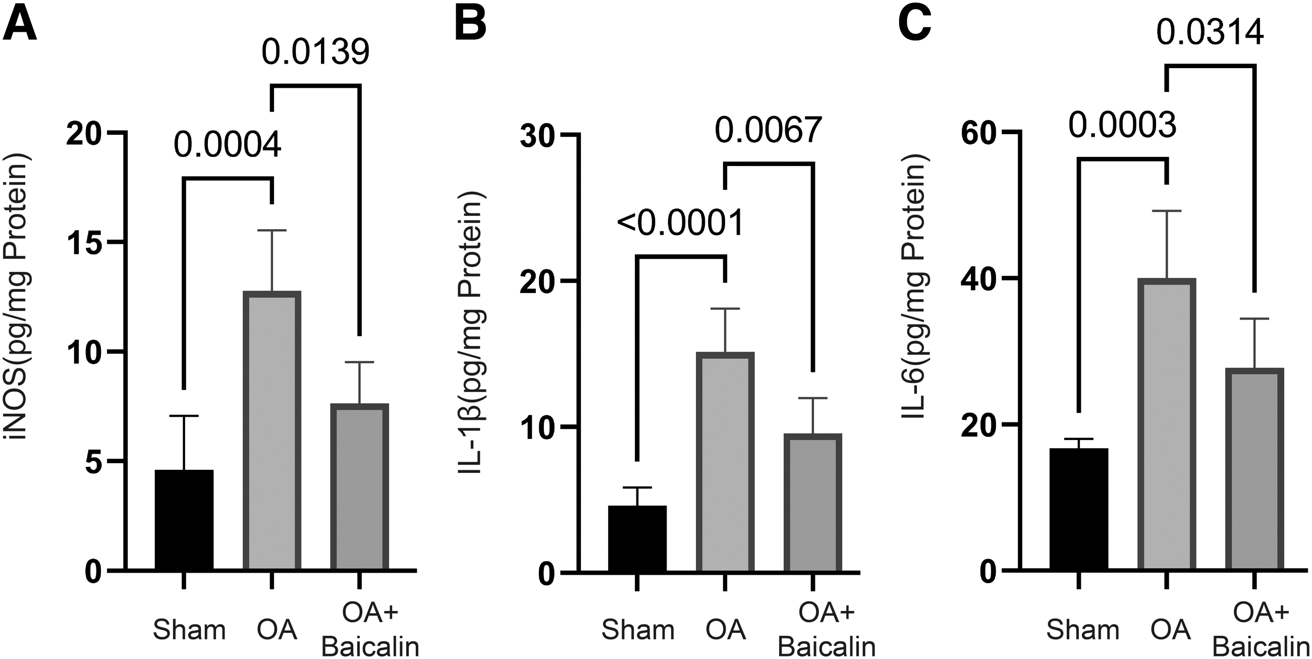
One hundred milligram per kilogram Baicalin attenuates inflammation levels in the CIOA model. The mice were randomly divided into three groups: sham operation group, OA group, and Baicalin group. The CIOA model was established by intra-articular injection of collagenase in the OA group and the Baicalin group. After operation, the Baicalin group was intraperitoneally injected daily with Baicalin (100 mg/kg), and the OA group was injected with vehicle (PBS) intraperitoneally for 8 weeks. The levels of iNOS
Low FOXO1 expression and autophagy levels in OA-affected human knee cartilage, and 20 μM Baicalin treatment reversed all
Subsequently, we investigated cartilage samples obtained from OA patients and categorized them into lesion and nonlesion areas. The cartilage samples from lesion areas were then treated with Baicalin for 1 month. Our observations revealed that compared to nonlesion explant tissues, the levels of FOXO1 and autophagy in lesion explant tissues were lower. However, upon treatment with Baicalin, the mRNA level of FOXO1 (Fig. 4A) and various autophagy-related indicators exhibited a significant increase (Fig. 4B–D). These findings suggest that in OA patients, knee cartilage tends to have low FOXO1 and low autophagy levels, and Baicalin demonstrates the potential to effectively alleviate the series of changes induced by OA.
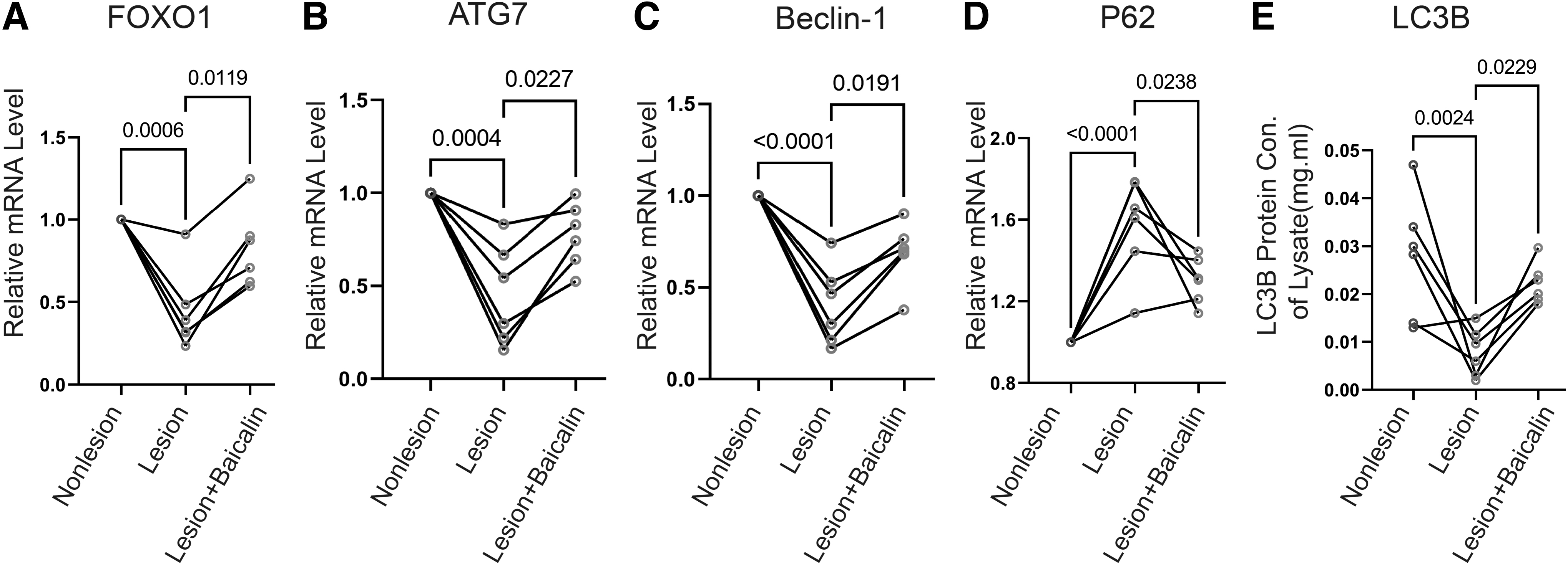
Twenty micromolar Baicalin reverses low FOXO1 expression and low autophagy levels in human OA samples. Human OA samples were divided into lesion area and nonlesion area, and then made into explants for culture. After treating with 20 μM Baicalin for 1 month, the mRNA levels of FOXO1
Effects of Baicalin on FOXO1 expression and autophagy-related indicators in CIOA animal models and IL-1β-induced human OA chondrocytes
In our previous discoveries, Baicalin at 20 and 30 μM had the ability to increase FOXO1, Beclin-1, and ATG7 protein levels, while suppressing the P62 accumulation. However, it was noted that 20 μM Baicalin had a more significant influence on the regulation of FOXO1 and autophagy (Supplementary Fig. S2A, C). Animal experiments indicated that the administration of either 100 or 150 mg/kg Baicalin improved the reduced levels of FOXO1, Beclin-1, and ATG7 proteins, as well as the elevated levels of P62 protein induced by CIOA. Notably, the regulation of FOXO1 was more pronounced with the administration of 100 mg/kg Baicalin (Supplementary Fig. S2B, D).
Figure 5A illustrates that when human OA chondrocytes were stimulated with IL-1β, there was a noteworthy decrease in FOXO1 mRNA levels compared to the control group. Conversely, administering 20 μM Baicalin caused an increase in the FOXO1 mRNA level. In addition, IL-1β resulted in a low level of autophagy, whereas 20 μM Baicalin exhibited an inhibitory effect on the IL-1β-induced decrease in autophagy levels (Fig. 5B, C). In contrast, the results also revealed that the introduction of 100 mg/kg Baicalin significantly amplified FOXO1 expression and the autophagy level in the CIOA model (Fig. 5D, F). These combined observations suggest that Baicalin functions as a potent activator of FOXO1 in both human OA chondrocytes and mice cartilage.
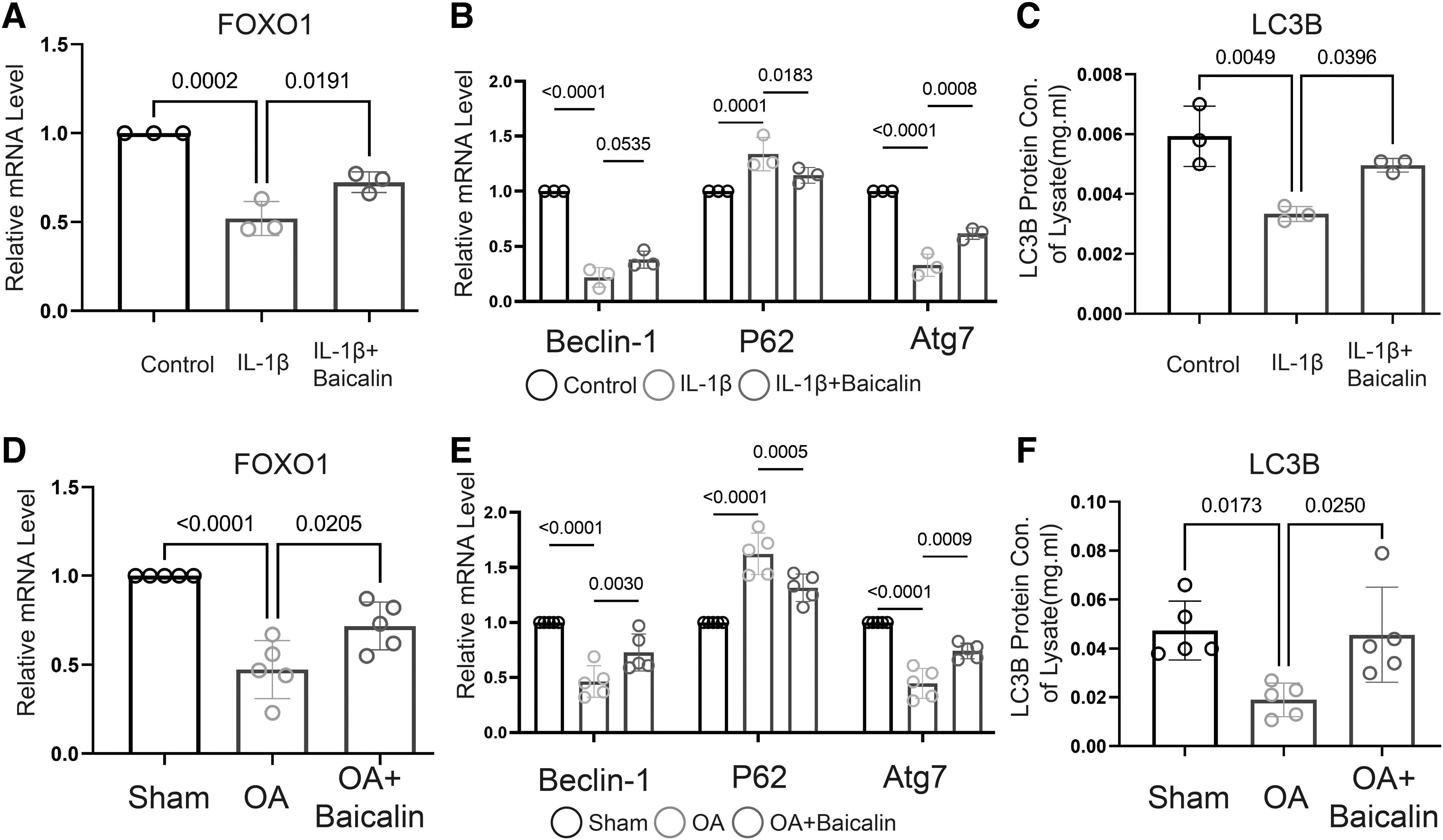
Baicalin enhances autophagic activity both in human primary chondrocytes and mice.
Baicalin exhibits chondroprotective effect through FOXO1
To investigate the expression of FOXO1 and its relationship with cartilage development, we performed qRT-PCR to detect the expression of FOXO1 in 10 mouse cartilage tissues. Pearson correlation analysis revealed a positive correlation between FOXO1 and anabolic indicators (Col2, Aggrecan), while a negative correlation was found with catabolic indicators (MMP-13) (Fig. 6A). These findings indicate that FOXO1 is closely related to cartilage metabolism.
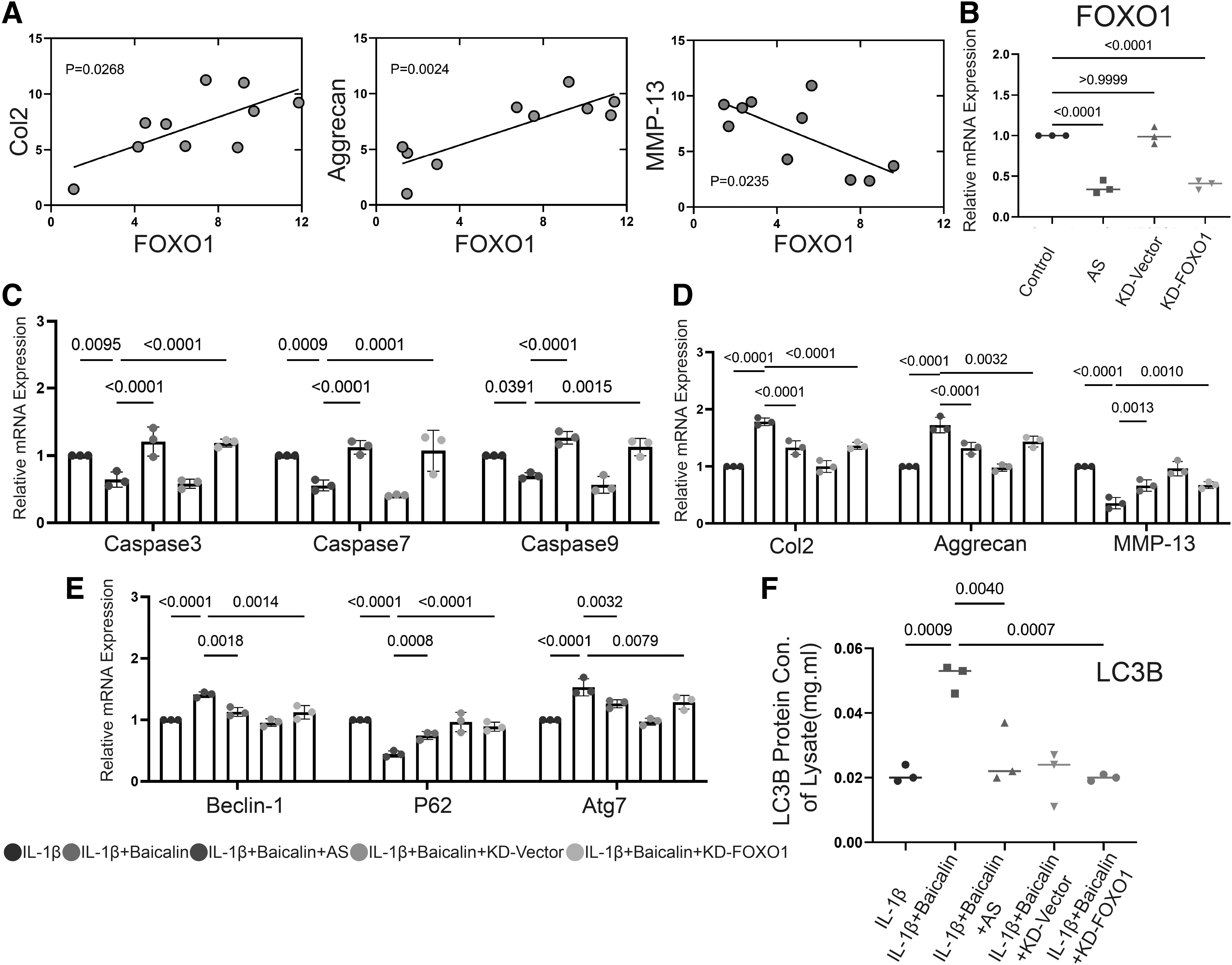
FOXO1 is necessary for Baicalin to exert its anti-OA function.
Next, we evaluated the effect of 20 μM Baicalin after using the FOXO1 inhibitor AS1842856 (AS) or knockout lentivirus. We first confirmed the effectiveness of AS and KD-FOXO1. Compared with the control group, both AS and KD-FOXO1 effectively inhibited the expression of FOXO1 (Fig. 6B).
Upon IL-1β stimulation, we observed that inhibiting the expression of FOXO1 significantly weakened the ability of 20 μM Baicalin to regulate chondrocyte apoptosis and autophagy. In addition, the ability of 20 μM Baicalin to regulate chondrocyte metabolism was also negatively affected. Based on above results, we propose that the protective effect of Baicalin mainly relies on FOXO1.
FOXO1 directly regulates Beclin-1 in human primary chondrocytes
Given that FOXO1 is a transcription factor, we speculated that it may directly regulate autophagy-related proteins. To confirm this, we predicted the possible binding site of FOXO1 in the Beclin-1 promoter region using JASPAR (Fig. 7A). We then conducted a CHIP experiment to verify the binding of FOXO1 to Beclin-1, and the results demonstrated that FOXO1 can indeed bind to the Beclin-1 promoter (Fig. 7B). These findings suggest that Baicalin may alleviate the low autophagy level of OA chondrocytes by promoting the expression of FOXO1 in chondrocytes, thereby enhancing the transcriptional activity of Beclin-1.

FOXO1 binds to the Beclin-1 promoter region.
DISCUSSION
OA is characterized by the presence of significant chondrocyte apoptosis and heightened degradation of the ECM. Prior research has demonstrated that the active progression of OA in cellular or animal models is influenced by the regulation of ECM synthesis and apoptosis through chemical and genetic mechanisms. 35,36 However, few studies have focused on the therapeutic potential of Baicalin in OA. 29,34,37 The objective of this investigation was to examine the impacts and molecular mechanisms of Baicalin in a mouse model of CIOA and the injury of human chondrocytes induced by IL-1β. The findings reveal that Baicalin possesses the capacity to decrease apoptosis of chondrocytes, degradation of ECM, and pathological damage caused by OA. Moreover, the outcomes offer proof that the molecular mechanism of Baicalin's effect is interconnected with the activation of FOXO1 and the induction of autophagy in chondrocytes.
The ability of IL-1β to promote degradation of the ECM, induce apoptosis in chondrocytes, and inhibit cell proliferation has been widely acknowledged in previous studies. 38,39 Therefore, to simulate the damage observed in OA cells, we treated human OA chondrocytes with IL-1β. As expected, the administration of IL-1β resulted in a significant increase in apoptosis and expression of matrix metalloproteinase, while also causing a notable decrease in chondrocyte viability and synthesis of the ECM.
Notably, treatment with Baicalin was found to effectively reverse these detrimental effects induced by IL-1β, indicating its potential as a protective agent against inflammatory damage in human OA chondrocytes. To further explore the impact of Baicalin, we created a mouse model of CIOA. The results from our experiment demonstrated that Baicalin, when administered through intraperitoneal injection, effectively inhibited cartilage surface defect and reduced the level of joint inflammation in the CIOA model. These findings provide additional affirmation that Baicalin possesses cartilage protective properties.
Autophagy functions as a cellular mechanism for maintaining homeostasis in various pathological occurrences. Numerous reports have indicated that modifying autophagy can effectively inhibit the apoptosis process. One noteworthy characteristic of patients with OA is the decreased level of autophagic activity. 40 Based on our findings, it is evident that the application of IL-1β results in compromised chondrocyte autophagy, whereas the administration of Baicalin enhances the autophagic flux. Similarly, our experiments conducted in vivo also consistently demonstrated similar outcomes. In comparison to the control group, the CIOA group exhibited a decrease in the levels of autophagy markers, including Beclin-1, ATG7, and LC3B, accompanied by an increase in the levels of P62. However, following Baicalin treatment, there was a significant enhancement in the autophagy level in CIOA group. These in vivo results further confirm that Baicalin exhibits chondroprotective properties by stimulating autophagic flux.
Research has shown that the loss of FOXO1 in chondrocytes after birth can result in the initial thickening of cartilage. This thickening eventually leads to the degeneration of the cartilage, which in turn may cause symptoms similar to OA. 19,41 FOXO1 regulates autophagy genes, such as Becn1 and Map1lc3b, at the transcriptional level. It has been observed that the increased expression of FOXO1 leads to higher levels of the LC3B protein. 41 Our study centered on the comparison of FOXO1 expression and the levels of autophagy in human OA samples, specifically focusing on the diseased and nonlesioned tissues. Our findings revealed a reduction in FOXO1 expression and a decrease in autophagy levels in the diseased tissues when compared to the nonlesioned tissues. These results led us to propose a hypothesis suggesting that Baicalin exerts its effects through the activation of FOXO1, thus potentially acting as the underlying mechanism.
Remarkably, a month later, following the treatment of human OA explants with Baicalin, the expression of FOXO1 was amplified, while there was also an augmentation observed in the level of autophagy. To investigate the association between Baicalin and FOXO1, we employed FOXO1 inhibitors alongside knockdown (KD) lentivirus. After suppressing FOXO1, the capability of Baicalin to hinder chondrocyte apoptosis was diminished, and the regulatory impact of Baicalin on autophagy indicators Beclin-1, Atg7, P62, and LC3B ceased to function. Furthermore, the capacity of Baicalin to regulate cartilage metabolism also vanished. This once again provided evidence that FOXO1 is intimately interconnected with chondrocyte autophagy, and Baicalin's regulation of chondrocytes occurs through FOXO1. The findings of the CHIP study indicate that FOXO1 has the direct ability to bind to the Beclin-1 promoter. This implies that Baicalin enhances chondroautophagy by amplifying the expression of FOXO1, subsequently resulting in the augmented transcription of Beclin-1.
Although our study highlights the crucial role of FOXO1 in mitigating damage to articular cartilage caused by OA, further investigations are necessary for a comprehensive understanding of its underlying molecular mechanism. Our experimental findings demonstrate the efficacy of Baicalin in ameliorating the OA phenotype, resulting from impaired signaling of FOXO1. Nevertheless, additional research is essential to elucidate the regulatory effects of Baicalin on chondrocyte apoptosis and autophagy in the articular joint. It is important to note that our results do not preclude the involvement of other signaling pathways, aside from FOXO1, in maintaining chondrocyte homeostasis within the joint. Nonetheless, our findings strongly suggest that the Baicalin-FOXO1 signaling pathway plays a pivotal role in modulating autophagy and preserving the homeostasis of articular cartilage. This provides compelling evidence for the chondroprotective properties of Baicalin in OA and supports the potential therapeutic application of selectively activating FOXO1 to treat OA.
In summary, this study showcases the ability of Baicalin to combat OA in human chondrocytes and CIOA mouse models. The breakdown of the ECM triggered by IL-1β or CIOA is effectively halted by Baicalin, leading to the inhibition of chondrocyte apoptosis and initiation of autophagy by the activation of the FOXO1 signaling pathway. In addition, Baicalin exhibits promising outcomes in terms of mitigating cartilage degeneration and inflammation in bone OA joints of mice. These findings strongly support Baicalin's potential as a viable treatment option for OA.
Footnotes
AUTHORs' CONTRIBUTIONS
Designing research studies: Q.W., conducting experiments: Q.W., Z.Y., P.Y., and X.C., acquiring data: Q.W., Z.Y., P.Y., anf X.C., analyzing data: Q.W., writing the article: Q.W.
ETHICAL APPROVAL
All the experimental procedures were performed in accordance with the ethical standards recommended by the International Committee of Medical Journal Editors.
AUTHOR DISCLOSURE STATEMENT
No competing financial interests exist.
FUNDING INFORMATION
This project has received funding support by Chongqing Rongchang District Science and Health Joint Medical Research Project (2023RCMSXM).
SUPPLEMENTARY MATERIAL
Supplementary Figure S1
Supplementary Figure S2