




Abstract
Acorus gramineus has a number of beneficial effects, including protective effects against age-related disorders. In this study, the effects of A. gramineus on testosterone production and andropause symptoms were evaluated. We first treated TM3 mouse Leydig cells, responsible for testosterone production, with A. gramineus aqueous extract at different concentrations. In TM3 cells, the testosterone concentration increased in a concentration-dependent manner compared with those in the control. In addition, at 400 μg/mL extract, the mRNA expression level of the steroidogenic enzyme CYP11A1 was increased. Subsequently, 23-week-old Sprague–Dawley (SD) rats exhibiting an age-related reduction in serum testosterone (approximately 80% lower than that in 7-week-old SD rats) were administered A. gramineus aqueous extract for 8 weeks. Serum total testosterone and free testosterone levels were higher and serum estradiol, prostate-specific antigen levels, and total cholesterol levels were lower in the AG50 group (A. gramineus aqueous extract 50 mg/kg of body weight/day) than in the OLD (control group). The AG50 group also showed significant elevations in sperm count, grip strength, and mRNA expression of StAR, CYP11A1, 17β-HSD, and CYP17A1 compared with those in the OLD group. In conclusion, A. gramineus aqueous extract facilitated steroidogenesis in Leydig cells, elevated testosterone levels, lowered serum estradiol and total cholesterol levels, and increased muscle strength and sperm count, thus alleviating the symptoms of andropause. These findings suggest that A. gramineus aqueous extract is a potentially effective therapeutic agent against various symptoms associated with andropause.
INTRODUCTION
The symptoms of aging in men, known as late-onset hypogonadism (LOH) or andropause, are attributed to reduced testosterone levels with advancement in age. Testosterone levels in men gradually decline after the age of 40 years by 0.4–2.6% per year. 1,2 Significant reductions in testosterone levels are accompanied by reduced sexual function, muscle mass, bone density, and physical functions, such as strength and endurance, and elevations in fatigue and depression. 3 Testicular function decreases naturally with age. Testosterone decline varies among individuals and is affected by chronic diseases, such as obesity, disease, stress, and drugs. 4 The symptoms of testosterone decline and aging have several similarities. 5 Therefore, the normalization of testosterone levels in aging men with low testosterone is expected to provide various benefits.
Approximately 95% of testosterone synthesis takes place in the Leydig cells of the testes. In Leydig cells, the luteinizing hormone (LH) activates the steroidogenic acute regulatory protein (StAR) and cholesterol side-chain cleavage enzymes (P450scc and CYP11A1) via the cAMP/PKA pathway. Cholesterol is translocated to the inner mitochondrial membrane of Leydig cells by StAR and pregnenolone is synthesized by CYP11A1. Testosterone is also synthesized from pregnenolone by steroidogenic enzymes, such as 3β-HSD, CYP17A1, and 17β-HSD. Synthesized testosterone can be converted to dihydrotestosterone (DHT) and estradiol (E2) by 5α-reductase2 and aromatase. 6 –8
In aged men, the LH concentration increases to maintain testosterone levels. However, despite an increase in LH, testosterone levels are markedly reduced by decreased testicular function. 9,10
Testosterone replacement therapy, where testosterone is directly injected, is a common method for treating LOH. However, periodic monitoring is necessary because it may increase the risk of prostate diseases, including prostate cancer, prostate enlargement, and the risk of heart disease. 11,12 Effective and safe natural plant materials are currently being studied as alternatives to testosterone replacement therapy.
Acorus gramineus Soland has traditionally been used as a sedative, anticonvulsant, and digestive agent in East Asia, including Korea, China, and Japan. It is widely used to improve cognitive problems in traditional Chinese medicine 13 and to enhance learning and memory in Korean medicine. 14,15 Several studies have shown that A. gramineus contains various phenylpropanoids. 16 –18 A. gramineus has various established bioactivities, including anti-obesity, 19 antitumor, anti-inflammatory, 20 antistress, 21 fatigue delay during exercise, 22 and antidementia effects through memory enhancement. 23 Recent reports suggest that its active compounds, such as α-asarone and β-asarone, have beneficial effects in age-related neurological disorders. 24 –27 However, research on the overall improvement in age-related symptoms in response to A. gramineus and their correlation with testosterone levels is lacking. Therefore, this study assessed the effects of A. gramineus extract on the symptoms of LOH and testosterone deficiency in vitro and in vivo.
MATERIAL AND METHODS
Sample preparation and liquid chromatography
Dried A. gramineus Soland was purchased from Donggwang General Corporation (Korea). A 15-fold volume of water was added and refluxed at 100 ± 5°C for 8 h twice. The extract was passed through a 70 mesh filter and evaporated to obtain an extract with approximately 20% solid content (or 20% brix) in vacuo at 75 ± 5°C. The product was spray-dried with dextrin and yielded 36.5 ± 5%. The dried powder was used in experiments. A. gramineus extract solution (10 mg/mL) was prepared for high-performance liquid chromatography (HPLC) analysis. In addition, 5-hydroxymethyl-2-furaldehyde (5-HMF; Merck, Darmstadt, Germany) was dissolved in water and used for analysis. HPLC analysis was conducted using an Agilent Technologies 1200 system (Agilent Technologies, Santa Clara, CA). An Eclipse Plus C18 column (4.6 × 250 mm, 5 μm) was used. The temperature was maintained at 30°C, with an injection volume of 10 μL at a flow rate of 0.7 mL/min. The mobile phase consisted of methanol (A) and water with 0.1% trifluoroacetic acid (B). The gradient elution conditions were as stated below: initial 0 min A:B (5:95, v/v), 40 min A:B (100:0), 43 min A:B (100:0), 45 min A:B (5:95), and 50 min A:B (5:95). The detection wavelength was 284 nm.
Cell culture
The TM3 mouse Leydig cell line was purchased from ATCC (Manassas, VA). TM3 cells were cultured in Dulbecco’s modified Eagle’s medium (DMEM)/Ham’s F12 media supplemented with 5% horse serum and 2.5% fetal bovine serum at 37°C with 5% CO2. hCG (0.05 IU/mL) was added to cells with serum-free media and cultured for 20 h. Then, cells were exposed to oxidative stress with 200 μM H2O2 and incubated for an additional 4 h.
Cell viability test
TM3 cells were seeded in 96-well plates (1 × 10⁴ cells/well) and cultured for 24 h. Then, A. gramineus aqueous extract (0, 100, 200, 400, 500, and 1000 μg/mL) was added. After 24 h, a thiazolyl blue tetrazolium bromide (MTT) solubilized in water was distributed into every well and left to incubate for 2 h. Subsequently, dimethyl sulfoxide was replaced in the well to make evenly formazan color. The absorbance at 570 and 630 nm was measured using an Epoch microplate reader (BioTek Instruments, Winooski, VT). The values were converted into percentages relative to values for the control.
Testosterone measurement in the cell culture supernatant
To assess the testosterone concentration in the culture media, TM3 Leydig cells (1 × 10⁴ cells/well) were cultured in 96-well plates. After 24 h, serum-free DMEM containing different concentrations of A. gramineus aqueous extract and hCG (0.05 IU/mL) replaced the cell medium, with incubation for 24 h. The cells were exposed to 200 μM H2O2 for 4 h. Cell supernatants were collected for enzyme-linked immunosorbent assays (ELISA) using an ELISA kit (Enzo Life Sciences, Farmingdale, NY). The absorbance was measured at 405 nm and calculated using Curve Expert 1.3 (Hyams Development, Starkville, MS).
Animals
Male Sprague–Dawley (SD) rats were purchased from ORIENT Bio (Seongnam, Korea). Animals at 23 weeks of age were used as an andropause in vivo model and 7-week-old animals were used as normal controls. Rats were kept under a controlled temperature of 22 ± 2°C and humidity of 55 ± 5% on 12 h light/dark cycles and allowed free access to feed and water. These animal experiments were approved by the Institutional Animal Care and Use Committee of the University of Suwon (approval number: USW-IACUC-2023-002). Rats were divided into four groups (eight animals per group): (1) OLD group, 23-week-old rats given saline by oral gavage; (2) AG25 group, 23-week-old rats given A. gramineus aqueous extract (25 mg/kg BW/day) by oral gavage; (3) AG50 group, 23-week-old rats given A. gramineus aqueous extract (50 mg/kg BW/day) by oral gavage; and (4) YOUNG group: 7-week-old rats given saline by oral gavage. Eight weeks after the experimental period, all animals were euthanized. The liver, testes, epididymal fat, and hindlimb muscles (tibialis anterior, extensor digitorum longus, soleus) were weighed and stored at −80°C. The epididymis was isolated from the testes and used for sperm counting.
Quantitative real-time PCR
Total RNA was extracted from decapsulated testes. Testis homogenate in 200 μL of chloroform per 1000 μL of RNAiso plus reagent was mixed thoroughly. After centrifugation at 12,000 g for 15 min at 4°C, supernatant was supplemented with 500 μL of isopropanol and mixed well. A cold 75% equivalent volume of ethanol was mixed with the supernatant. The RNA precipitate was dried for several minutes and dissolved in RNase-free water. cDNA was synthesized utilizing a T100 Thermal Cycler (Bio-Rad Laboratories Ltd., Hercules, CA). Real-time quantitative polymerase chain reaction was performed using a Roche Light Cycler (Roche, Basel, Switzerland) to determine mRNA expression levels of steroidogenic enzymes and internal control, GAPDH. The primer sequences are presented in Table 1.
Oligonucleotide Primers Used for Quantitative RT-PCR
RT-PCR, reverse transcription-polymerase chain reaction.
ELISA for the detection of steroid hormones
Serum was collected blood at after centrifuging blood samples at 3000 g for 20 min. Total testosterone, free testosterone, and E2 concentrations in serum were measured using an ELISA kit (Cusabio, Wuhan, China). The assays were executed in accordance with the manufacturer’s recommendations. The absorbance was measured at 450 nm; the equation was formulated using Curve Experts 1.3 (Hyams Development) and the values derived.
Prostate-specific antigen measurement
Serum prostate-specific antigen (PSA) levels were measured using a rat glandular kallikrein ELISA kit (Cusabio) following the manufacturer’s specifications. PSA levels were calculated from the absorbance values using Curve Expert 1.3 (Hyams Development).
Immunohistochemical staining
Immunohistochemical (IHC) staining of the testis tissues and Leydig cells was performed to detect CYP11A1 expression. The testis samples were embedded in paraffin blocks. The sections were then dehydrated and washed to remove the xylene. Antigen retrieval and peroxidase blocking were performed. Samples were incubated with the primary antibody (CYP11A1) at 4°C overnight. A DAB (3,3'Diaminobenzidine) kit was used to visualize the antibody‐antigen complexes, and positive labeling of Leydig cells via brown cytoplasmic staining was performed. Mayer’s hematoxylin was applied as the counterstaining reagent. Leydig cells with staining were counted.
Biochemical analysis
The serum levels of alanine transaminase (ALT), aspartate transaminase (AST), as well as the lipid profile, including serum triglyceride, total cholesterol, High-density lipoprotein (HDL) cholesterol, and Low-density lipoprotein (LDL) cholesterol, in the rats were analyzed using a biochemical device (FUJI DRY CHEM 500i; FUJI Photo Film Co., Ltd., Tokyo, Japan).
Sperm count
The cauda epididymis was incised and chopped in phosphate-buffered saline. The method used for sperm count measurement has been described in a previous study. 28 Sperms were counted under an optical microscope (×400 magnification).
Grip strength test
To evaluate improvements in muscle strength, the forelimb grip strength of 23-week-old rats was measured. The grip strength test method has been described in a previous study. 28 The test was conducted weekly at predetermined time points.
Statistical analysis
All values are expressed as the means ± standard deviations (SDs). Statistical significance was analyzed by the Mann–Whitney U test using SPSS Statistics 22 (International Business Machines Corp., Armonk, NY). Results were considered statistically significant at P < 0.05.
RESULTS AND DISCUSSION
A. gramineus increases testosterone production via Cyp11a1 in TM3 Leydig cells
An MTT assay was conducted to evaluate the toxicity of the A. gramineus aqueous extract on Leydig cells. As shown in Figure 1A, there was no significant decrease in cell viability with extract concentrations of up to 500 µg/mL. A. gramineus aqueous extract treatment at concentrations of 0, 100, 200, and 400 µg/mL resulted in a concentration-dependent increase in testosterone levels in TM3 cells (Fig. 1B). At a concentration of 400 µg/mL, the testosterone concentration was more than three times higher than that in the control (P < 0.01). These results showed that the extract promoted an increase in testosterone levels in TM3 Leydig cells.
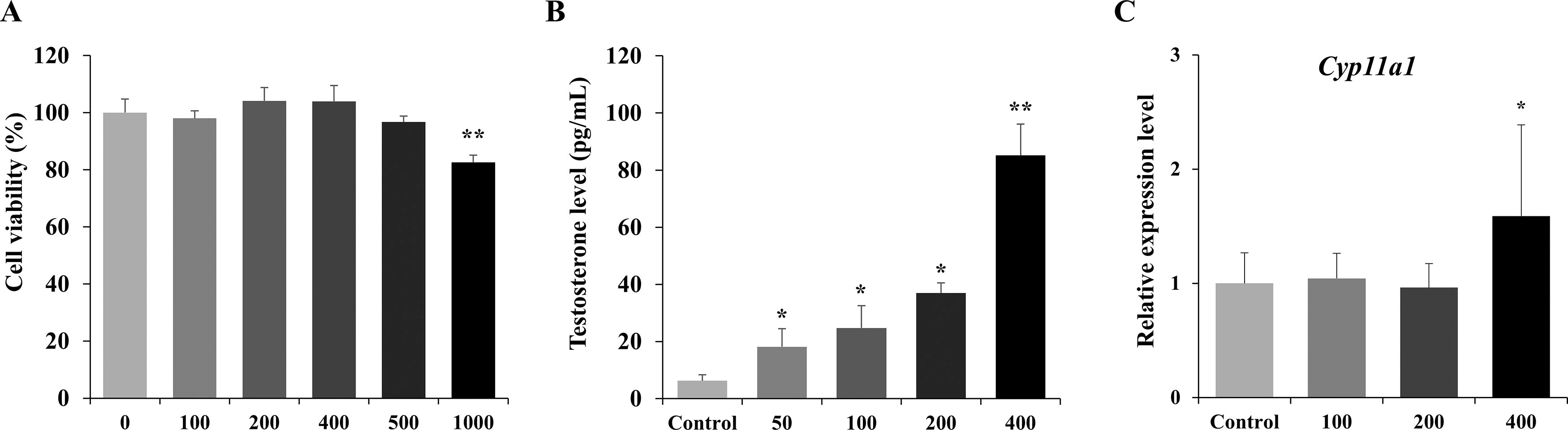
Testosterone (T) concentration and Cyp11a1 mRNA expression levels in TM3 Leydig cells treated with A. gramineus aqueous extracts.
Cytoplasmic cholesterol translocated across the inner mitochondrial membrane is converted to pregnenolone via catalysis with the first enzyme in the steroidogenesis pathway, CYP11A1. A. gramineus aqueous extract treatment increased the mRNA expression level of Cyp11a1 significantly at a concentration of 400 µg/mL in cells (P < 0.05) (Fig. 1C). Pregnenolones produced by CYP11A1 are subsequently converted to testosterone via enzymes such as 3β-HSD, CYP17A1, and 17β-HSD. Increased CYP11A1 expression initiates steroidogenesis, thereby promoting testosterone biosynthesis. 29 Therefore, the increase in Cyp11a1 gene expression following treatment with A. gramineus aqueous extract appears to have contributed to testosterone production. For aromatase (CYP19A1), which converts testosterone to estrogen, we found a significant reduction in mRNA expression at concentrations of 100, 200, and 400 μg/mL of A. granineus aqueous extract (Supplementary Fig. S2). 30
This appears to have contributed to the elevation of testosterone levels at concentrations below 400 µg/mL of A. gramineus aqueous extract by reducing the buffering action of steroidogenesis-mediated by estrogen. 31
HPLC analysis of 5-Hydroxymethyl-2-furaldehyde in A. gramineus extracts
To identify marker compounds separated from A. gramineus extracts, the retention times of each peak were examined by HPLC analysis (Supplementary Fig. S3). As a result, 5-HMF was detected between 10 and 11 min, displaying retention times and absorbance consistent with the A. gramineus extracts standard solution.
Effects of A. gramineus extract on serum testosterone, free testosterone, and E2 levels
To establish an andropause animal model, serum testosterone levels in 23-week-old SD rats (OLD group) were measured before treatment. Following the confirmation of a significant decrease in serum testosterone levels in 23-week-old SD rats (OLD group) compared with those in 7-week-old SD rats (YOUNG group), A. gramineus aqueous extract was administered (Supplementary Fig. S1).
After an 8-week administration period, the AG50 group (treated with 50 mg/kg A. gramineus aqueous extract) exhibited a twofold increase in the serum testosterone levels compared with that of the OLD group (P < 0.05) (Fig. 2A). These results were similar to those in the YOUNG group. In the AG50 group, there was a significant increase of approximately 43.65% in serum-free testosterone levels compared with that in the OLD group, which was equivalent to that in the YOUNG group (P < 0.05) (Fig. 2B). However, serum E2 levels in the AG50 group were significantly lower by approximately 25.54% than those in the OLD group (Fig. 2C).
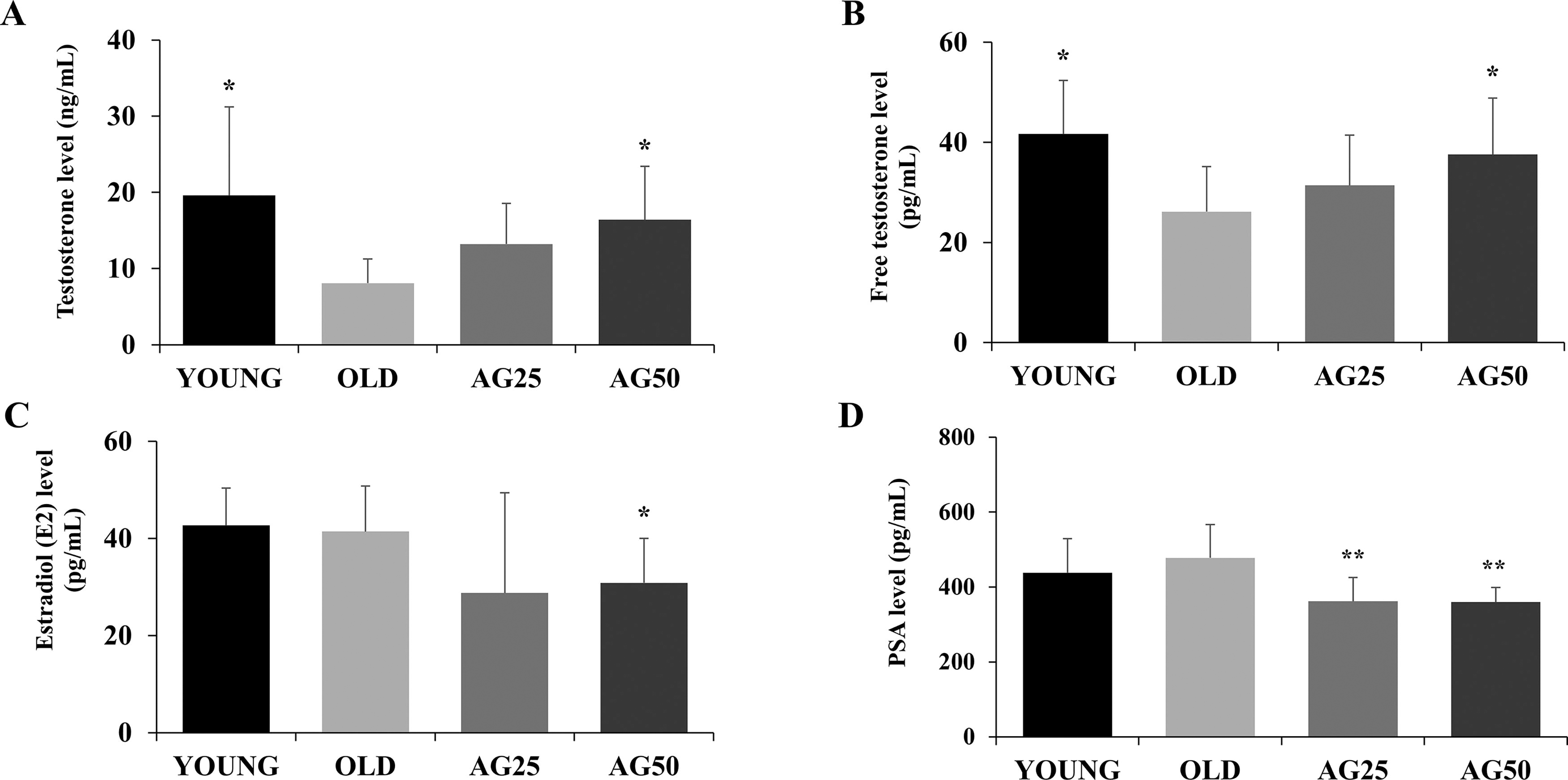
Effects of A. gramineus on steroid hormone and PSA levels in the serum of rats.
Approximately 98% of circulating testosterone is tightly bound to sex hormone-binding globulins and albumin. 32 Only free testosterone, which is weakly bound to albumin, is bioavailable to tissues. The A. gramineus aqueous extract not only restored the decreased testosterone levels associated with aging but also increased biologically active free testosterone. These results indicate that A. gramineus may improve testosterone production in older men experiencing andropathy. E2 is generated from testosterone through aromatase conversion. Estrogen inhibits androgen production during steroidogenesis and suppresses testosterone synthesis. 31 Therefore, the decrease in serum estradiol levels in the AG50 group may have contributed to the increase in testosterone levels.
Effects of A. gramineus on serum PSA levels in rats
Despite the potential advantages of testosterone replacement therapy in elderly men, meta-analyses have demonstrated associations with an increase in PSA levels and a higher risk of prostate disease, suggesting the need for regular monitoring. 33 A. gramineus aqueous extract increased serum testosterone levels but did not elevate PSA levels (Fig. 2D). Serum PSA levels decreased significantly by 24.27% in the AG25 group and 24.72% in the AG50 group (P < 0.01). The decrease in PSA levels indicated that the potential for prostate complications because of A. gramineus aqueous extract was very low, suggesting that it might be safe for human use.
A. gramineus improves sperm counts and muscle strength in rats
Elderly males exhibit histologically reduced spermatogenesis compared with that in young males, inevitably resulting in a decrease in both sperm count and sperm motility. 34 Sperm counts were measured to assess the improvement in the fertilization capacity because of A. gramineus in men experiencing andropause symptoms. A significant increase of approximately 46.19% in the sperm count in the AG50 group compared with that in the OLD group was detected (Fig. 3A; P < 0.05). The A. gramineus aqueous extract may assist in sperm production, improving reproductive function in older men experiencing andropause.

Effects of A. gramineus on sperm count and grip strength in rats.
Diminished testosterone levels during aging are associated with physiological changes, such as reduced muscle mass and strength. 35,36 To evaluate the enhancement in muscle strength following the administration of the A. gramineus aqueous extract for 8 weeks, a grip strength test was conducted. From the 2nd week onward, both the AG25 and AG50 groups exhibited improvements in grip strength compared with that in the OLD group (Fig. 3B; P < 0.05). The increase in grip strength in the group treated with A. gramineus aqueous extract persisted until the 8th week. The grip strength in the AG25 group improved by 14.01% over that in the OLD group, whereas in the AG50 group, it improved by 13.27% in the 8th week (P < 0.05). In elderly men, testosterone replacement therapy offers various benefits, including improvements in muscle strength. 37 Increased testosterone levels enhance muscle mass and performance through anabolic effects. 38 The improvement in muscle strength attributed to the A. gramineus aqueous extract could be attributed to elevated serum testosterone levels. According to some studies, Acorus tatarinowii Schott, a member of the Acoraceae family, reduces exercise-induced fatigue. 39 This suggests that A. gramineus aqueous extract exerts a positive effect on muscle strength in older men experiencing andropause.
Effects of A. gramineus on ALT, AST, and lipids in rats
To evaluate hepatic dysfunction caused by A. gramineus aqueous extract, liver weight and serum ALT and AST levels were measured. There were no elevations in liver weight, ALT, or AST in response to A. gramineus. A significant reduction in the AST levels was observed in the AG25 group (Table 2). Lee et al. evaluated the subchronic toxicity of A. gramineus rhizomes and reported no issues related to body weight, food consumption, blood parameters, reproductive function, or other aspects at a concentration of 2000 mg/kg/day. 18 No signs of liver toxicity were observed following the administration of the extract.
Effect of Acorus Gramineus on Liver Weight and the Levels of Serum ALT and AST in Rats
Values are presented as means ± SD. Asterisks indicate a significant difference from the OLD group, denoted by *P < 0.05, **P < 0.01, and ***P < 0.001.
ALT, alanine transaminase; AST, aspartate transaminase.
To observe the physiological alterations induced by A. gramineus aqueous extract, the serum lipid profile was examined. Treatment with A. gramineus aqueous extract did not result in changes in serum triglyceride levels, but total cholesterol was significantly decreased. The AG50 group exhibited a significantly lower total cholesterol (by ∼12.79%) than that in the OLD group (P < 0.05) (Table 3). Therefore, A. gramineus appears to offer the benefits of testosterone supplementation in aging men, while concurrently reducing total cholesterol in serum.
Effect of Acorus Gramineus on Lipid Profile in Serum of Rats
Values are presented as means ± SD. Asterisks indicate a significant difference from the OLD group, denoted by *P < 0.05, **P < 0.01, and ***P < 0.001.
HDL, high-density lipoprotein; LDL, low-density lipoprotein; SD, standard deviation.
A. gramineus enhances mRNA expression levels of steroidogenic genes
Quantitative reverse transcription-polymerase chain reaction was performed to investigate the expression of steroidogenic enzymes catalyzing testosterone synthesis in the Leydig cells of the testes. In the AG50 group, Star gene expression was higher than that in the OLD group (P < 0.05) (Fig. 4A). The mRNA expression of Cyp11a1 in the AG50 group was also higher than that in the OLD group (P < 0.01) (Fig. 4B). The mRNA expression levels of Hsd17b3 increased significantly in all groups administered A. gramineus extracts (Fig. 4C). As shown in Figure 4D, there was a significant increase in Cyp17a1 expression in the AG50 group compared with the OLD group (P < 0.05).
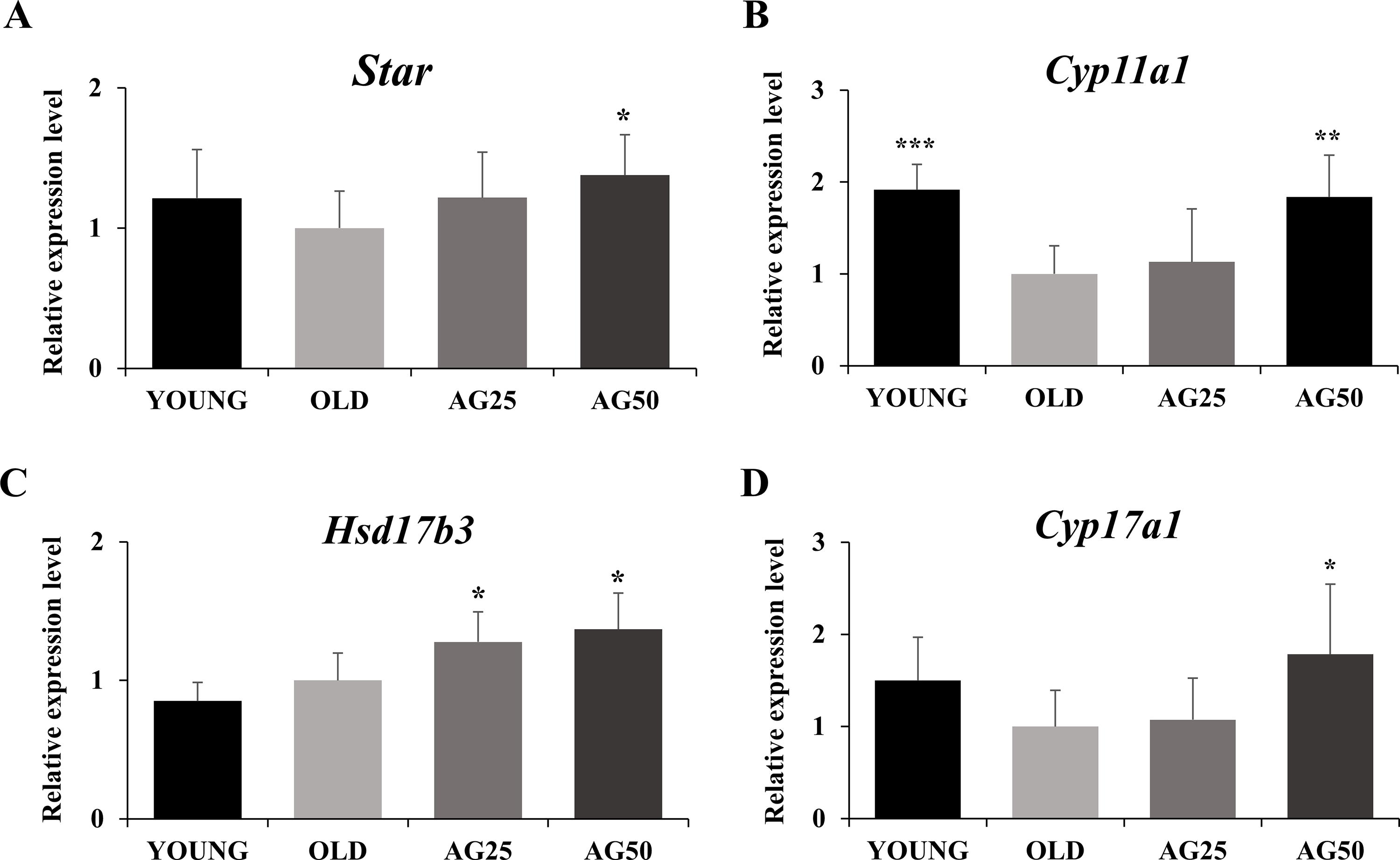
The mRNA expressions of testicular steroidogenic genes in rats. Values are presented as means ± SD. Asterisks indicate a significant difference from the OLD group, denoted by *P < 0.05, **P < 0.01, and ***P < 0.001. SD, standard deviation.
In addition, immunohistochemical staining was performed to count CYP11A1-positive Leydig cells in rat testes. Staining of CYP11A1-positive Leydig cells in the OLD group decreased significantly to 31.05% compared with that in the YOUNG group (P < 0.01) (Fig. 5). Moreover, the levels of CYP11A1 in testicular Leydig cells were significantly higher in the group treated with A. gramineus aqueous extract at a concentration of 50 mg/kg compared with those in the OLD group (P < 0.01).

IHC analysis of CYP11A1 in testis tissues of rats.
StAR facilitates the inner mitochondrial membrane transport of cholesterol and regulates transcription factors that are essential for the expression of steroidogenic genes. 40 Testosterone synthesis occurs through a series of actions involving various steroidogenic enzymes that are regulated by interactions with StAR. 41 The increased mRNA expression of Star because of the extracts seemed to play a pivotal role in testosterone synthesis during testicular steroidogenesis. CYP11A1 converts cholesterol into the initial steroid precursor pregnenolone, which is crucial for testosterone synthesis. Pregnenolone is converted to progesterone by 3β-HSD, and the resulting progesterone is hydroxylated by CYP17A1, serving as a catalyst for transformation into androstenedione, the precursor of testosterone. 29 Subsequently, 17β-HSD converts androstenedione into testosterone. Therefore, the increased expression of each enzyme appears to activate the production of precursor molecules for testosterone and facilitate testosterone synthesis. Consequently, the A. gramineus aqueous extract is thought to enhance steroidogenesis by increasing Cyp11a1 expression, initiating steroid production, and upregulating Cyp17a1 and Hsd17b3 expression, thereby promoting testosterone synthesis.
CONCLUSION
A. gramineus aqueous extract elevated testosterone levels and mRNA expression levels of the steroidogenic enzyme Cyp11a1 in TM3 mouse Leydig cells. In addition, it increased serum testosterone and free testosterone levels in 23-week-old SD rats and decreased estradiol, PSA, and total cholesterol levels. It augmented sperm count and grip strength and upregulated the expression of testicular steroidogenesis-related genes, such as Star, Cyp11a1, Hsd17b3, and Cyp17a1. These results indicate positive effects of A. gramineus aqueous extract for managing andropause.
Footnotes
ACKNOWLEDGMENTS
This work was supported by “Food Functionality Evaluation Program “under the Ministry of Agriculture, Food and Rural Affairs and partly by the Korea Food Research Institute.
AUTHORS’ CONTRIBUTIONS
Y-H.L. oversaw conceptualization, project management, supervision, resourcing, funding acquisition, formal analysis, validation, visualization, writing, review, and editing. J.Y.L., S.K., and H.K. were responsible for data organization, formal analysis, methodology, and writing. S-H.Y., S-Y.K., R.H.S., and C.L.P. were in charge of investigation, supervision, and visualization.
AUTHOR DISCLOSURE STATEMENT
No competing financial interests exist.
FUNDING INFORMATION
This work was supported by “Food Functionality Evaluation Program” under the Ministry of Agriculture, Food and Rural Affairs, and partly by the Korea Food Research Institute.
SUPPLEMENTARY MATERIAL
Supplementary Figure S1
Supplementary Figure S2
Supplementary Figure S3
References
Supplementary Material
Please find the following supplemental material available below.
For Open Access articles published under a Creative Commons License, all supplemental material carries the same license as the article it is associated with.
For non-Open Access articles published, all supplemental material carries a non-exclusive license, and permission requests for re-use of supplemental material or any part of supplemental material shall be sent directly to the copyright owner as specified in the copyright notice associated with the article.
