




Abstract
Metabolic dysfunction-associated steatotic liver disease (MASLD) is a complex and multifactorial disease. Dark tea exhibits great potential for various bioactivities for metabolic health. In this study, we aimed to evaluate therapeutic effects and the underlying mechanisms of dark tea wine (DTW) on MASLD with obesity. A rat model of MASLD was established by high-fat diet and administered with different doses of DTW as an intervention. The biomarkers of lipid metabolism and oxidative stress in rats were tested. The weight of organs and adipose tissues and the expressions of nuclear factor erythroid 2-like 2 (Nrf2) and heme oxygenase-1 (HO-1) were investigated based on the pathology and western blot analysis. We found that DTW enhanced antioxidant capacity via activating the Nrf2/HO-1 signaling pathway, further markedly triggering inhibition of weight gain, reduction of lipid dysfunction, and improvement of pathological characteristics to ameliorate MASLD induced by high-fat diet. These results suggest that DTW is a promising functional supplement for prevention and treatment of MASLD and obesity.
INTRODUCTION
Metabolic dysfunction-associated steatotic liver disease (MASLD) is considered as a high incidence chronic liver disease throughout the world. 1,2 It could progress to other liver diseases, including liver cirrhosis and hepatocellular carcinoma. 3 –5 Most people with MASLD have different degrees of overweight/visceral obesity, dyslipidemia, and elevated liver enzymes. 6,7 Weight loss and alleviation of hepatic steatosis are beneficial for preventing and treating MASLD. 8 Many investigations have aimed at the development of strategies for treating MASLD with obesity; nevertheless, there is still a lack of clear pathogenesis and satisfactory therapeutic outcomes. 9 Therefore, the research and development of novel and effective therapeutic approaches with high efficacy and low toxicity are necessary.
Pathologically, oxidative stress is closely related to liver injury and disease progression of MASLD. 10 –12 Inhibition of excessive oxidation involving the nuclear factor erythroid 2-like 2 (Nrf2)/heme oxygenase (HO)-1 signaling pathway, which is a key regulator of oxidative stress injury, leads to therapeutic effects on MASLD. 13 –15 Nrf2, as a critical transcription protein, controls various antioxidant factors and the glutathione REDOX system to improve the oxidative stress state of the cells. Moreover, activation of Nrf2 leads to enhanced level of HO-1, which exhibits inhibitory effects on oxidative stress. The Nrf2/HO-1 signaling pathway is key for the control of oxidative stress and lipid metabolism in MASLD.
Chinese dark tea is one of the post-fermentation teas produced by the special fermentation of microorganisms, possessing a wide range of interesting biological activities. 16 –20 In particular, it has been extensively studied for its antioxidation, which could be attributed to the effects of the complex mixture. 21 –28 Traditionally, dark tea products have long been a necessity of people living in the minority areas of southwest and northwest China. More interestingly, they prepared dark tea wine (DTW) with alcoholic content as a functional supplement for inhibiting the physical damage caused by high-fat and high-protein diets. DTWs such as the herbal wines of the traditional Chinese medicines is becoming increasingly popular. 29 –31 DTW involves rich metabolites, which are from Chinese liquors’ abundant nonalcoholic components and co-fermentation of dark tea and fungal communities, which has the potential to treat MASLD. 32 –34 However, the protective effects and the underlying mechanisms of DTW on MASLD with obesity are unknown. Therefore, a rat model of MASLD was induced by high-fat diet to evaluate therapeutic effect of DTW on MASLD. The biomarkers of lipid metabolism and oxidative stress in rat were tested. The weights of organs and adipose tissues and the expressions of Nrf2 and HO-1 were studied based on the pathology and western blot analysis. Furthermore, Nrf2/HO-1 signaling pathway-related mechanism of action was analyzed. This study indicated the potential of proper consumption of DTW used as effective functional supplements for treatment of MASLD and obesity.
MATERIALS AND METHODS
Materials and chemicals
The DTW (52%, v/v) was provided by Hezhou Tianzhou Tea Industry Co. LTD, which was produced by Jinyuantai Wine Industry Co., LTD. Maotai Town, Renhuai City, Guizhou Province; Simvastatin was from Hangzhou MSD Pharmaceutical Co., Ltd; Primary antibodies (anti-Nrf2 and anti-HO-1) were from Abcam (Cambridge, UK); Detection kits, including total cholesterol (TC), triglyceride (TG), high-density lipoprotein cholesterol (HDL-C), low-density lipoprotein cholesterol (LDL-C), the total antioxidant capacity (T-AOC), aspartate aminotransferase (AST/GOT), glutamic-pyruvic transaminase (ALT/GPT), super oxide dismutase (SOD), catalase (CAT), glutathione-peroxidase (GSH-Px), and malonaldehyde (MDA), were from the Nanjing Jiancheng Bioengineering Institute (Nanjing, Jiangsu, China).
Animals and experimental design
Sprague-Dawley male rats (180–200 g) were from the Jinan Pengyue Experimental Animal Breeding Co., Ltd. with license number SCXK (Lu) 2019–0003. Rats were housed in plastic cages and allowed food and water freely under control conditions (20°C–25°C, 40–70% humidity, and light/dark cycles). After adaptive feeding 1 week, rats were administered with high-fat diets, inducing hyperlipidemia with MASLD. The high lipid profile (TG and TC) changes of these rats before and after model establishment were recorded. After 4 weeks, these rats were used. The procedures were designed according to the method described via Qiu et al., 2020 with slight modifications. Briefly, rats were randomly divided into six groups. Normal control (NC) group with standard diet was administered with distilled water. Hyperlipidemic control (HC) group with high-fat diet was treated by administering distilled water. The hyperlipidemic rats were treated with DTW that was diluted with distilled water to half-concentration at 1.3 mL/kg/day (WL), 2.7 mL/kg/day (WM), and 5.4 (WH) mL/kg/day. Hyperlipidemic rats were also treated with suspension solutions of simvastatin tablets (20 mg/tablet) that were ground and dissolved in distilled water to obtain a 0.4 mg/mL as the positive control (ST). After 4 weeks, overnight-fasted rats were treated by intraperitoneal administration of urethane (20%). Blood samples from each group were harvested via abdominal vein. Then, the rats were euthanized, and liver, heart, fat of abdominal and epididymal tissues were collected, washed with precooled saline solution, and weighed after drying with filter paper.
All animal experimental procedures followed the National Institutes of Health guide for the care and use of laboratory animals and were performed in accordance with protocols approved by the Institutional Animal Care and Use Committee of Anhui University of Chinese Medicine (2023027).
Physiological and biochemical analyses
Body weights were recorded once every 4 days. Serum was obtained from the blood by centrifuging (LX-165T2R, Haier Biomedical, China) at 3,000 r/min for 15 min. Each rat’s liver (1 g) was excised with a scalpel and homogenized via a glass homogenizer with 0.9% pre-cooled normal saline in an ice water bath. After centrifugation at 12000 g for 15 min, supernatants were obtained. The biomarker levels of serum or liver tissue from all groups were measured using an Infinite F50 enzyme micro-plate reader (Tecan, Switzerland).
Histopathology and immunohistochemistry analysis
The livers were collected, fixed with 12% paraformaldehyde for 24 h, and then embedded in paraffin. About 5 μm slices were dewaxed and stained using hematoxylin & eosin. Liver histopathological changes were visualized on a microscope (Nikon DS-U3, Japan). The expression levels of Nrf2 were studied according the reported method using the biotin-streptavidin horseradish peroxidase (HRP) detection system. 35 The tissues were incubated with primary antibodies (1: 2 000 and 1: 4 000) maintaining at 4°C. Thereafter, the tissues were washed with phosphate buffered saline (PBS) for three times. Then secondary antibody (HRP-labeled) and diaminobenzidine (DAB) complex were added, with incubation at 37°C for 15 min. The tissues were counterstained with hematoxylin stain solution and visualized using a light microscope (Nikon DS-U3, Japan) to identify target proteins in the liver tissues.
Western blotting
The expression of protein in rat liver was detected via Western blotting. The frozen liver tissues were homogenized with radio immunoprecipitation assay (RIPA) lysis buffer with phenylmethylsulfonyl fluorid (PMSF) complex. Total protein samples were acquired from the fully cracked samples by centrifuging at 12000 g for 10 min to remove the precipitates. The samples were then separated via sodium dodecyl sulfate-polyacrylamide gel electrophoresis (SDS-PAGE), transferred to a polyvinylidene difluoride (PVDF) membrane (Immobilon ®-P Transfer Membrane, IPVH00010), enclosed in 5% skim milk for 2 h, and then incubated with the primary antibody at the ratio of 1: 1000 overnight. The membrane was incubated with appropriate secondary isotype-specific antibody for 1 h. Finally, the quantity of immunoreactive protein was determined by using a chemiluminescence detection system. Protein density values were analyzed by Image J software. 36
Statistical analysis
The data were presented as the means ± standard deviations. All values were analyzed using a one-way analysis of variance and LSD-t test by IBM SPSS Statistics 23.0. All statistical graphs were made in Graph-Pad Prism 8.0 software. P values < .05 were considered statistically significant.
RESULTS
Body weights
After 4 weeks of treatment, rats were photographed on a platform to evaluate their shapes. As shown in Figure 1A, the body length and body width of the HC rats were obviously higher than the others. Among all groups, WM and WH rats were remarkably smaller than NC rats in size, and NC, ST, and WL rats were similar. Furthermore, the body weight was recorded (Fig. 1B). After the experiment, weights of rats on the high-fat diet of rats in HC showed an increase of 52.7%, whereas the NC group had a 33.4% increase. Moreover, compared with the HC group, there were statistically significant differences in body weights of rats administrated with 4 mg/kg of ST or 1.35, 2.7, and 5.4 mL/kg/day of DTW. The results represented that appropriate DTW could inhibit the body weight gain of rats induced via a high-fat diet.

DTW inhibited the body weight gain of rats induced via high-fat diet.
Organ indexes and liver histopathology
Organs and adipose tissues could directly show changes in the pathological process of MASLD. The weights of organs and adipose tissues were measured and shown in Table 1. Significant analysis was performed in all the experiments groups. A significant difference in liver weight was observed between rats of the HC and the NC, but not in the weights of the hearts. Interestingly, prophylactic administration of DTW at 5.4 mL/kg/day or 2.7 mL/kg/day resulted in an effective reduction of liver weight, as well as abdominal fats comparing with the HC group. The weights of heart and adipose tissues were effectively reduced in treating with DTW at 5.4 mL/kg/day compared with that of HC group (P < .05). Moreover, liver morphology (Fig. 2A) exhibited that HC rats possessed obvious larger livers than the others. Furthermore, the color of the livers in HC rats was red ochre. After 28 days of prophylaxis with DTW, the appearance of liver in the WH (5.4 mL/kg/day) group was similar to the NC group (dark red), and the WL, WM, and TC groups were close to the NC group. In addition, the histopathology H&E staining for liver sections revealed (Fig. 2B) that the hepatocyte cord was arranged neatly, and the structure of hepatic lobule was complete in NC rats. Compared with NC rats, vacuolation and microvascular fat droplets were observed in the central hepatic vein and the edge of the hepatic lobule indicating that serious steatosis occurred in the liver tissue of the HC group. Fortunately, vacuolation and microvascular fat droplets also appeared in the liver tissues of the WL and ST groups, but the degree of liver steatosis was less than that in HC rats. As the dose of DTW increased, liver histopathology of the WH group tended to be closer to the NC group. These results showed that DTW could improve the liver pathological damage and inhibit the production of abdominal fat and epididymal fat of MASLD rats.

DTW prevented the gain of liver weight and the damage to the livers of MASLD rats.
Weight of Organs and Adipose Tissues in Sprague-Dawley Male Rats
Values are expressed as mean ± standard deviations (n = 8).
P < .05.
P < .01 compared with NC.
P < .05.
P < .01 compared with HC.
NC, normal control group; HC, hyperlipidemic control group; ST, hyperlipidemic rats treated with simvastatin; WL, hyperlipidemic rats treated with DTW at 1.3, mL/kg/day; WM, hyperlipidemic rats treated with DTW at 2.7 mL/kg/day; WH, hyperlipidemic rats treated with DTW at 5.4 mL/kg/day.
Lipid and liver function biomarkers
After 4 weeks of the high-fat diet, rats were shown to have markedly high serum cholesterol levels (Table 2). After treatment with DTW and ST, statistical analyses were conducted on the experiment and control groups (Fig. 3). Compared with NC rats, the serum levels of TC and TG in HC rats were higher (Fig. 3A and 3B); the former was higher 55.9%, and the latter was 80.8%, respectively. Compared with the HC group, the DTW and ST would significantly reduce the serum of TC and TG levels induced by high-fat diet. Particularly, it was clear that the WH group was able to achieve superior properties in comparison with that of ST. Moreover, the serum levels of LDL-C, HDL-C, AST/GOT, and ALT/GPT in HC rats were significantly higher than those of NC rats. After 4 weeks of preventive protection with DTW or simvastatin, the serum levels of LDL-C and HDL-C decreased and showed statistical significance, except for the group with DTW dose of 1.35 mL/kg/day (Fig. 3C and 3D). As shown in Figure 3E and F, DTW could inhibit serum levels of AST/GOT and ALT/GPT in dose-dependent manners. These results demonstrated that DTW prevented abnormal lipid metabolism and improved liver function in rats fed high-fat diets.
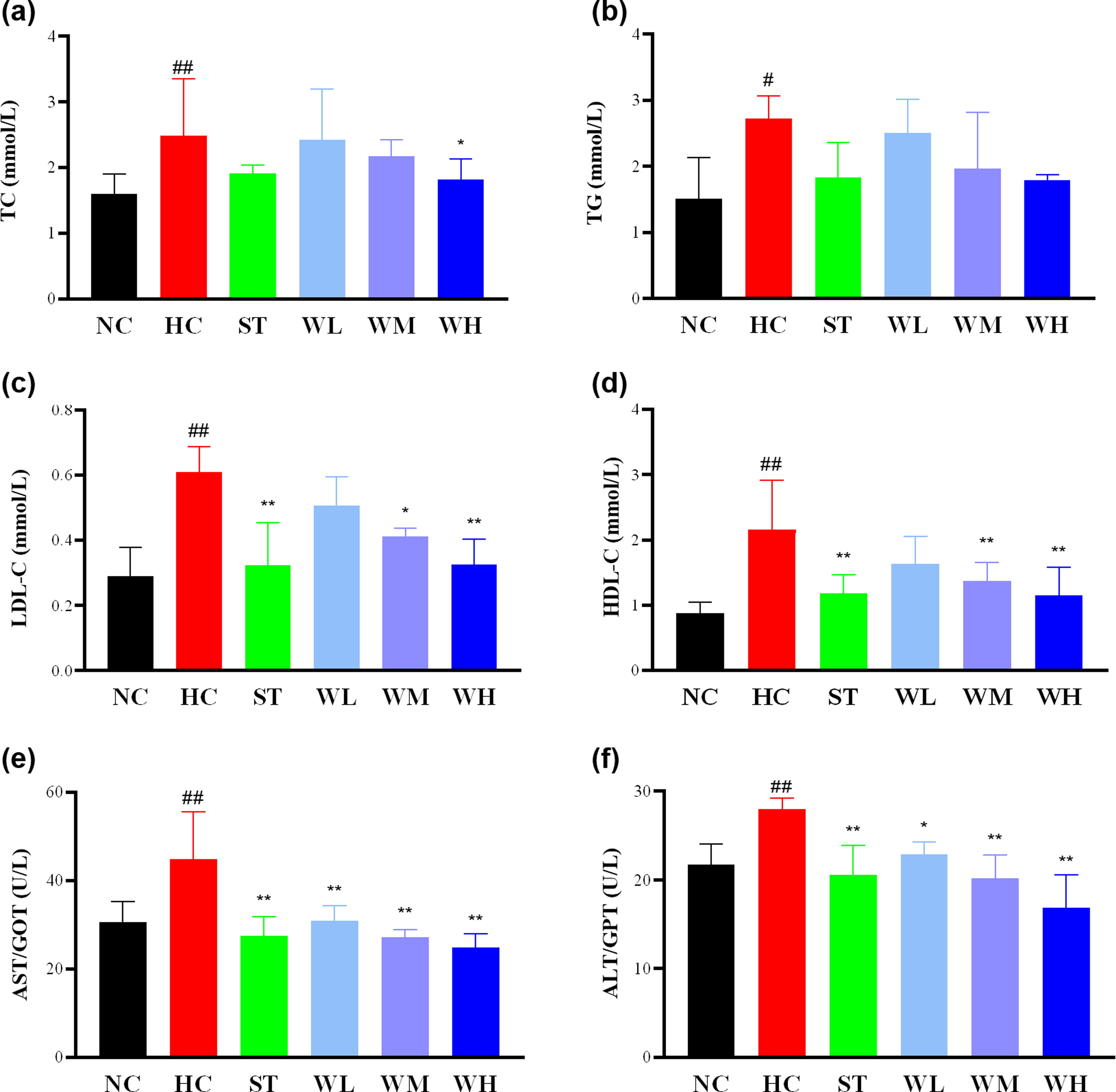
DTW ameliorated levels of TC
Blood Lipid Levels in Sprague-Dawley Male Rats Before and After Rat Modeling
Values are expressed as means ± standard deviations (n = 8).
P < .05 compared with before modeling of the HC group.
TC, total cholesterol; TG, triglycerides.
Oxidative stress indices
The results exhibited that the serum levels of antioxidants were decreased in HC rats (Fig. 4A, 4B, 4C and 4D), whereas the serum SOD level was without significance compared with NC rats (P > .05). DTW could improve concentrations/activities of antioxidants in serum of high-fat diet rats, and the serum levels of SOD and T-AOC in rats treating with DTW at 5.4 mL/kg/day were distinctly enhanced compared with HC rats (P < .01). The liver levels of antioxidants were decreased in the HC group (Fig. 4F, 4G, 4H, and 4I), and the activities of SOD and CAT were significantly lower than those in NC rats (P < .05, .01). The DTW increased the levels of SOD and T-AOC in liver tissue of rats with high-fat diet (Fig. 4F and 4G). MDA, a lipid damage product, was increased in the HC group (Fig. 4E and 4J) compared with the NC group, only in serum were there significant differences (P < .01). After the experiment, the contents of MDA in serum were decreased in ST and WH rats compared with HC rats. Further analysis of the results revealed that the effects of DTW on oxidative factors in serum and liver tissues of high-fat fed rats were basically consistent with those of ST.
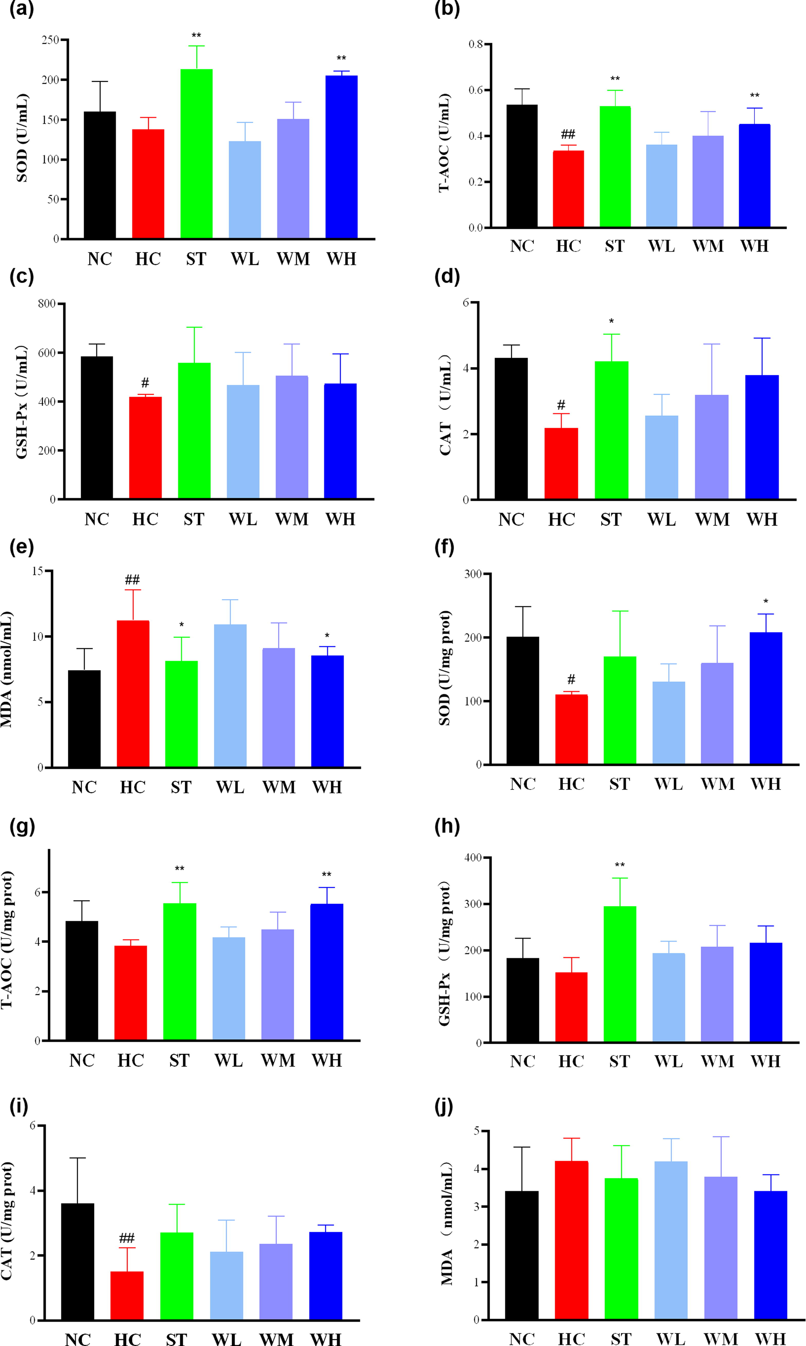
DTW exerted ameliorative effects on oxidative stress indices SOD, T-AOC, GSH-Px, CAT, and MDA
Immunohistochemical analysis
The Nrf2 pathway is critical to regulate oxidative stress. To further analyze the antioxidant mechanism of DTW, immunohistochemistry analysis was selected to determine the Nrf2 expression cells in the liver. In this work, the result exhibited a significant decrease of Nrf2-positive expression cells in liver of HC rats as compared with NC rats (P < .01). Compared with HC rats, Nrf2 showed an increased expression in cells of WH rats (P < .01). The number of Nrf2-positive expression cells in the DTW at 1.35 mL/kg/day group, 2.7 mL/kg/day group, and 5.4 mL/kg/day group were directly proportional to the dose of the DTW (Fig. 5). Therefore, DTW may be resistant to oxidative damage by activating Nrf2 antioxidant pathway.
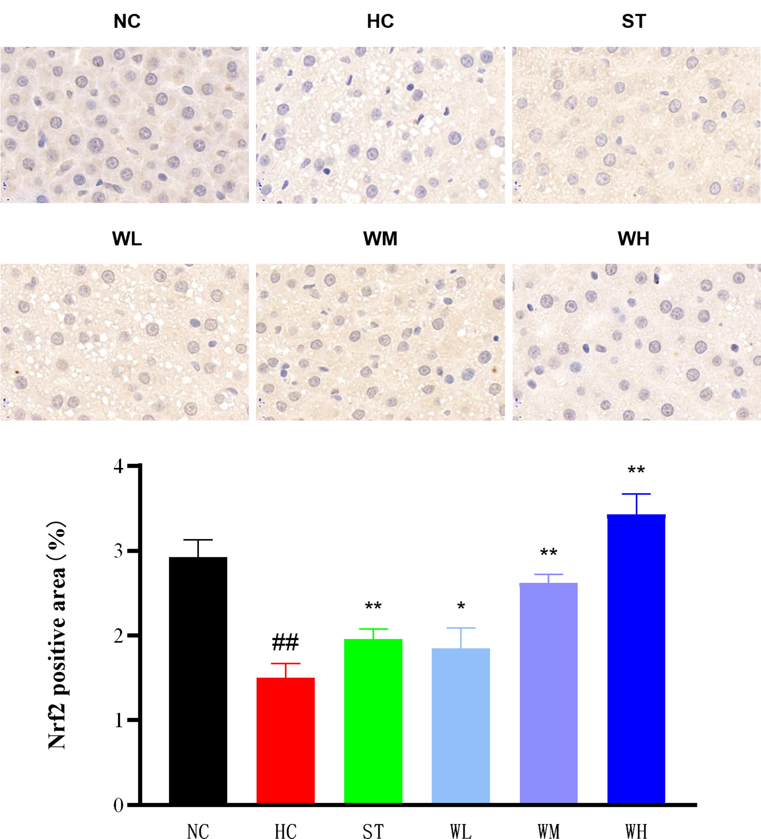
DTW was resistant to oxidative damage by activating Nrf2 antioxidant pathway (80 × magnification). DTW, dark tea wine; HC, hyperlipidemic control group; NC, normal control group; Nrf2, nuclear factor erythroid 2-like 2; ST, hyperlipidemic rats treated with simvastatin; WH, hyperlipidemic rats treated with DTW at 5.4 mL/kg/day; WL, hyperlipidemic rats treated with DTW at 1.3, mL/kg/day; WM, hyperlipidemic rats treated with DTW at 2.7 mL/kg/day. Values were means ± standard deviations (n = 4). ## P < .01 compared with control; *P < .05, **P < .01 compared with HC.
Western blot analysis
The proteins of Nrf2/HO-1 antioxidant signaling pathway in liver were detected via western blotting assay. Compared with NC rats, the expression of Nrf2 and HO-1 in HC rats was greatly reduced (P < .01). Compared with HC rats, expressions of Nrf2 and HO-1 in DTW at 2.7 mL/kg/day group and 5.4 mL/kg/day group were obviously increased (P < .01), but there was no significant difference in the 1.35 mL/kg/day DTW group (Fig. 6 and Supplementary Fig. S1-S3). DTW has a protective effect on MASLD induced via high-fat diet, and the mechanism may be related to the enhanced expression of Nrf2/HO-1 signaling pathway.

The expression levels of Nrf2 and HO-1 in rat liver tissues were significantly increased. The protein expression level of Nrf2 in nucleus and cytoplasm, as well as the protein expression levels of HO-1, was analyzed by western blot. The relative expression levels of Nrf2 and HO-1 were quantified. HC, hyperlipidemic control group; HO-1, heme oxygenase-1; NC, normal control group; Nrf2, nuclear factor erythroid 2-like 2; ST, hyperlipidemic rats treated with simvastatin; WH, hyperlipidemic rats treated with DTW at 5.4 mL/kg/day; WL, hyperlipidemic rats treated with DTW at 1.3, mL/kg/day; WM, hyperlipidemic rats treated with DTW at 2.7 mL/kg/day. Values were means ± standard deviations (n = 3). # P < .05, ## P < .01 compared with control; *P < .05, **P < .01 compared with HC.
DISCUSSION
Our present studies support the hypothesis that a moderate intake of DTW would ameliorate MASLD in rats by regulating the generation of obesity, dyslipidemia, and oxidative stress. We found that DTW markedly triggered inhibition of weight gain. Furthermore, reduction of lipid dysfunction and improvement of pathological characteristics were observed in the results. In addition, DTW obviously enhanced antioxidant capacity via activating the Nrf2/HO-1 signaling pathway.
MASLD is a complex, multifactorial disease, which is closely related to obesity, dyslipidemia and related metabolic syndrome. At present, the harm of MASLD to human health is increasing, and the cure of this kind of disease has become a new challenge in contemporary medical field. Epidemiological studies showed that MASLD was closely correlated to changes in lifestyle, obesity is one of the risk factors. Patients with obesity are prone to developing MASLD, and patients with MASLD are also prone to obesity, and they go hand in hand. In our study, the effect of DTW, prepared by a special process of a post-fermentation tea and Chinese liquor, on obesity was examined in high-fat diet fed Sprague-Dawley male rats. The established method of high-fat diet was used in the present study. 37,38 After the modeling, the body weight in the HC rats was significantly higher than that in the NC rats, the volume of the liver swelled and increased, and the color of the liver turned yellow, which was consistent with the characteristics of fatty lesions in the liver.
In addition to obesity, patients with MASLD often have other manifestations of metabolic syndrome, such as hyperlipidemia. Serum lipid indexes are closely related to MASLD. The levels of TC, TG, LDL-C, and HDL-C show lipid status in body, and AST/GOT and ALT/GPT reflect the liver status in body. 39 –41 MASLD is characterized by excessive lipid accumulation and abnormal lipid metabolism in the serum. Prophylactic administration of DTW significantly reduced the levels of serum lipids and liver functional biomarkers in rats with MASLD. In addition to blood test indicators, liver tissue section staining can directly show changes in the pathological process of MASLD. In this study, HE staining showed that lipid droplets were permeated into hepatocytes in the HC group, indicating successful replication of the rat model of MASLD. Less vacuolation and micro vesicular fat droplets were also seen in the treatment groups. The results in this study indicate that the right amount of DTW has preventive and therapeutic effects on MASLD.
Oxidative stress plays an important part in the process of steatosis. Oxidative stress is a major factor leading to liver injury and disease progression in MASLD. At high concentrations, ROS can cause the accumulation of cell-damaged macromolecules, thereby causing liver damage, which further leads to the disorder of liver lipid metabolism. 11 The biomarkers of oxidative stress were decreased in most people with MASLD. In this study, the levels of SOD, T-AOC, GSH-Px, CAT, and MDA in serum and liver tissues were measured. MDA is a product of lipid peroxidation, which was selected to reflect the degree of lipid peroxidation in liver tissue. The antioxidant enzyme levels in vivo were tested to reveal the antioxidant effect, including SOD, GSH-PX, T-AOC, and CAT. DWT enhanced the antioxidant activity with significant differences with HC group data. The Nrf2/HO-1 signaling pathway is an important pathway to regulate the oxidation–antioxidant axis balance and plays a key role in the pathological process of MASLD. The results of this experiment showed that DTW showed potential antioxidant ability for the expression of oxidative stress markers in serum and liver of rats and enhanced the expression of Nrf2/HO-1 signaling pathway in liver.
CONCLUSION
In summary, we studied our hypothesis and have revealed that DTW enhances the antioxidant activity via activating Nrf2/HO-1 pathway, further markedly triggering the inhibition of weight gain, reduction of lipid dysfunction, and improvement of pathological characteristics to effectively protect the liver against MASLD induced via high-fat diet. Overall, DTW was shown to be a promising intervention as an effective functional supplement from dark tea for the treatment of MASLD and obesity that deserves further development.
Footnotes
ACKNOWLEDGMENT
The authors are grateful to the participants for their contribution and support throughout the experiment.
AUTHORS’ CONTRIBUTIONS
X.Z.: Investigation, data analysis, visualization, validation, and writing. S.L.: Investigation and visualization. T.W.: Investigation and statistical analysis. J.B.: Pathological diagnosis, investigation, and validation. F.X.: Project administration, validation, supervision, funding acquisition, and review. W.Z.: Funding acquisition, original writing draft, and supervision.
AUTHOR DISCLOSURE STATEMENT
No competing financial interests exist.
FUNDING INFORMATION
This work was supported by National Natural Science Foundation of China (82304324), Open Fund of State Key Laboratory of Tea Plant Biology and Utilization (SKLTOF20190109, 20160109), Guangxi Innovation-driven Development Project of Hezhou (CX1907007), Research Foundation of Education Bureau of Anhui Province (2022AH050472), and High-level Talents Support project of Anhui University of Chinese Medicine (2022rczd011).
SUPPLEMENTARY MATERIAL
Supplementary Figure S1
Supplementary Figure S2
Supplementary Figure S3
References
Supplementary Material
Please find the following supplemental material available below.
For Open Access articles published under a Creative Commons License, all supplemental material carries the same license as the article it is associated with.
For non-Open Access articles published, all supplemental material carries a non-exclusive license, and permission requests for re-use of supplemental material or any part of supplemental material shall be sent directly to the copyright owner as specified in the copyright notice associated with the article.
