




Abstract
Skin aging, accelerated by oxidative stress from environmental factors, results in the breakdown of collagen and elastin, causing visible signs like wrinkles and inflammation. Natural antioxidants such as nicotinamide mononucleotide (NMN), decursin, and
INTRODUCTION
The aging process of the skin, known as skin senescence, is influenced by various factors. 1 Oxidative stress accelerates the breakdown of collagen and elastin fibers, leading to wrinkles, and loss of skin elasticity. 2 In contrast, studies have explored the potential of various natural antioxidants to mitigate damages caused by oxidative stress and to delay aging process. 3,4 Among many natural products, nicotinamide mononucleotide (NMN) participates in the production of nicotinamide adenine dinucleotide (NAD+) 5 that is essential for activating sirtuin and poly (ADP-ribose) polymerase, 6 all of which maintain genomic stability within cells and prevent aging. 7 Deficiency of NAD+ can be compensated for by supplementing NAD+ precursors like NMN that present higher bioavailability than directly consuming the NAD+, which is considered impermeable to the plasma membrane, and NAD+ administration cannot efficiently increase NAD+ levels. 8
Decursin is a natural coumarin present in Angelica gigas Nakai. 9 Previous research has shown that decursin has various biological activities, including anti-inflammatory, 10 anticancer, 11 and inhibition of melanogenesis. 12 In the context of skin-aging, decursin effectively mitigated ultraviolet B-induced photoaging, 13 lowered allergic reaction (that mimicked atopic dermatitis [AD]) by reducing histamine secretion, 14 and exhibited anti-inflammatory through suppressing nitric oxide production in raw 264.7 cells. 15 However, it is not fully understood how this naturally occurring coumarin derivative will elicit protection against skin-aging phenotypes when combined with NMN.
Last,
MATERIALS AND METHODS
Cell culture
The human skin keratinocyte (HaCaT) cell line was obtained from the American Type Culture Collection (PCS-200-011; Manassas, VA). The cells were cultured following a method largely adapted from previously published studies. 18
3-(4,5-dimethylthiazol-2-yl)−2,5-diphenyltetrazolium bromide assay
The 3-(4,5-dimethylthiazol-2-yl)-2,5-diphenyltetrazolium bromide (MTT) assay was performed to evaluate sample cytotoxicity as described in previous studies with slight modifications. 19,20
Dichlorodihydrofluorescein diacetate assay: Intracellular oxidative stress assessment
To assess intracellular oxidative stress, we used a dichlorodihydrofluorescein diacetate (DCF-DA) cellular ROS assay kit (Abcam; Cambridge, UK) per manufacturer’s instruction.
Wound healing assay
The wound healing potential was evaluated using a scratch assay as described elsewhere 21 with slight modifications.
Immunohistochemical staining and confocal fluorescence microscopy
HaCaT cells were treated with 10 mM 2,2’-azobis (2-amidinopropane) dihydrochloride (AAPH) (Sigma-Aldrich; St. Louis, MO) for 24 h, alone or with sample mixtures. Fixed cells were stained with tri-methyl-histone H3 primary (Cell Signaling Technology; Danvers, MA) and Alexa Fluor 488-conjugated secondary (Invitrogen; Carlsbad, CA) antibodies for senescence-associated heterochromatin foci (SAHF) detection. Nuclei were counterstained with 4′,6-diamidino-2-phenylindole (DAPI) (Sigma-Aldrich), and images were captured using a confocal microscope (Zeiss LSM 900; Oberkochen, Germany), showing SAHF as green fluorescence and nuclei as blue.
Quantitative real-time PCR
Total RNA was extracted using RNeasy Plus Kits (Qiagen; Hilden, Germany) with minor manufacturer protocol adjustments. The mRNA expression levels were determined using a SYBR Green quantitative real-time polymerase chain reaction assay with PowerUp SYBR Green Master Mix (Thermo Scientific), following the manufacturer’s instructions. It was performed using the QuantStudio 3 Real-Time PCR System (Applied Biosystems; Foster City, CA, USA), and data were analyzed with QuantStudio Design and Analysis Software v1.5.1. Primer details are available in Supplementary Table S1.
Dinitrochlorobenzene-induced dermatitis model in vivo
Male BALB/c mice (6–8 weeks, 20–25 g) were used to model AD through dinitrochlorobenzene (DNCB) application. A sample size of n = 10 per group was accepted based on a statistical power calculation (80% power, significance level [α] = 0.05). All animal experiments were conducted in accordance with the Institutional Guidelines for the Use and Care of Laboratory Animals and were approved by the
Assessment of ear and epidermal thickness
Ear thickness was measured to quantify the degree of inflammation in the AD model. A hand-held caliper was used to measure ear thickness, ensuring consistent application pressure. The epidermal thickness measurement method was adapted with slight modifications based on previously published methods. 22
Measurement of scratch behavior
After a 24-h acclimation in observation cages, scratching bouts, defined as continuous hind limb movements toward irritated areas, were recorded for 10 min and analyzed independently by two researchers.
Measurement of transepidermal water loss and erythema index
The transepidermal water loss (TEWL) and erythema index were measured weekly, with dorsal skin shaved prior to each assessment. TEWL was measured using the SKIN-O-MAT (Cosmomed; Ruhr, Germany), while the erythema index was assessed using the Mexameter® MX18 (Courage + Khazaka electronic GmbH; Cologne, Germany). Each site was measured three times, and averages were recorded for accuracy. Dermatitis scores were calculated according to Scoring Atopic Dermatitis Index (SCORAD) criteria, evaluating erythema, scarring, edema, and excoriation on a scale of 0 (none) to 3 (severe), following manufacturer protocols.
Assessment of plasma IgE level
Approximately 500 µL of whole blood per animal was drawn into ethylenediaminetetraacetic acid-containing tubes to prevent coagulation. Samples were placed on ice and centrifuged at 3,000 rpm for 10 min at 4°C to separate plasma. The plasma was carefully collected and used to quantify immunoglobulin E (IgE) levels using a commercial kit (BD Bioscience; Franklin Lakes, NJ).
Mast cell count
The dorsal and ear skin tissues were processed into sections for mast cell analysis. Slides were stained with 0.5% Toluidine Blue O (Sigma-Aldrich), highlighting mast cells as metachromatically purple-blue due to their granules. After slides were washed with distilled water, dehydrated through graded ethanol, cleared with xylene, and mounted using synthetic medium. Mast cells were identified under a light microscope (LEICA DM500; Leica Microsystems, Germany) at 200× magnification, using Leica Application Suite (LAS) version 4.12 for imaging. They appeared as purple-stained cells with granular cytoplasm. Five random fields per section were analyzed, and mast cells were manually counted to ensure regional variability was accounted for.
In silico prediction of target genes: Network pharmacology
The potential targets of the active compounds in the mixture (i.e., NMN, decursin, and
Gene ontology and Kyoto Encyclopedia of Genes and Genomes pathway enrichment analysis
Using ClueGO 2.5.8, a Cytoscape plug-in, gene ontology (GO) analysis was conducted to investigate functions across three categories: biological process, molecular function (MF), and cellular component (CC). A bubble chart illustrating the results of the GO and Kyoto Encyclopedia of Genes and Genomes enrichment analyses was generated using an online R package (http://www.ehbio.com/ImageGP/index.php/Home). In the chart, the X-axis represents the percentage of associated genes, while the Y-axis corresponds to the description of the terms. The significance threshold for identifying relevant terms and pathways was set at P value <0.001.
Statistical analysis
Statistical evaluations were conducted using GraphPad Prism version 8.0.2 software (Boston, MA). We assessed whether the data followed a normal distribution using the Shapiro–Wilk test. When the data did not fit a normal distribution, the Mann–Whitney test was applied. Conversely, for data that conformed to a normal distribution, we used a two-tailed unpaired Student’s t-test with Welch’s correction. We presented the data as mean ± standard deviation or standard error of means where appropriate. The results were considered statistically significant if P values were <0.05.
RESULTS AND DISCUSSION
Determination of NMN, decursin, and l -cysteine cytotoxicity
The cytotoxicity of NMN, decursin, and
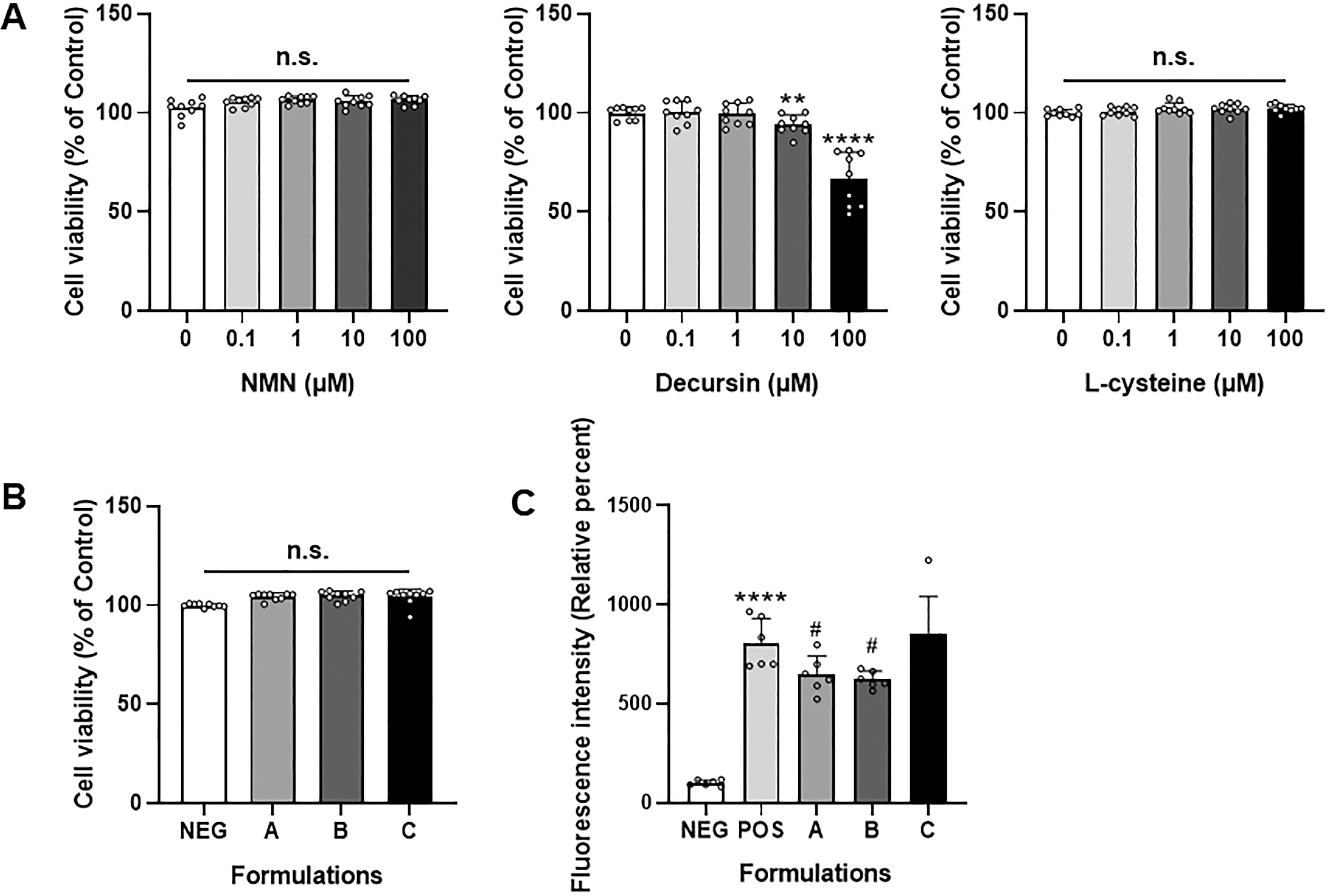
Effect of nicotinamide mononucleotide, decursin, and
The Composition of Nicotinamide Mononucleotide (NMN)-Based Natural Product Mixture
Protective effects of the mixtures against AAPH-induced intracellular ROS
The antioxidant effects of NMN, decursin, and
ROS play a critical role in skin aging by damaging CCs and accelerating senescence, leading to visible aging signs (e.g., wrinkles and reduced elasticity).
23
While previous studies highlighted the antioxidative roles of NMN, decursin, and
Wound healing efficacy of the mixtures in the HaCaT cells
Following the creation of a uniform scratch to simulate a wound, the HaCaT cells were treated with each mixture and incubated for 11 h to observe wound closure (Fig. 2). Quantitative analysis of the wound healing area revealed differences in wound closure among the groups. In this, the mixtures B and C showed enhancement in wound closure, with a statistically significant increase in wound healing area compared to the NEG (P < 0.05 for both; Fig. 2). No difference was noted with mixture A.

Effects of mixtures of nicotinamide mononucleotide, decursin, and
Inhibitory effects of mixtures on the formation of senescence-associated heterochromatin foci induced by AAPH in HaCaT
The effects of mixtures of NMN, decursin, and
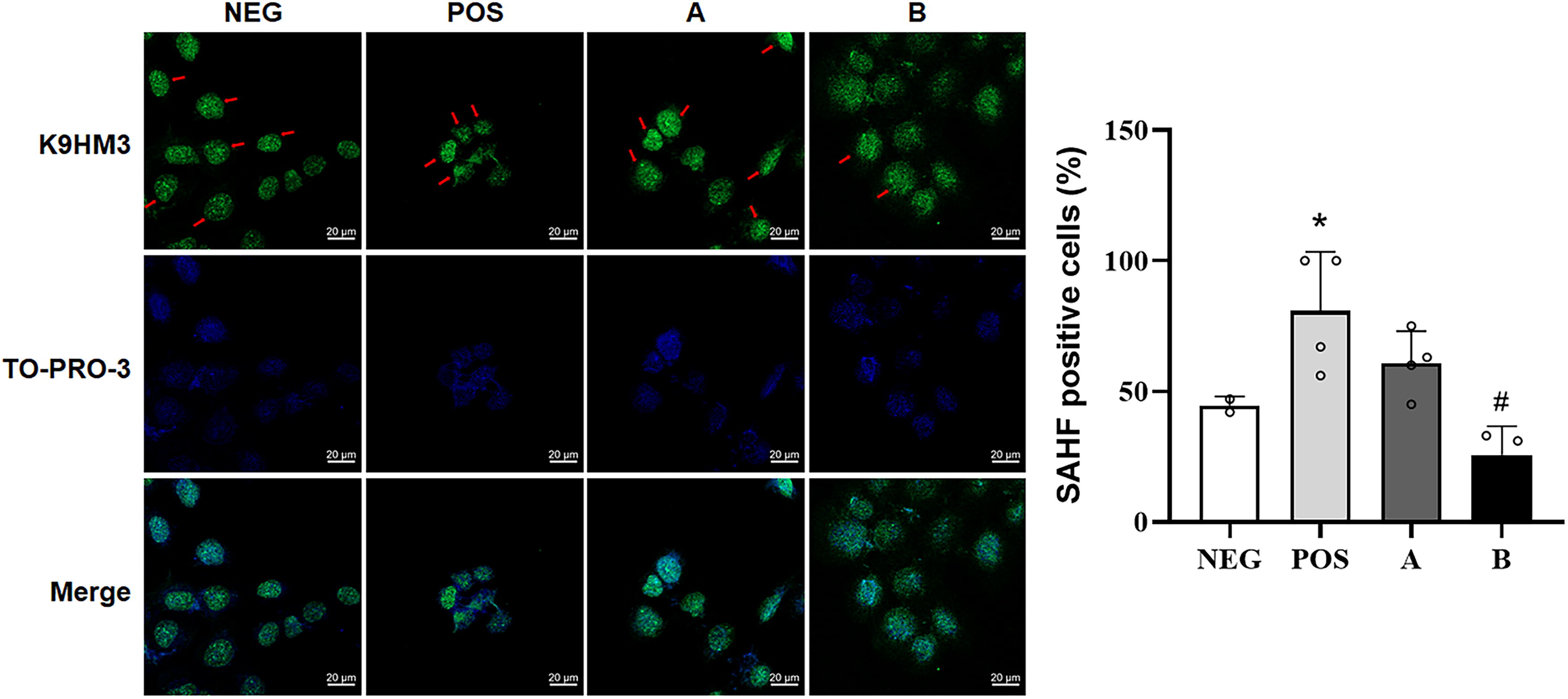
Inhibitory effects of mixtures of nicotinamide mononucleotide, decursin, and
Effects of mixture B on the expressions of inflammatory genes in HaCaT
The protective effects of mixture B against AAPH-induced SAHF formation and ROS were confirmed by analyzing the expression of key inflammation and oxidative stress-related genes: interleukin-6 (IL-6), tumor necrosis factor-α (TNF-α), and nuclear factor erythroid 2-related factor 2 (NRF2). We found a significant increase in IL-6 and TNF-α in the POS group compared to the NEG, indicating successful ROS induction and activation of inflammatory pathways (P < 0.01 for both genes; Fig. 4). Treatment with mixture B, however, significantly reduced the genes’ expression compared to the POS group (P < 0.0001 for IL-6 and P < 0.01 for TNF-α), suggesting an anti-inflammatory effect under oxidative stress conditions. Furthermore, NRF2 gene expression, a key regulator of antioxidant responses, was notably elevated in the POS group relative to the NEG while no difference was noted in the mixture B treatment group (Fig. 4). Excessive ROS levels in the skin contribute to cellular senescence and the degradation of structural proteins, leading to visible aging signs. Elevated IL-6 and TNF-α levels are typical markers of inflammation in skin cells subjected to oxidative stress, promoting collagen degradation and impairing skin barrier function, both of which accelerate the aging process. 26 In this study, on the other hand, mixture B significantly downregulated IL-6 and TNF-α mRNA levels in AAPH-treated HaCaT cells, suggesting its potential to reduce oxidative stress-induced inflammation, which is crucial for slowing skin aging.
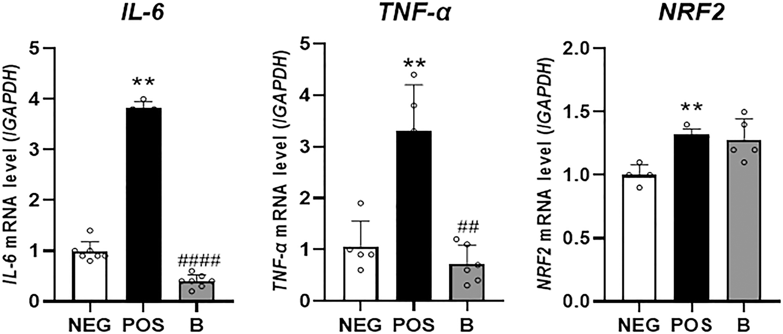
Quantification of target gene mRNA expressions following reactive oxygen species induction in human skin keratinocyte (HaCaT) cell lines. HaCaT cells were seeded in 60 × 15 mm plates and treated with the mixture B (see the main text for the composition) for 24 h, followed by stimulation with 2,2’-azobis (2-methylpropionamidine) dihydrochloride (AAPH, 3 mM) or not for 1 h. The cells were harvested, and total RNA was extracted for quantitative PCR analysis of interleukin-6 (IL-6), tumor necrosis factor-α (TNF-α), and nuclear factor erythroid 2-related factor 2 (NRF2) mRNA levels, with glyceraldehyde-3-phosphate dehydrogenase (GAPDH), as an internal reference. Results are expressed as means ± standard deviations (n = 4–6). Normality was assessed using the Shapiro–Wilk test. Differences between groups were evaluated using Welch’s t-test or Mann–Whitney U test for independent samples. ** denotes P < 0.001 compared to the negative control (NEG) while ## and #### denote P < 0.01 and P < 0.0001, respectively, compared to the positive control (POS).
Effects of oral administration of mixture B on DNCB-induced AD: Primary phenotypes
The DNCB-induced AD model visualized in Figure 5(A) showed that the POS group displayed typical AD symptoms, including erythema, dryness, and crust formation 27 as well as significantly increased spleen weight, ear thickness, and scratching behavior compared to the NEG group Figure 5(B), (C), (D). Spleen analysis, indicative of immune involvement in AD, 28 showed significantly increased spleen weights in the POS group compared to the NEG, while the reference control (i.e., Pred group) exhibited significant reductions and the mixture groups (low and high doses) showed non-significant reductions Figure 5(C). Ear thickness was significantly greater in the POS group compared to the NEG, with no significant improvement noted in the mixture or Pred groups Figure 5(C). However, mixture and Pred groups reduced scratching frequency compared group to the POS Figure 5(D).
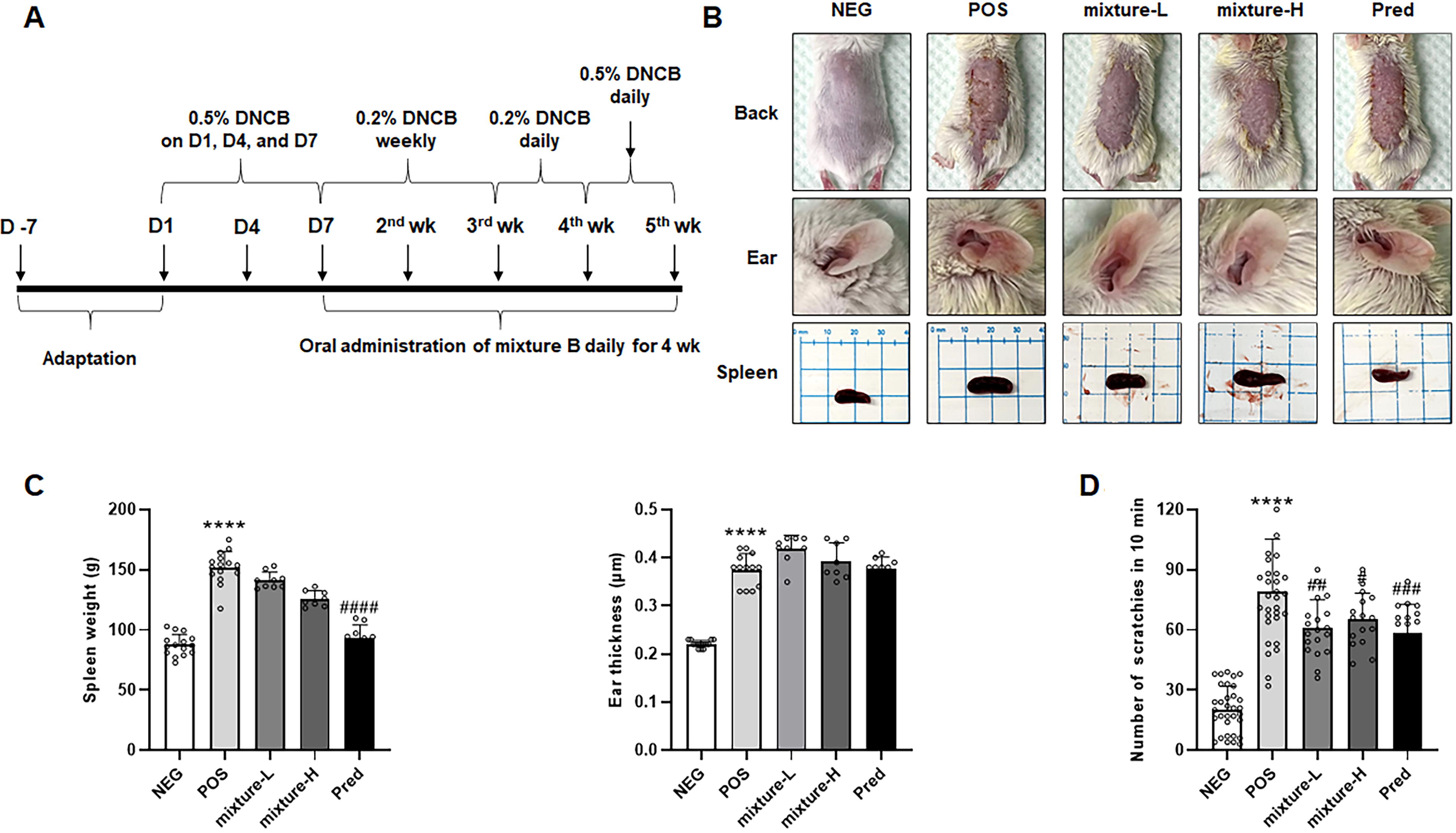
Effects of mixtures of nicotinamide mononucleotide, decursin, and
Histological analysis showed substantial increases in epidermal and dermal thickness and elevated mast cell counts in the POS versus NEG groups Figure 6(A) and (B). The mixture groups significantly reduced epidermal thickness, while the mixture-L decreased dermal thickness and mast cell infiltration; the reference group (i.e., Pred group) effectively improved all markers. Further, the mixture groups significantly improved skin barrier function, as evidenced by reduced TEWL, though benefits in SCORAD scores and erythema were less pronounced Figure 6(C). Repeated DNCB application caused systemic immune activation and chronic inflammation, as evidenced by spleen enlargement and various skin manifestations.
28
Mixture groups, particularly mixture-L, demonstrated decreases in scratching behavior, epidermal thickness, TEWL, dermal thickness, and mast cell counts, with similar patterns observed in ear tissues Figure 6(D, E). These results highlight the potential efficacy of NMN, decursin, and

Effects of nicotinamide mononucleotide, decursin, and
Oral administration of mixture B decreased proinflammatory gene expressions in DNCB-induced AD
To explain the mechanisms, a few key anti-inflammatory genes were assessed. In the POS group, as expected, the levels of TNF-α, IL-1β, IL-4, IL-6, and IL-13 were all markedly elevated compared to the NEG group (Fig. 7). Although each sample group showed a trend toward reduced cytokines gene expressions, the results varied depending on the specific cytokine. In particular, TNF-α in the Pred and mixture-H groups demonstrated significant decreases compared to the POS group (P < 0.05 for Pred and P < 0.001 for mixture-H), while mixture-L showed no effect. For IL-1β, mixture-L achieved a statistical decline (P < 0.05), whereas Pred and mixture-H did not show statistical relevance. In the case of IL-4 and IL-13, both mixture groups exhibited substantial reductions while the Pred group failed to recapitulate the effects. Last, the IL-6 decreased in the mixture-L and Pred groups but, unexpectedly, it increased in the mixture-H. These findings indicate that proinflammatory cytokine expression, heightened during DNCB-induced acute and chronic inflammatory responses, 29 is effectively modulated. Last, we did not find statistical significance in serum IgE protein levels between the mixture and the POS (data not shown) although our DNCB protocol dramatically increased the serum IgE level in the POS compared to the NEG (P < 0.0001).
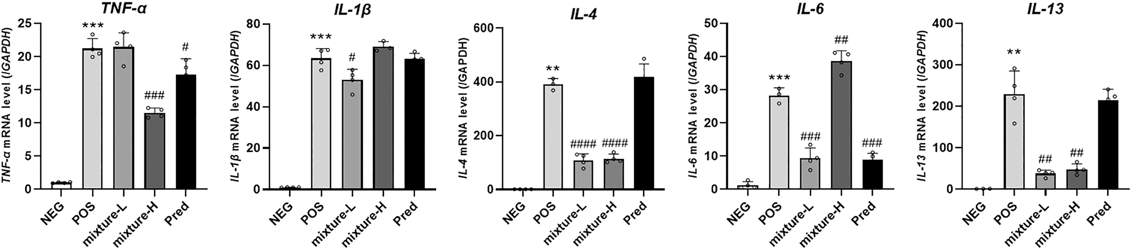
Quantification of proinflammatory cytokine mRNA expression in 2,4-dinitrochlorobenzene (DNCB)-induced atopic dermatitis BALB/c mouse skin tissues. Effect of nicotinamide mononucleotide, decursin, and
An application of network pharmacology
Network pharmacology (NP) is an emerging approach that integrates bioinformatics tools to explore the complex interactions between biological networks,
30
enabling comprehensive prediction and mapping of potential targets for active compounds. By using computational databases (e.g., PharmMapper), we identified 61 target genes of NMN, decursin, and
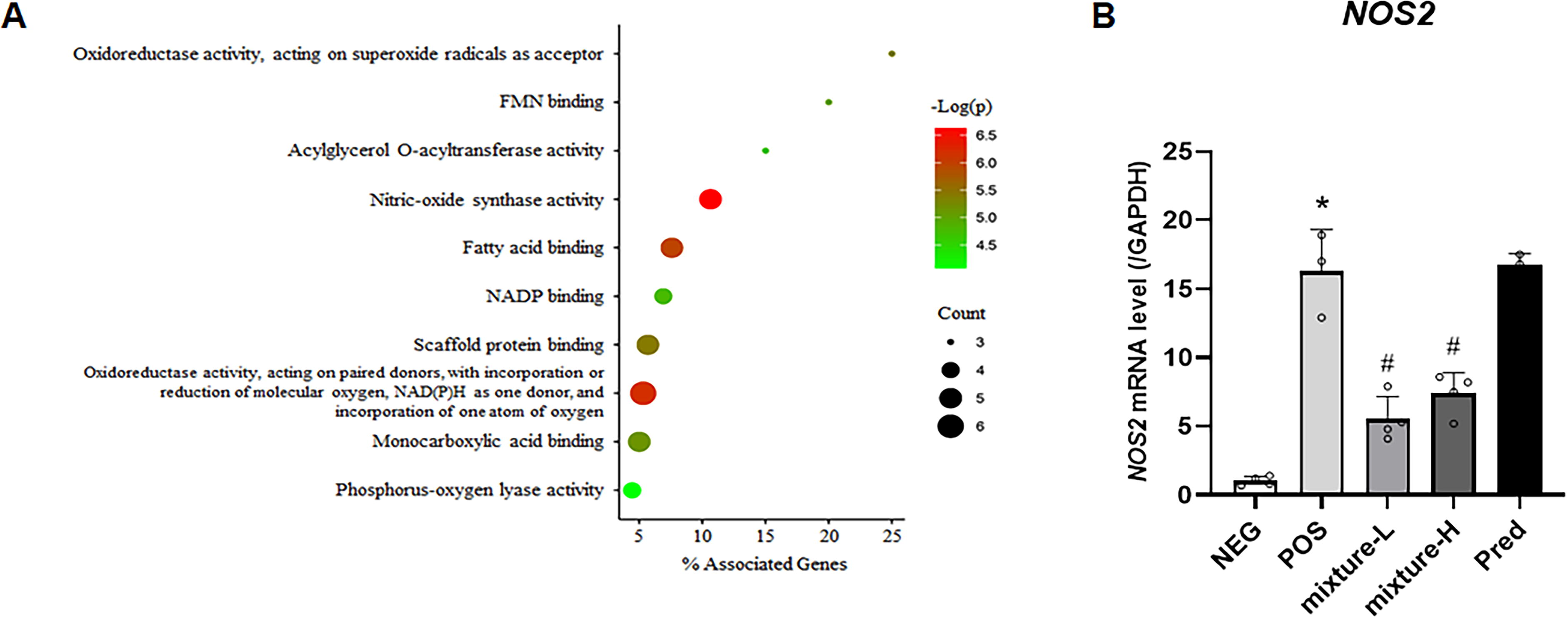
Gene ontology (GO) enrichment analysis of molecular functions associated with the predicted gene set.
Together, the study highlights the efficacy of NMN, decursin, and
Footnotes
AUTHORS’ CONTRIBUTIONS
H.R.S.: Writing—original draft (lead); formal analysis (lead); and validation (support). L.Y.C.: Writing—original draft (support); formal analysis (lead); and validation (lead). S.H.B.: Writing—original draft (support); formal analysis (support); and visualization (lead). T.G.K.: Funding acquisition (lead) and project administration (support). H.T.C.: Formal analysis (support) and funding acquisition (support). T.J.C.: Resources (support) and review and editing (support). N.S.O.: Resources (support) and review and editing (support). Y.Y.S.: Methodology (support) and review and editing (support). M.J.T.R.: Methodology (support) and review and editing (support). J.H.P.: Investigation (lead) and review and editing (support). J.-O.S.: Conceptualization (equal). E.-C.S.: Conceptualization (equal). S.-G.K.: Methodology (lead). J.P.: Investigation (support) and resources (support). Y.H.: Investigation (support) and resources (support). K.-C.C.: Review and editing (support). Y.J.K.: Supervision (support) and project administration (support). W.H.: Methodology (lead) and review and editing (support). K.L.: Writing—review and editing (equal); formal analysis (lead); and supervision (lead). J.K.K.: Writing—review and editing (equal) and project administration (lead).
AUTHOR DISCLOSURE STATEMENT
No competing financial interests exist.
FUNDING INFORMATION
This work was supported by a Korea University Grant (K2317281) and Regional Innovation Strategy (RIS) through the National Research Foundation of Korea (NRF) funded by the Ministry of Education (MOE; 2024C0701006).
SUPPLEMENTARY MATERIAL
Supplementary Figure S1
Supplementary Table S1
Supplementary Table S2
Supplementary Table S3
References
Supplementary Material
Please find the following supplemental material available below.
For Open Access articles published under a Creative Commons License, all supplemental material carries the same license as the article it is associated with.
For non-Open Access articles published, all supplemental material carries a non-exclusive license, and permission requests for re-use of supplemental material or any part of supplemental material shall be sent directly to the copyright owner as specified in the copyright notice associated with the article.
