




Abstract
This study investigated the effects of Humulus japonicus extract supplementation on bone growth and microstructural properties in male and female rats, with a particular focus on the JAK2/STAT5/IGF-1 signaling pathway. Three-week-old Sprague–Dawley rats were assigned to different groups receiving a normal diet (ND), growth hormone (GH), or varying doses of H. japonicus extract: low (L; 73 mg/kg body weight [bw]/day), medium (M; 146 mg/kg bw/day), and high (H; 292 mg/kg bw/day) for four weeks. The results demonstrated that the H group exhibited significant increases in femur and tibia lengths, trabecular and cortical bone mineral density, and growth plate thickness compared with the ND group. Furthermore, the H group demonstrated elevated serum and hepatic IGF-1 and IGFBP-3 levels, as well as enhanced phosphorylation of JAK2 and STAT5. The findings suggest that H. japonicus supplementation promotes longitudinal bone growth by stimulating growth plate activity and modulating the JAK2/STAT5-IGF-1 signaling pathway. This research indicates that H. japonicus extract could potentially be used as a natural therapeutic agent to support skeletal development and maintain bone health.
INTRODUCTION
Bone growth and development are fundamental physiological processes regulated by complex interactions among genetic, nutritional, and hormonal factors. During the growth phase, longitudinal bone growth primarily occurs through endochondral ossification in the growth plate, a cartilage structure located at the ends of long bones. 1,2 Proper regulation of chondrocyte function within the growth plate is necessary for healthy bone elongation.
Among key growth regulators, the insulin-like growth factor (IGF) system—particularly IGF-1 and its binding protein IGF binding protein-3 (IGFBP-3)—plays a crucial role in promoting chondrocyte activity and differentiation. 3 IGF-1 serves as a major anabolic hormone that stimulates chondrocyte proliferation, differentiation, and matrix production, thereby accelerating bone growth. Dysregulation of the IGF-1/IGFBP-3 pathway has been associated with growth impairments and developmental disorders, emphasizing its central role in bone development. 4,5
Natural compounds have long been widely studied for their potential benefits in supporting bone health and development. Among these, Humulus japonicus has attracted attention for its diverse bioactive compounds, including flavonoids and phenolic acids, which exhibit antioxidant, anti-inflammatory, and hormone-modulating properties. Previous research demonstrated that a mixture of H. japonicus and garlic/watermelon powder (at an 8:2 ratio) positively influenced longitudinal bone growth in Sprague–Dawley (SD) rats, suggesting its potential role in bone development. 6 However, this mixture showed significant growth-promoting effects, although the specific role and underlying mechanisms of H. japonicus alone in regulating bone growth remain largely unexplored. Initial findings indicate that bioactive compounds in H. japonicus may positively influence the IGF-1/IGFBP-3 axis, a critical regulatory pathway in endochondral ossification and bone elongation. 6 Further investigation into the direct effects of H. japonicus on bone growth and its molecular mechanisms is warranted to elucidate its full potential as a natural growth-promoting agent. Therefore, we investigated the effects of H. japonicus supplementation on bone growth in normal male and female SD rats. By examining key markers of bone development, including IGF-1 and IGFBP-3, we aimed to clarify the potential of H. japonicus as a natural agent for promoting skeletal growth. This research provides a novel understanding of the significance of natural compounds in skeletal health and highlights the therapeutic potential of H. japonicus in promoting longitudinal bone growth.
MATERIALS AND METHODS
Preparation of H. japonicus extracts
The manufacturing process of H. japonicus extracts involves several sequential steps to ensure product quality and efficacy. H. japonicus was extracted in hot water at 80∼90°C for 8 hours. The extracted solution is then filtered using an 80-mesh filter to remove impurities. Subsequently, the filtrate is subjected to vacuum concentration at temperatures below 70°C to enhance the extract’s potency. The concentrated extract is then freeze dried (FD) to preserve its bioactive properties. The standardized compound content in H. japonicus extracts was determined to be 0.1505 ± 0.006 mg/g of vitexin. This standardization ensures consistency in the extract’s composition and potency.
Animals
Three-week-old male and female SD rats were maintained under controlled environmental conditions, including a temperature of 22 ± 2°C, humidity levels of 50–60%, and a 12-h light/dark cycle. Initially, they were fed a standardized AIN-93G diet for a seven-day adaptation period. Following this, they were randomly assigned to one of five groups (eight rats per group): (1) normal diet (ND) control group receiving only the standardized AIN-93G diet, (2) recombinant growth hormone (GH) group supplemented with an intraperitoneal injection of 0.37 mg/kg body weight (bw)/day recombinant GH, (3) low-dose (L) H. japonicus group receiving 73 mg/kg bw/day of H. japonicus extract, (4) medium-dose (M) H. japonicus group supplemented with 146 mg/kg bw/day, and (5) high-dose (H) H. japonicus group receiving 292 mg/kg bw/day. The feeding regimen was maintained for four weeks, after which the rats were euthanized through cervical dislocation. The study was conducted in accordance with ethical guidelines and approved by the Animal Care and Use Review Committee of Kyung Hee University (KHGASP-24-571).
Levels of IGF-1, IGFBP-3, osteocalcin, and alkaline phosphatase in the serum
The levels of IGF-1 and IGFBP-3 were determined using enzyme-linked immunosorbent assay (ELISA) kits (R&D Systems, Minneapolis, MN, USA), while osteocalcin and bone alkaline phosphatase (ALP) were assessed with ELISA kits from Mybiosource Inc. (San Diego, CA, USA). All assays were conducted according to the manufacturer’s instructions, utilizing an ELISA reader form Bio-Rad (Hercules, CA, USA).
Real-time PCR
Total mRNA was extracted from rat liver tissues using the QIAGEN’s RNeasy Mini Kit (Germantown, MD, USA). The extracted RNA was then reverse transcribed into cDNA using the iScript™ cDNA Synthesis Kit (Bio-Rad). The study utilized primers targeting GAPDH, IGF-1, and IGFBP-3. The PCR amplification methodology and data analysis were conducted as detailed in previous study. 6
Western blotting
Liver tissues were homogenized using Tissue Lysis Buffer (Cell Biologics, Chicago, IL, USA). After protein loading, the transfer, blocking, and antibodies’ dilutions were conducted as detailed in a previous study. 6 Chemiluminescent detection was performed using EzWestLumi plus (ATTO, Tokyo, Japan), and protein bands were visualized using Ez-Capture II equipment (ATTO). Quantitative protein band analysis was conducted using both the CS Analyzer 3.0 software (ATTO) and Image J software (NIH, Bethesda, MD, USA).
Micro-computed tomography bone analysis
Bone microarchitecture and mineral content of the femur and tibia were assessed using high-resolution micro-computed tomography (micro-CT). The Scanco Medical AG vivaCT 80 scanner was employed with optimized parameters, including an 18 μm voxel size, 70 kVp energy, and 114 μA intensity, with a 31.9 mm field of view and 200 ms integration time. This advanced imaging technique allowed for detailed analysis of various bone characteristics, such as femur and tibia lengths, bone mineral density (BMD), trabecular architecture (including number, thickness, and separation), and bone volume fraction. The formalin-fixed bone specimens were scanned to generate three-dimensional images, enabling comprehensive evaluation of structural and density-related properties of the skeletal tissue, which is crucial for understanding bone health and the effects of interventions on bone integrity.
Histological and immunohistochemical analysis
Rat tibial specimens from the study underwent a series of preparatory steps for histological analysis. After extraction, the samples were fixed in a paraformaldehyde solution and subsequently decalcified using an EDTA-based method. The processed tissues were then embedded in paraffin and cut into thin sections. These slices were treated with various staining and labeling techniques, including H&E for general morphology and BrdU incorporation for determining cellular proliferation. In addition, immunohistochemical methods were employed to detect the presence of specific bone-related proteins such as bone morphogenetic protein 2 (BMP-2), IGF-1, and IGFBP-3. To quantify cellular proliferation in the growth plate, two independent observers calculated the density of BrdU-positive cells, expressed as the number of labeled nuclei per unit area. This comprehensive approach allowed for a detailed examination of bone structure, growth dynamics, and the expression of key regulatory factors in the tibial samples.
Statistical analysis
The study results are presented as mean values with standard deviations (means ± standard deviations). Statistical evaluation was performed using one-way analysis of variance with SPSS PASW Statistics 24.0 software (SPSS Inc., Chicago, IL, USA). To determine significant differences between groups, Duncan’s multiple range test was applied. The threshold for statistical significance was established at P < .05.
RESULTS
Effect of H. japonicus extracts on body length and weight gain
This study investigated the effects of H. japonicus extract supplementation on body growth parameters in both male and female SD rats. Growth assessments included nose-to-anus and nose-to-tail lengths and weight gain, as shown in Table 1. In both male and female rats, the GH group demonstrated the most significant increases in these growth parameters compared with the ND group. Among the H. japonicus extract-treated groups, the H group exhibited significantly greater nose-to-anus and nose-to-tail length gains than compared with the ND group. However, no significant difference in weight gain was observed between the ND and H groups for either male or female SD rats (P < .05).
Effect of Supplementation with H. japonicus Extracts on Increases in Nose-to-Anus and Nose-to-Tail Lengths, and Weight Gain in SD Rats
GH, recombinant growth hormone group (intraperitoneal injection of 0.37 mg/kg body weight/day recombinant GH); H, high-dose H. japonicus group (diet supplemented with 292 mg/kg body weight/day H. japonicus extracts); L, low-dose H. japonicus group (diet supplemented with 73 mg/kg body weight/day H. japonicus extracts); M, medium-dose H. japonicus group (diet supplemented with 146 mg/kg body weight/day H. japonicus extracts); ND, normal diet control group; SD, Sprague-Dawley. Values are presented as means ± standard deviations, and different superscript letters (a > b > c) indicate significance at P < .05.
Effect of H. japonicus extracts on levels of IGF-1, IGFBP-3, osteocalcin, and bone ALP expression and activation of the JAK2/STAT5 pathway in the liver
The study evaluated hepatic mRNA and protein expression levels of IGF-1 and IGFBP-3, along with their serum concentrations in male and female rats. Both the GH group and H. japonicus-treated groups showed significantly increased hepatic mRNA expression, serum concentrations, and hepatic protein levels of IGF-1 and IGFBP-3 compared with the ND group (Fig. 1A–D, H–K). In addition, serum levels of osteocalcin and bone ALP, values of bone formation markers, were significantly higher in the GH group and H. japonicus-treated groups compared with the ND group in both male and female SD rats (Fig. 1E, F, L, M). Furthermore, the hepatic protein levels of phosphorylation of JAK2 and STAT5 were significantly higher in the GH group and H. japonicus-treated groups compared with the ND group in both male and female SD rats (Fig. 1). These results suggest that H. japonicus supplementation enhances the IGF-1/IGFBP-3 axis in both liver and serum through the activation of the JAK2/STAT5 signaling pathway, thereby potentially promoting longitudinal bone growth.
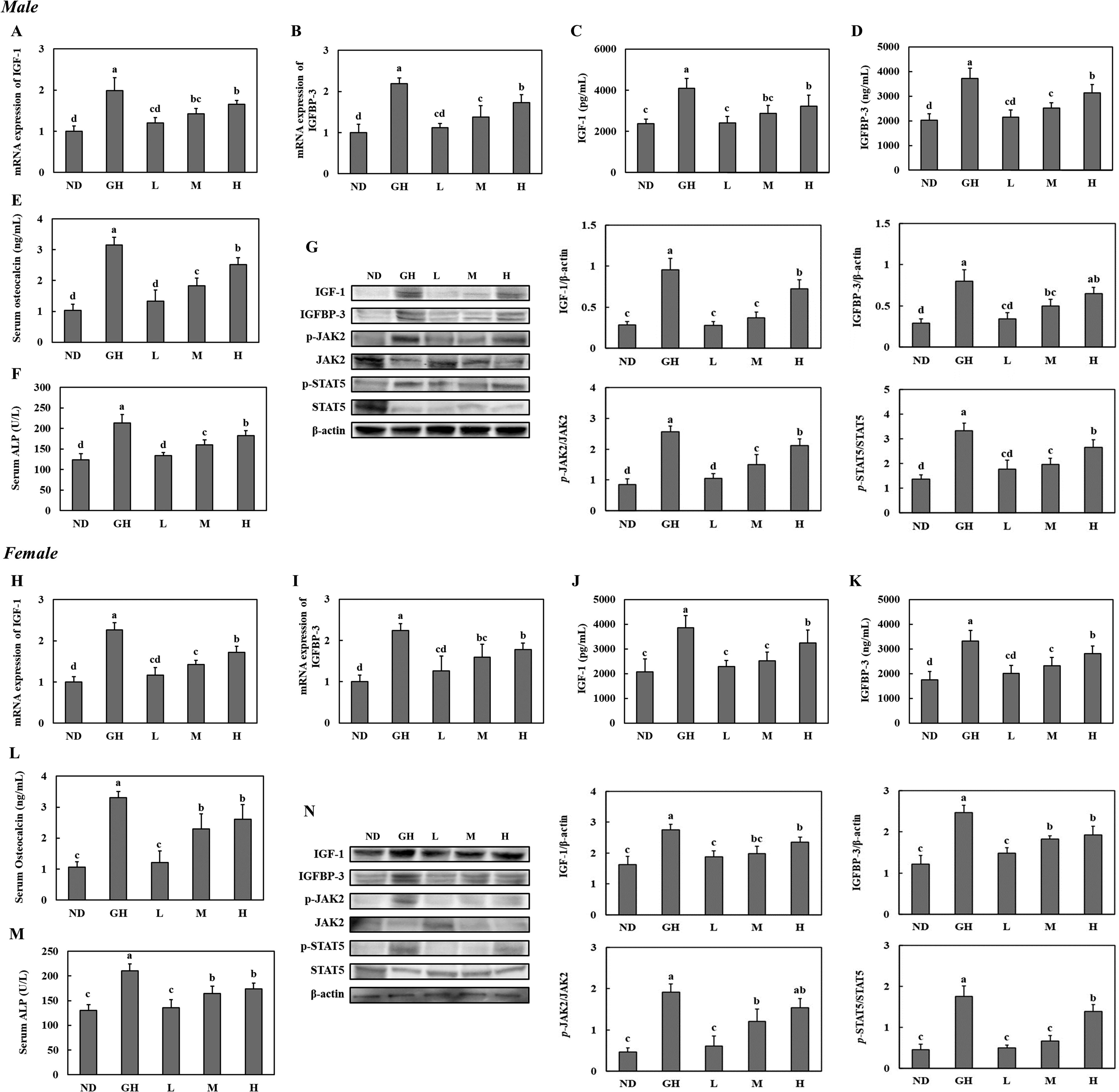
Effect of supplementation with H. japonicus extracts on levels of IGF-1, IGBBP-3, osteocalcin, and ALP in the liver and serum of SD rats. mRNA expression of IGF-1 in the liver
Effect of H. japonicus extracts on BMP-2, IGF-1, and IGFBP-3 expression in the bone
The expression levels of BMP-2, IGF-1, and IGFBP-3 in bone tissue were evaluated using immunohistochemistry in both male and female SD rats (Fig. 2). The GH and H groups exhibited the strongest expressions of BMP-2, IGF-1, and IGFBP-3 in bone tissue compared with the ND group, in both male and female SD rats (Fig. 2). These findings suggest that H. japonicus supplementation enhances the expression of IGF-1, IGFBP-3, and BMP-2 in bone tissue, suggesting their potential roles in promoting bone growth and development.
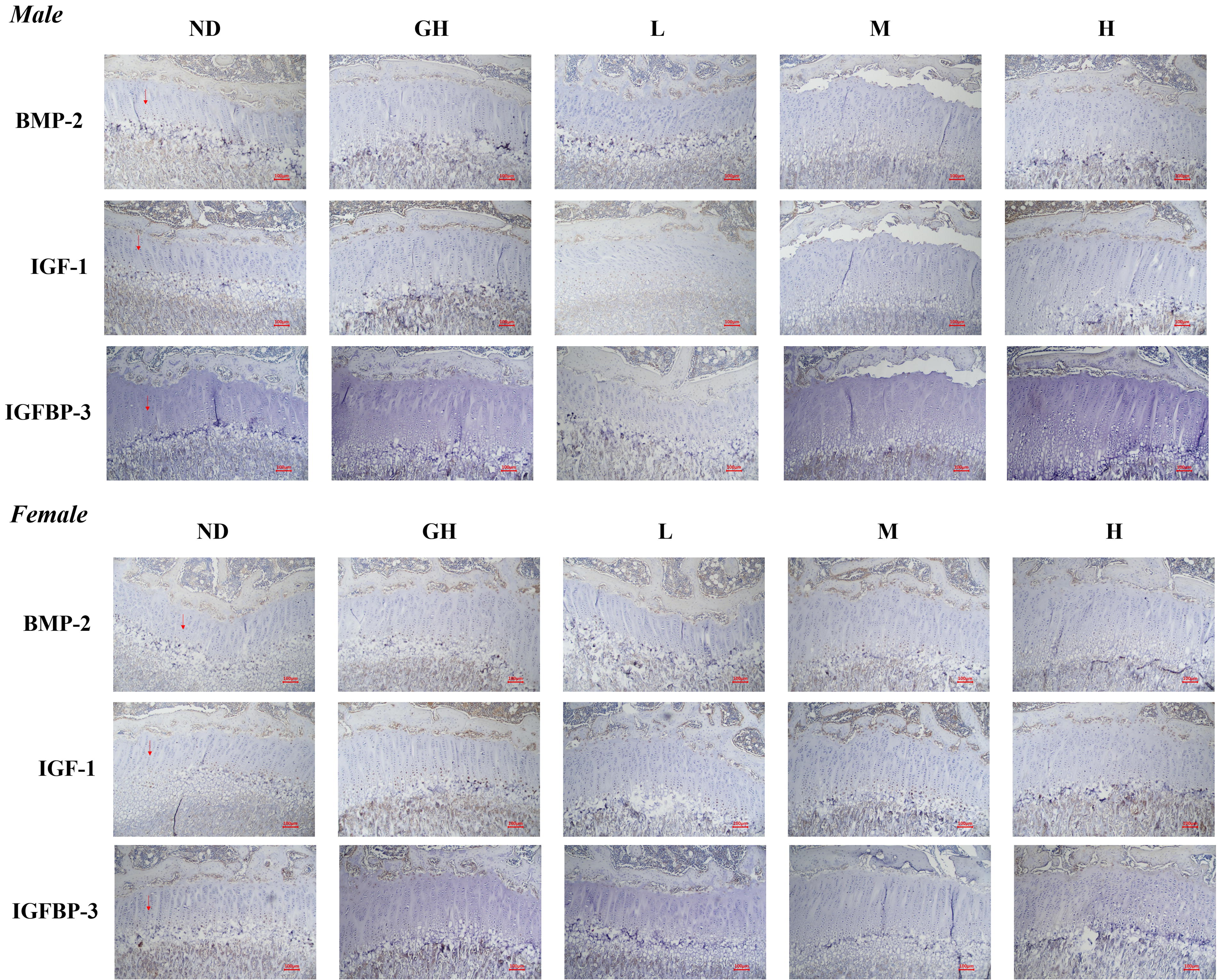
Effect of supplementation with H. japonicus extracts on bone morphogenetic protein 2, IGF-1, and IGFBP-3 expression in the bone from SD rats.
Effect of H. japonicus extracts on femur and tibia lengths
The lengths of the femur and tibia were measured to assess the effect of H. japonicus supplementation on bone growth in male and female SD rats. In both sexes, the H group exhibited significantly increased femur and tibia lengths compared with the ND group (Fig. 3). These results indicate that dietary supplementation with H. japonicus extracts, particularly at a high dose, promotes longitudinal bone growth in both male and female SD rats, suggesting its potential role in supporting growth.

Effect of supplementation with H. japonicus extracts on the lengths of the femur and tibia in SD rats. Values are presented as means ± standard deviations, and different superscript letters (a > b > c) indicate significance at P < .05.
Effect of H. japonicus extracts on mineralization parameters
Micro-CT analysis was performed to assess trabecular and cortical BMD and structural parameters in male and female SD rats, as presented in Figure 4 and Tables 2 and 3. In both male and female SD rats, the H group showed significant increases in BMD-apparent, bone volume/total volume, trabecular number, and trabecular thickness in the trabecular bone compared with the ND group (Table 2). In addition, in the cortical bone of male SD rats, the H group exhibited notable increases in BMD-apparent, BMD-material, cortical bone area, total cross-sectional area, cortical bone area/total cross-sectional area, and cortical thickness relative to the ND group, while female rats also demonstrated improvements in BMD-apparent, BMD-material, cortical bone area, total cross-sectional area, and cortical thickness in the H group compared with the ND group (Table 3; P < .05). These results indicate that H. japonicus supplementation significantly improves trabecular and cortical bone microarchitecture, demonstrating its potential role in enhancing bone quality and strength.

Effect of supplementation with H. japonicus extracts on the on architectural changes in cortical and tibia trabecular bone in SD rats.
Effect of Supplementation with H. japonicus Extracts on Trabecular Mineralization Parameters in SD Rats
Values are presented as means ± standard deviations, and different superscript letters (a > b > c > d) indicate significance at P < .05. ns, not significant.
Effect of Supplementation with H. japonicus Extracts on Cortical Mineralization Parameters in SD Rats
Values are presented as means ± standard deviations, and different superscript letters (a > b > c > d) indicate significance at P < .05.
Effect of H. japonicus extracts on proliferation of chondrocytes
BrdU staining was used to evaluate the proliferation of chondrocytes in the growth plates of male and female SD rats. The GH group exhibited the highest number of proliferating chondrocytes for both sexes, significantly higher than the ND group. Among the H. japonicus extract-treated groups, the H group showed an increase in chondrocyte proliferation comparable with that observed in the GH group, with significantly higher numbers than the ND group (Fig. 5). These findings suggest that dietary supplementation with H. japonicus promotes chondrocyte proliferation in the growth plates of male and female SD rats, supporting its potential to stimulate longitudinal bone growth.
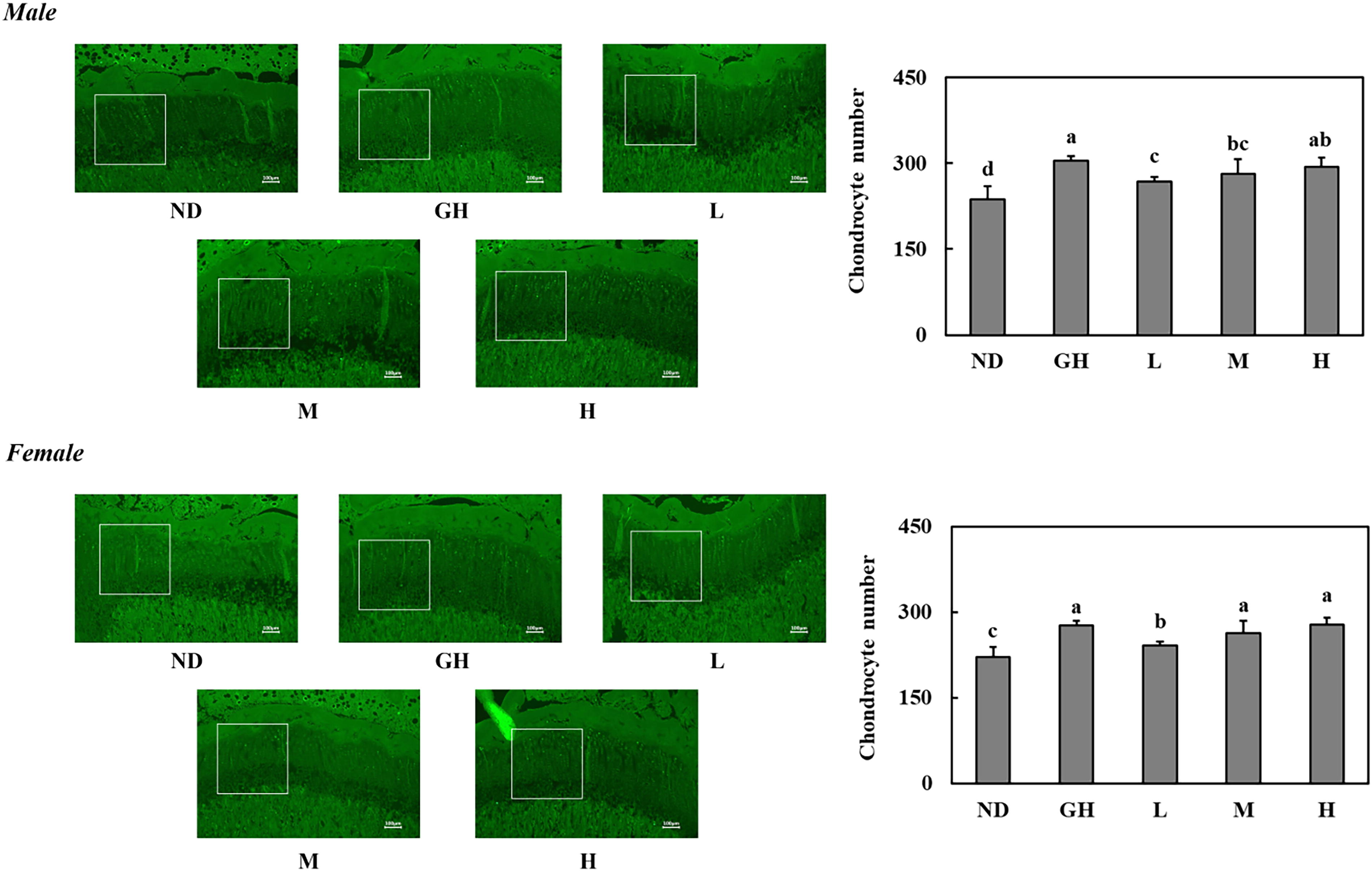
Effect of supplementation with H. japonicus extracts on the number of chondrocytes in SD rats. Values are presented as means ± standard deviations, and different superscript letters (a > b > c > d) indicate significance at P < .05.
Effect of H. japonicus extracts on growth plates
Histological analysis of the growth plates, stained to examine their structural zones, revealed notable differences among the experimental groups. The GH group exhibited the greatest thickness across the growth plate, resting zone, proliferating zone, and hypertrophic zone in both male and female SD rats. In male SD rats, the H group demonstrated significantly increased thicknesses in the growth plate, resting zone, and hypertrophic zone compared with the ND group, while in female SD rats, the H group showed significantly increased thicknesses in all four zones (the growth plate, resting zone, proliferating zone, and hypertrophic zone) compared with the ND group (Fig. 6). These findings suggest that H. japonicus supplementation positively influences the growth plate structure, highlighting its potential role in promoting bone growth.

Effect of supplementation with H. japonicus extracts on the growth plate in SD rats. Values are presented as means ± standard deviations, and different superscript letters (a > b > c > d) indicate significance at P < .05.
DISCUSSION
This study provides compelling evidence that dietary supplementation with H. japonicus extracts significantly enhances bone growth and structural integrity in both male and female rats. The results obtained from biochemical assays, micro-CT analysis, and histological evaluation indicate that H. japonicus positively influences trabecular and cortical bone microarchitecture, as well as the expression of essential bone growth regulators such as IGF-1, IGFBP-3, and BMP-2.
The JAK2/STAT signaling pathway plays a critical role in regulating IGF-1 expression in the liver, which contributes to bone growth via both systemic and local mechanisms. 7,8 In this study, hepatic proteins of phosphorylated JAK2 and STAT5 were significantly increased in the H. japonicus-treated groups, correlating with elevated IGF-1 levels in both the liver and serum. IGF-1 is a pivotal factor in promoting chondrocyte proliferation and hypertrophy, essential processes for endochondral ossification and bone elongation. Systemically, IGF-1, produced primarily in the liver, acts on growth plate chondrocytes through endocrine mechanisms. In addition, locally produced IGF-1 enhances paracrine signaling within the growth plate, further amplifying chondrocyte activity and matrix turnover. 9 Furthermore, while IGF-1 is known to regulate chondrocyte proliferation and hypertrophy, the current study did not directly assess IGF-1 activity within the growth plate itself, leaving some gaps in understanding its localized effects. 10 Measuring IGF-1 receptor activation, local IGF-1 synthesis, and chondrocyte-specific markers would provide deeper insights into the mechanistic role of H. japonicus in promoting longitudinal bone growth. Additional histological and molecular analyses could further clarify how H. japonicus modulates bone elongation at the cellular level. Despite these limitations, the significant increases in trabecular BMD, bone volume fraction, trabecular number, and trabecular thickness observed in the high-dose H. japonicus group suggest that this supplement promotes trabecular bone health. Similarly, improvements in cortical bone parameters, including cortical bone area, cross-sectional area, and cortical thickness, suggest that H. japonicus strengthens cortical bone, reinforcing skeletal integrity. These findings are consistent with previous studies demonstrating the importance of natural products in maintaining skeletal health. Although our findings demonstrate a correlation between increased hepatic JAK2/STAT5 activation and elevated IGF-1 levels, we acknowledge that this does not provide direct evidence of a causal relationship with bone growth. The cited references supporting the link between hepatic JAK2/STAT5 signaling and skeletal development primarily highlight its role in IGF-1 regulation rather than establishing a direct mechanistic connection to bone growth. 11 Further studies using targeted inhibition of JAK2/STAT5 signaling in the liver or IGF-1 knockout models would be necessary to confirm whether the observed improvements in bone parameters are directly mediated by this pathway. In addition, future research should examine the activation of IGF-1 receptors and downstream signaling components such as AKT and ERK within the growth plate to strengthen the link between systemic IGF-1 elevations and localized bone growth effects. Beyond IGF-1 signaling, other molecular pathways may contribute to the observed improvements in bone parameters. H. japonicus contains various bioactive compounds, including polysaccharides and peptides, which have been reported to influence osteogenic differentiation and bone remodeling. 12,13
The elevated expression of growth factors within the growth plate suggests that H. japonicus may promote longitudinal bone growth by directly targeting the signaling pathways responsible for growth plate regulation. These findings align with prior research, including our earlier study, which demonstrated the beneficial effects of a mixture of H. japonicus and garlic/watermelon powder on bone growth. Notably, this study provides additional insights into the specific effects of H. japonicus alone in both male and female rats.
Bone microarchitecture is a key determinant of bone strength and resilience. 14 The significant increases in trabecular BMD, bone volume/total volume, trabecular number, and trabecular thickness observed in the H group highlight the ability of H. japonicus to promote trabecular bone health. Similarly, improvements in cortical bone parameters, including cortical bone area, cross-sectional area, and cortical thickness, suggest that H. japonicus enhances the structural integrity of cortical bone, further contributing to overall bone strength. These findings are consistent with previous studies showing the importance of natural products in maintaining skeletal health.
The growth plate, or epiphyseal plate, is a specialized cartilage structure essential for longitudinal bone growth during development. 15 It comprises of distinct zones—resting, proliferative, and hypertrophic zones—that regulate endochondral ossification, the primary process responsible for bone elongation. 16,17 The observed increases in the thickness across all zones and the higher number of chondrocytes in the H. japonicus-supplemented groups, in both male and female rats, suggest enhanced chondrocyte proliferation, hypertrophy, and extracellular matrix production. These results further support the role of H. japonicus in promoting endochondral ossification and accelerating longitudinal bone growth. These findings suggest enhanced growth plate activity and indicate that H. japonicus exerts its effects through multiple mechanisms, including stimulation of chondrocyte proliferation and hypertrophy, as well as the maintenance of progenitor cell populations.
From a practical perspective, these findings suggest that H. japonicus could be a promising natural supplement for promoting bone health and growth and skeletal health, particularly in individuals at risk of growth deficiencies or bone fragility. The ability of H. japonicus to improve bone mineralization and promote longitudinal bone elongation indicates its potential use in pediatric growth support and the prevention of age-related bone loss.
In conclusion, dietary supplementation with H. japonicus extracts significantly enhances bone growth and structural integrity by modulating critical molecular pathways involved in bone formation and remodeling. These findings provide a strong foundation for further research to elucidate the precise mechanisms underlying the effects of H. japonicus and support its potential development as a natural therapeutic agent for improving skeletal health.
Footnotes
AUTHORS’ CONTRIBUTIONS
Conceptualization: S.-H.P., J.P., M.L., and O.-K.K. Methodology: S.-H.P., J.P., M.L., and O.-K.K. Data curation: S.-H.P., J.P., M.L., and O.-K.K. Investigation: S.-H.P., J.P., J.-Y.Y., H.-S.K., M.L., and O.-K.K. Writing—original draft: S.-H.P., J.P., M.L., and O.-K.K. All authors have read and approved the final version of the article.
AUTHOR DISCLOSURE STATEMENT
The authors declare no conflicts of interest.
FUNDING INFORMATION
This work was supported by the Korea Institute of Planning and Evaluation for Technology in Food, Agriculture and Forestry through High Value-added Food Technology Development Program (or Project) (121023-3), funded by the Ministry of Agriculture, Food and Rural Affairs and Korea Institute of Startup & Entrepreneurship Development through Startup Scale-Up Package 2023 Program (or Project; No. 20165902), funded by Ministry of SMEs and Startups.