
Abstract
Abstract
Purpose:
To evaluate the effects of intravitreal moxifloxacin with or without dexamethasone on experimental Bacillus cereus endophthalmitis.
Methods:
The right eyes of 21 New Zealand albino rabbits weighing 2–3 kg were used in this study. Each eye was inoculated with 1×10⁶ colony-forming units of B. cereus microorganisms via intravitreal injection into the vitreous cavity, and an experimental model of B. cereus endophthalmitis was formed. The rabbits were separated into 3 groups: Group 1 was given 0.1 mL of balanced saline solution intravitreally, group 2 was given 50 μg of moxifloxacin, and group 3 was given 50 μg of moxifloxacin plus 400 μg of dexamethasone 24 h after the inoculation. Vitreous aspirates were taken for microbiological examination on the 3rd day. Clinical inflammation scores were evaluated on days 1, 7, and 14. The rabbits were killed on the 14th day, and the eyes were enucleated for histopathological examination.
Results:
On the 7th day, only the vitreous scores of the treatment groups were significantly low compared with those of the control group (P<0.05). On day 14, the clinical scores of vitreous inflammation were 2.43±0.79, 1.43±0.53, and 1.29±0.49 in Groups 1, 2, and 3, respectively. The clinical scores of the treatment groups were significantly lower compared with those of the control group on day 14 (P<0.05). Histopathological scores were 2.43±0.79, 1.43±0.53, and 1.43±0.79 for the iris and 2.14±0.69, 1.57±0.53, and 1.14±0.38 for the vitreous in Groups 1, 2, and 3, respectively. Apart from the conjunctiva, the histopathological scores of the other tissues in the treatment groups were significantly lower compared with those of the control group (P<0.05). No significant differences were found in the histopathological or clinical scores among the treatment groups (P>0.05). Microbiological scores at day 14 were 151±6.43, 125.43±13.44, and 131.14±16.99 for Groups 1, 2, and 3, respectively. The microbiological scores of the treatment groups were significantly lower compared with those of the control group (P<0.05).
Conclusions:
Intravitreal moxifloxacin injection is effective in experimental B. cereus endophthalmitis. The addition of intravitreal dexamethasone may not significantly affect treatment efficacy.
Introduction
Clindamycin, vancomycin, ampicillin, ciprofloxacin, and dexamethasone have been used in the treatment of B. cereus endophthalmitis,11,16–18 although a standard treatment has not yet been defined. Some studies have recommended the use of fluoroquinolones in B. cereus endophthalmitis. 17 To the best of our knowledge, no previous study has applied moxifloxacin, a fourth-generation fluoroquinolone with broad antibacterial activity,19,20 to treat B. cereus endophthalmitis. The current study evaluated the effects of intravitreal moxifloxacin treatment alone or moxifloxacin together with dexamethasone in experimental B. cereus endophthalmitis.
Methods
Animals
Twenty-one New Zealand albino rabbits weighing 2–3 kg were used in the study. All experimental procedures were performed in accordance with the Association for Research in Vision and Ophthalmology Statement for the Use of Animals in Ophthalmic and Vision Research and complied with the Declaration of Helsinki. Approval was obtained from the local ethics committee before the study. Before each procedure, the rabbits were anesthetized with an intramuscular combination of 30 mg/kg ketamine hydrochloride (Ketalar; Pfizer, Istanbul, Turkey) and 10 mg/kg xylazine hydrochloride (Rompun; Bayer, Istanbul, Turkey). Mydriasis was achieved with topical 0.5% tropicamide (Tropamid; Bilim, Istanbul, Turkey) and 1% cyclopentolate hydrochloride (Sikloplejin; Abdi Ibrahim, Istanbul, Turkey). For topical anesthesia, 0.5% proparacaine hydrochloride (Alcaine; Alcon, Fort Worth, TX) was used.
Intraocular inoculations and experimental endophthalmitis
The Bacillus organism was obtained as a result of 24-h agar cultivation of B. cereus cell culture in isotonic sodium chloride, thereby creating a 0.098 (108 cells/mL) optic density. This solution was then diluted to obtain 1×10⁶ colony-forming units (CFU) in 0.1 mL. This was confirmed by quantitative measurements of the microorganisms covering the blood agar. A 30-gauge needled tuberculin syringe was used for intravitreal injections. Before intravitreal injections, 0.1 mL aqueous humor was aspirated from the anterior chamber to prevent an increase in intraocular pressure. The right eyes of the rabbits were inoculated with the B. cereus microorganism (0.1 mL) by an injection into the mid-vitreous cavity, 2–3 mm behind the limbus. The eyes were examined 24 h after inoculation of the bacteria. All eyes were observed to have similar levels of endophthalmitis with findings such as medium or severe conjunctival hyperemia and chemosis, vitreous opacity, non-visualization of the retina, and partial or definite corneal opacity. The eyes were randomly distributed into 3 equal groups.
Treatment
The moxifloxacin and dexamethasone used in the treatment were diluted with an aseptic balanced saline solution to obtain the required dosage. The eyes in group 1 (control group) received no treatment and were only given an intravitreal injection of balanced saline solution. The eyes in group 2 were given an intravitreal injection of 50 μg/0.1 mL moxifloxacin only. The eyes in group 3 were given an intravitreal injection of 50 μg/0.1 mL moxifloxacin plus 400 μg/0.1 mL dexamethasone. The moxifloxacin and dexamethasone were administered by separate injections in group 3.
Clinical evaluation
Clinical examinations were performed using slit-lamp biomicroscopy and indirect ophthalmoscopy on the 1st, 7th, and 14th days. Examiners were masked in all examinations. Blinded to the groups, the ocular findings were classified from 0 to 3 for conjunctiva, cornea, iris, and vitreous. The clinical classification system described by Peyman et al. was used (Table 1). 21
Microbiological evaluation
For microbiological evaluation, the vitreous was aspirated with a 30-gauge tuberculin syringe on day 3. The samples were inoculated to blood agar and incubated at 35°C for 48 h. After incubation, B. cereus was defined, and the surface colonies were counted.
Histopathological evaluation
For histopathological examination, the rabbits were killed on the 14th day, and the eyes were enucleated. The eyes were fixed in 10% buffered formalin and then embedded in paraffin blocks. Sections of 5 μm were taken and stained with hematoxylin–eosin. Blinded to the groups, the same pathologist evaluated all samples by light microscopy. The scale given in Table 2 was applied for histopathological grading.
Statistical analysis
SPSS version 15.0 (SPSS, Inc., Chicago, IL) was used for statistical evaluations. Data were given as the mean±standard deviation. The Kruskal–Wallis test was used to compare the clinical and histopathological scores between the groups. Double comparisons between the groups were made by using the Mann–Whitney U-test. A value of P<0.05 was accepted as statistically significant.
Results
Clinical results
The clinical scores from post-treatment days 1, 7, and 14 are shown in Table 3. No significant difference was seen in day 1 clinical scores between the eyes given only moxifloxacin and the eyes treated with moxifloxacin plus dexamethasone (P>0.05). On day 7, the vitreous inflammation scores of the treated eyes were significantly lower than those of the untreated eyes (P<0.05). However, on day 7, no significant differences were found in the conjunctiva or iris clinical scores between the treated and untreated eyes (P>0.05).
Data are given as mean±SD.
P<0.05, statistically significant compared with control group.
SD, standard deviation.
On day 7, no significant difference in clinical scores was observed between the eyes treated with moxifloxacin alone and those treated with moxifloxacin plus dexamethasone. On day 14, the clinical scores of the treated eyes were significantly lower compared with those of the untreated eyes (P>0.05). The clinical scores on days 1, 7, and 14 showed no significant difference between the eyes treated with moxifloxacin alone and the eyes treated with moxifloxacin and dexamethasone (P>0.05).
Histopathological results
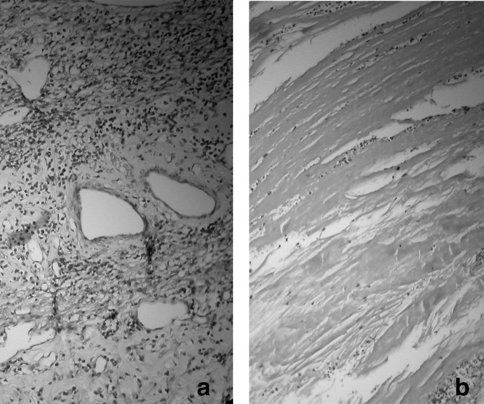
The evaluation of histopathological scores on day 14 showed no significant difference in conjunctiva inflammation scores of groups 2 and 3 compared with those of the control group (P>0.05). However, compared with the control group, the iris, choroid, and retina scores of groups 2 and 3 were significantly lower (P<0.05). In group 1, extreme edema with vasculitis and inflammatory cells (predominantly polymorphonuclear leukocytes) were observed in the iris, and a vast number of inflammatory cells occurred in the vitreous (Fig. 1). Mild or moderate inflammation of the vitreous and iris was detected in groups 2 and 3 (Figs. 2 and 3). Although the vitreous scores of group 3 were found to be significantly lower compared with those of the control group (P<0.05), no significant difference was determined between group 2 and the control group (P>0.05). No significant difference in histopathological scores was observed between groups 2 and 3 (P>0.05). Histopathological evaluation scores are shown in Table 4.

H&E, 200×.
H&E, 200×.

H&E, 200×.
Data are given as mean±SD.
P<0.05, statistically significant compared with control group.
Microbiological results
The mean number of colonies was 151±6.43 CFU/mL in group 1, 125.43±13.44 CFU/mL in group 2, and 131.14±16.99 CFU/mL in group 3. The difference was significant (Kruskal–Wallis, P<0.05). A significant difference was noted when the treated groups (Groups 2 and 3) were compared with the control group (P<0.05). No significant difference was detected between the treated groups (P>0.05).
Discussion
Several drugs for B. cereus endophthalmitis have been investigated, and the majority have been reported as effective.11,16–18 The widespread use of antibiotics, however, has engendered the development of resistance to many antibiotics, and those with strong bactericidal effects and relatively low resistance have come into current use. Moxifloxacin is a fourth-generation fluoroquinolone with a broad-spectrum bactericidal effect against both gram-positive and -negative bacteria.22–26 Moxifloxacin, which contains a methoxy group in the eighth position of the quinolone circle, simultaneously inhibits both DNA gyrase and topoisomerase II. This configuration increases not only the potency of the medication, but also the necessary number of mutations for the development of resistance.27,28 Moxifloxacin shows higher activity compared with ciprofloxacin against the resistance chain of Staphylococcus aureus. 29 In addition, moxifloxacin has better penetration into the cornea and aqueous humor. 26 However, intravenous, oral, or topical application of moxifloxacin has not been shown to reach an effective concentration in the vitreous.18,30–33 Intravitreal injection of moxifloxacin should be recommended in the treatment of endophthalmitis. 2 Therefore, moxifloxacin was administered by intravitreal injection in this study. One of the concerns of intravitreal administration of antibiotics is retinal toxicity. Studies have reported that concentrations up to 150 μg/mL were not toxic in rabbits,34,35 and the dosage of moxifloxacin used in this study, 50 μg, was well below the defined toxic dose.
In this study, on the 7th day, the clinical scores of groups 2 and 3 were lower when compared with those of the control group. However, the vitreous inflammation only in group 2 was significantly lower compared with that in the control group, and no significant difference was observed between the treatment groups. Evaluation of the 14th-day clinical scores showed that compared with the control group, groups 2 and 3 eyes had milder iritis and less vitreous opacity and retinitis. No significant difference was seen between the treatment groups. Histopathological examination revealed that groups 2 and 3 showed milder iritis, less corneal edema, fewer vitreous cells, and significantly less retinitis when compared with the control group. Thus, one can conclude that intravitreal moxifloxacin treatment is significantly beneficial in cases of B. cereus endophthalmitis.
No significant difference was determined between eyes given only moxifloxacin and moxifloxacin plus dexamethasone for clinical and histopathological scores. The use of corticosteroids in the treatment of endophthalmitis is still a controversial topic, as the course of endophthalmitis may be affected by resultant debris and immunogenic particles of bacteria. Bacterial enzymes that appear in B. cereus endophthalmitis increase inflammation; thus, B. cereus causes a faster and more serious endophthalmitis compared with other types of endophthalmitis.16,36–38 Therefore, some studies have defended the use of corticosteroids alongside antibacterial treatment to eliminate bacteria,18,39–42 whereas other studies have shown no significant effect on the progress of endophthalmitis.43–45 In this study, the addition of dexamethasone to the treatment brought good results in both clinical and histopathological scores, but this benefit did not reach a significant level. Although a few studies have described the use of moxifloxacin for endophthalmitis caused by infectious agents other than B. cereus, to the best of our knowledge, none had used intravitreal moxifloxacin treatment for B. cereus endophthalmitis. Thus, this is the first study reporting on the efficiency of moxifloxacin.
In conclusion, intravitreal injection of moxifloxacin may be an alternative treatment choice for B. cereus endophthalmitis. Further studies are required to determine an efficient dosage and side effects. The addition of dexamethasone to treatment did not significantly affect the course of endophthalmitis.
Footnotes
Author Disclosure Statement
The authors had no additional financial support or national funding for this study. No competing financial interests exist. None of the authors has any proprietary interests or conflicts of interest related to this submission.