
Abstract
Abstract
Purpose:
The purpose of this study was to investigate the protective effects of sodium hyaluronate eyedrop against corneal epithelial disorders caused by antiglaucomatous eyedrops using an electrophysiological method.
Methods:
Three kinds of antiglaucomatous eyedrops, including benzalkonium chloride (BAC) as an ophthalmic preservative, a BAC-free antiglaucomatous eyedrop, and a sodium hyaluronate eyedrop, were used in this study. Eyedrops were applied to excised rabbit corneas, and the electrophysiological property of the cornea was monitored using an Ussing chamber with a turnover system that mimics human tear turnover. With this system, changes in transepithelial electrical resistance (TER) in the corneal surface were recorded.
Results:
The corneal TER after applying antiglaucomatous eyedrops tended to decrease concomitantly with increasing the concentration of the BAC included as a preservative. On the other hand, there was no significant change in the corneal TER for the initial 60 min after applying sodium hyaluronate eyedrop compared with those of the control. Moreover, the pretreatment with a sodium hyaluronate eyedrop reduced the extent of decrease in the corneal TER observed after application of antiglaucomatous eyedrops alone.
Conclusion:
Those results indicate that a sodium hyaluronate eyedrop has the potential to protect the corneal surface from antiglaucomatous eyedrops, including BAC as an ophthalmic preservative.
Introduction
BAC is widely used as an ophthalmic preservative at concentrations between 0.005% and 0.02% in commercial eyedrops because of its bacteriostatic and bacteriocidal efficacy. 4 However, numerous studies have revealed the deleterious corneal effects of BAC that include destabilization of the tear film, death of corneal epithelium cells, disruption of superficial cell membranes, loss of microvilli, and the reduction of the corneal epithelial barrier function.5–7 Since glaucoma is a chronic condition requiring long-term therapy, potential deleterious effects to the corneal surface from repeated exposures to antiglaucomatous eyedrops including BAC as an ophthalmic preservative need to be minimized.
On the other hand, sodium hyaluronate is a naturally occurring glycosaminoglycan of the extracellular matrix that plays an important role in development, wound healing, and inflammation. 8 It has been used in the treatment of dry eyes because of its protective effect on the cornea and long ocular surface residence time.9,10 Moreover, recent experiments in corneal and conjunctival cell cultures have shown that sodium hyaluronate has no cytotoxicity but possesses antioxidant properties and tends to reduce the toxic effects of BAC.11–13 These results may allow the use of sodium hyaluronate eyedrops, not only in dry eyes but also in corneal epithelial disorders caused by eyedrops, including BAC. However, their reports did not take into account drug dilution by tears. The ocular effects of drugs should be evaluated with methods considering tear flow, because topically instilled drugs become rapidly diluted with tears.
In a previous study, we developed a new electrophysiological method mimicking human tear flow to quantitatively measure the transepithelial electrical resistance (TER) of the rabbit cornea. 14 TER is the most sensitive electrophysiological parameter for changes occurring with the exposure of eyedrops to the corneal epithelium. This new system for examining the clinical instillation of eyedrops has revealed that eyedrops including BAC caused corneal epithelial disorders. 15
Using this new system, we investigated the effect of antiglaucomatous eyedrops and sodium hyaluronate eyedrops on corneal epithelium. In addition, we investigated the protective effects of sodium hyaluronate eyedrops against corneal epithelial disorders caused by antiglaucomatous eyedrops.
Methods
Animals
Male white Japanese rabbits (KBT: JW; KBT Oriental Co., Ltd., Tosu, Japan), 2.0–2.5 kg, were individually housed in cages in an air-conditioned room and maintained on a standard laboratory diet (ORC4; Oriental Yeast Co., Ltd., Tokyo, Japan). The rabbits had free access to food and water. All experiments in the present study conformed to the Guiding Principles in the Care and Use of Animals (DHEW Publication, NIH 80-23), the Association for Research in Vision and Ophthalmology Resolution on the Use of Animals in Research, and the Declaration of Helsinki.
Eyedrops
Three kinds of antiglaucomatous eyedrops including BAC as an ophthalmic preservative, an antiglaucomatous eyedrop including boric acid and zinc chloride as an ophthalmic preservative, and a sodium hyaluronate eyedrop were used in this study. Xalatan® (commercial preparation of 0.005% latanoprost, a prostaglandin-related agent, with 0.02% BAC) was obtained from Pfizer Japan Inc. (Tokyo, Japan). Rescula® (commercial preparation of 0.12% isopropyl unoprostone, a prostaglandin-related agent, with 0.01% BAC) was obtained from Santen Pharmaceutical Co., Ltd. (Osaka, Japan). Timoptol® (commercial preparation of 0.5% timolol maleate, a nonselective β-blocker, with 0.005% BAC) was obtained from Banyu Pharmaceutical Co., Ltd. (Tokyo, Japan). Travatan Z® (commercial preparation of 0.004% travoprost, a prostaglandin-related agent, with boric acid and zinc chloride) was obtained from Alcon Japan, Ltd. (Tokyo, Japan). Hyalein® Mini (commercial preparation of 0.3% sodium hyaluronate, an essential extracellular matrix component) was obtained from Santen Pharmaceutical Co., Ltd.
Electrophysiological experimental system and drug treatment
Figure 1A–C shows photographs and the scheme of the electrophysiological experiment system mimicking tear flow using excised rabbit corneas.14,15 The electrophysiological experiments were performed using a Ussing chamber CHM1 (World Precision Instruments, Sarasota, FL) with a donor-phase turnover system to mimic the rate of human tear turnover at 16%/min.16,17 Ag/AgCl half-cells were screwed into short tubes, which were plugged firmly into place in the chamber luer ports. Rabbits were placed in a restraint box and sacrificed by injecting an overdose of sodium pentobarbital into a marginal ear vein. The corneas were dissected and washed with warm glutathione-bicarbonate Ringer's solution (GBR). 18 GBR contained 106 mM NaCl, 4.8 mM KCl, 0.66 mM NaH2PO4·2H2O, 29.2 mM NaHCO3, 0.78 mM CaCl2·2H2O, 0.78 mM MgCl2·6H2O, 5.0 mM D-glucose, and 0.15 mM oxidized glutathione. The freshly excised corneas were immediately mounted on the Ussing chamber, using rubber supporters and O-rings. Then, the chambers were filled with GBR. Aeration and circulation in the tissue bath was provided by means of bubbling with a mixture of 95% and 5% CO2. All experiments were performed at 37°C, using a constant-temperature bath connecting a jacket of an Ussing chamber. A 0.53 cm2 area of tissue was exposed to the donor and receiver compartments, having a volume of 6 and 7 mL, respectively. The electrical output of the Ag/AgCl electrodes was fed to an automatic voltage-clamp unit (CEZ-9100; Nihon Koden, Tokyo, Japan). The spontaneous potential difference (PD) was measured with 2 matched Ag/AgCl electrodes. A direct current was sent across the tissue with a pair of matched Ag/AgCl electrodes whose tips were positioned away from the tissue surfaces at the far end of 2 reservoirs. The current flowing in the bath-tissue-bath circuit under short-circuit conditions was monitored. The short-circuit current (Isc) was measured as the current passing through the cornea under zero-voltage clamp conditions. At 60-s intervals, a 10-mV voltage pulse was imposed for 1 s across the short-circuited tissue to estimate corneal TER as the surface area normalized ratio of applied voltage pulse to observed deflection in the resultant current flow on top of Isc.

Photographs
Figure 1D shows an equivalent circuit model for corneal epithelium and electrophysiological parameters (PD, Isc and TER). A cornea is composed of 5 layers: the epithelium, Bowman's membrane, stroma, Descemet's membrane, and endothelium. Among the 5 layers, the barrier function for foreign matter mostly depends on the epithelium, which is composed of superficial cells, wing cells, and basal cells. Superficial cells existing in a tight junction and desmosomes play the main role in epithelial barrier function.
TER was described as follows: TER=RL×(Ra+Rb)/(RL+Ra+Rb). Since the electrical resistance of apical and basolateral cell membranes (Ra and Rb) was much higher than that of tight junctional resistance (Ra+Rb >> RL), TER reflects tight junctional conductance (reciprocal value of TER); therefore, TER, in general, is sensitive to changes occurring in the paracellular permeability of epithelial tissues. 19
We previously reported the electrophysiological characteristics and permeability of fluorescein isothiocyanate dextran (FD-4, an average molecular weight of 4,400), a hydrophilic and a highmolecular-weight compound, in the excised rabbit cornea. 7 Corneal TER reached about 1 k-ohm·cm2 within 80 min after mounting the cornea, followed by an extremely low permeability of FD-4. The sample for FD-4 was determined with a spectrofluorophotometer (FP-770; Jasco, Tokyo, Japan) at an excitation wavelength of 489 nm and an emission wavelength of 515 nm. FD-4 is well known to be an indicator of the epithelial paracellular pathway.20,21 Based on these results, the time at which an eyedrop was applied into the donor phase was decided at 80 min after mounting the cornea. In addition, we found 2 significant correlations between the cytotoxicity of preservatives and paracellular permeability of FD-4, and between the cytotoxicity of preservatives and conductance (reciprocal value of TER). 7 Therefore, the lower corneal TER values are indicative of the penetration of greater amounts of electrical current through the damaged superficial cells and tight junctions that exist in the epithelium.
After the eyedrop had been applied into the donor phase, the solution in the donor-phase was immediately perfused with a peristaltic pump at a rate of 0.96 mL/min with fresh GBR for 100 min. Subsequently, electrophysiological parameters were monitored at 5- or 10-min intervals for 100 min. In the use of sodium hyaluronate eyedrops, it was applied first followed by the treatment of a certain antiglaucomatous eyedrop at intervals of 10 min.
Decrement in corneal TER
Analysis of decrement in corneal TER was performed based on our previous study. 15 Decrement in the corneal TER after applying the eyedrops used in this study was calculated from the sum of the decrease in the corneal TER from its initial value (100%) for 60 min after their application.
Statistical analysis
Results are expressed as the mean±S.D. of at least 3 experiments. Statistical analysis was performed using Scheffe's test and Student's t-test. P<0.05 was considered to indicate significance.
Results
Figure 2A show the corneal TER (%) profile as a control monitored using the electrophysiological experimental system with donor-phase turnover. The corneal TER was ∼1 k-ohm·cm2 at a steady state after an 80-min incubation.
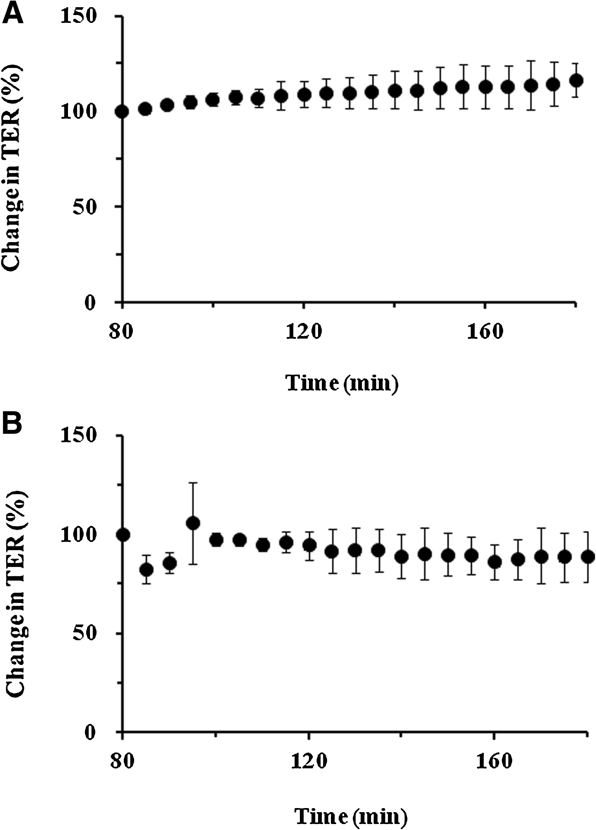
The corneal transepithelial electrical resistance (TER) profile as a control
The effects of Hyalein Mini on the electrophysiological property in the corneal epithelium were monitored using the same system, and the results are shown in Fig. 2B. The corneal TER after applying Hyalein Mini slightly decreased from the initial value (100%) and recovered after 15 min. Decrements in the corneal TER for the initial 60 min after applying Hyalein Mini (483%±336%·min) were not significantly different from those of the control (0%±0%·min) (P=0.633).
The effects of 3 kinds of antiglaucomatous eyedrops, including BAC as an ophthalmic preservative, and an antiglaucomatous eyedrop without BAC were monitored on the electrophysiological property in the corneal epithelium using the same system, and the results are shown in Fig. 3. The most extensive reduction on the corneal TER was observed after applying Xalatan (0.005% latanoprost ophthalmic solution with 0.02% BAC), and did not recover the initial level of corneal TER for 100 min. Rescula (0.12% isopropyl unoprostone ophthalmic solution with 0.01% BAC) and Timoptol (0.5% timolol maleate ophthalmic solution with 0.005% BAC) showed a moderate reduction on the corneal TER. On the other hand, the corneal TER was not decreased after applying Tavatan Z (0.004% travoprost ophthalmic solution with boric acid and zinc chloride).

Effects of antiglaucomatous eyedrops on the corneal TER in the electrophysiological experimental system with the donor-phase turnover.
Decrement in the corneal TER for the initial 60 min after applying the eyedrops was calculated, and the results are summarized in Table 1. The rank order is as follows: Xalatan (5,456%±73%·min)>Rescula (2,263%±648%·min)>Timoptol (1,732%±77%·min)>Travatan Z (20%±22%·min). Xalatan, Rescula, and Timoptol significantly reduced the corneal TER as compared with that of the control (P<0.001, P<0.001 and P<0.001, with Xalatan, Resclua, and Timoptol, respectively).
Each value represents the mean±S.D. of 3 experiments.
P<0.001 compared with control (Scheffe's test).
BAC, benzalkonium chloride.
Figure 4 shows the effects of the pretreatment with Hyalein Mini before applying 3 antiglaucomatous eyedrops, which showed a significant decrease in the corneal TER as compared with the control. In the case of Xalatan pretreated with Hyalein Mini, an irreversible reduction in the corneal TER was observed as well as an application of Xalatan alone (Fig. 4A). On the other hand, in the case of Rescula pretreated with Hyalein Mini, a reversible reduction in the corneal TER was observed (Fig. 4B). A similar result was observed in the case of Timoptol pretreated with Hyalein Mini (Fig. 4C). The pretreatment with Hyalein Mini significantly reduced the extent of decrease in the corneal TER observed after an application of each antiglaucomatous eyedrop alone (P<0.01, P<0.05 and P<0.01, with Xalatan, Resclua and Timoptol, respectively).
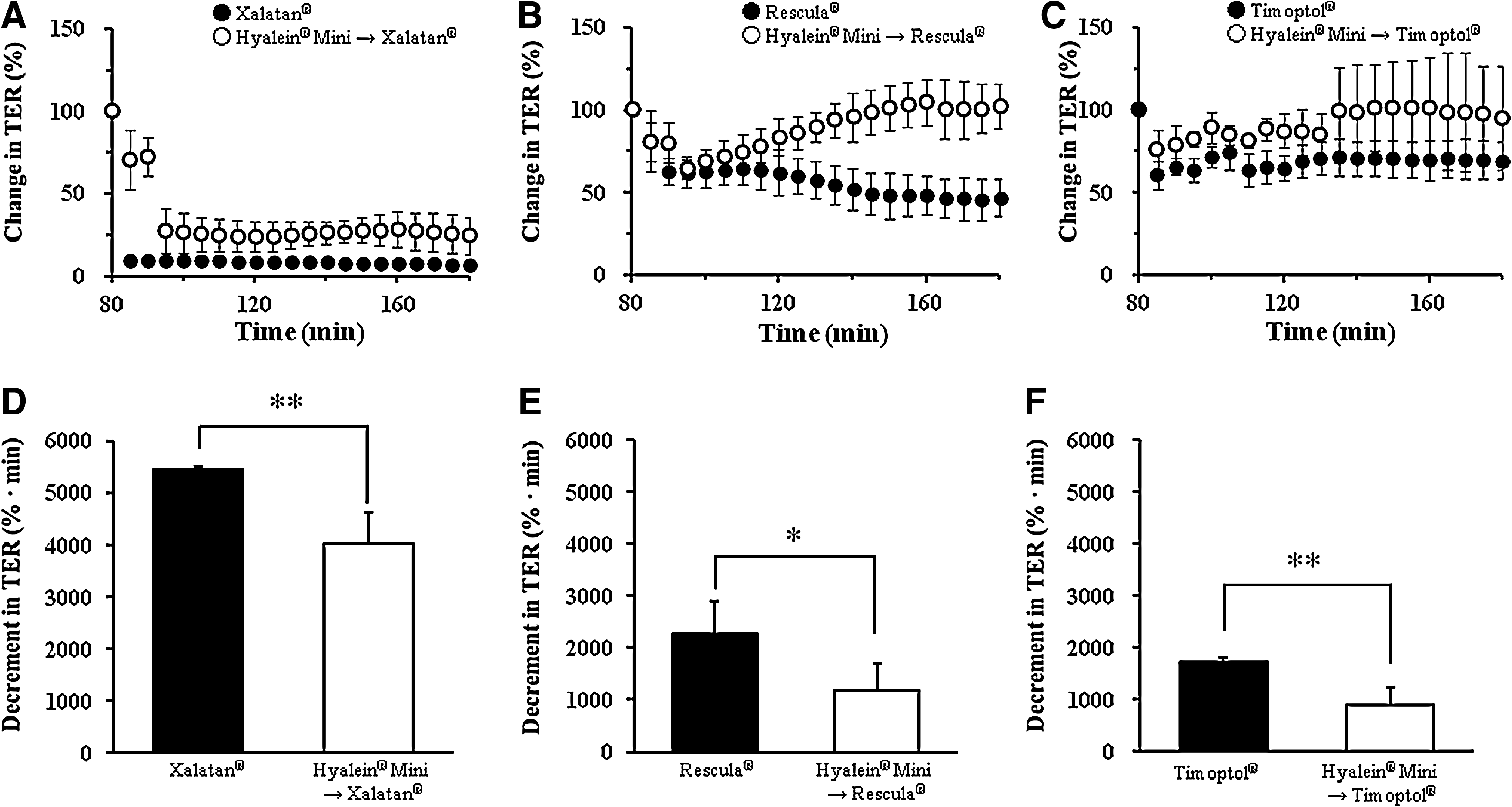
Effects of the pretreatment with Hyalein Mini (0.3% sodium hyaluronate ophthalmic solution) before application of antiglaucomatous eyedrop on the corneal TER in the electrophysiological experimental system with the donor-phase turnover.
Discussion
The corneal epithelium, which may be in direct contact with topically instilled drugs, is generally recognized as the primary source of the corneal barrier function.22,23 The barrier function of the epithelium is often evaluated by using an electrophysiological method. The TER was the most sensitive electrophysiological parameter for changes occurring with the exposure of eyedrops to the corneal epithelium. 19 The lower corneal TER values are indicative of the penetration of greater amounts of electrical current through the damaged superficial cells and tight junctions that exist in the epithelium.6,7,14,15,24 In addition, these electrical changes can be measured continuously and quantitatively. Therefore, measurement of corneal TER is a suitable method for evaluating corneal permeability and irritancy.25–27
The eye has protective mechanisms for serving its primary function of ensuring proper vision that are responsible for cleaning the ocular surface and for eliminating foreign matter. 28 Among the protective mechanisms, tear turnover, which is reported to be ∼16%/min in humans, is very important. Most of the instilled drugs are rapidly eliminated from the precorneal area due to drainage by tear turnover.16,17 We previously developed that new electrophysiological method mimicking tear flow in which the precorneal solution was exchanged for a buffer with a peristaltic pump at the rate of human tear flow and excised rabbit corneas. 14 The method for evaluating eyedrop-induced corneal toxicity considering tear flow is invaluable, as tears dilute the eyedrops immediately after instillation. By using this method, we were able to determine that the TER of the excised rabbit corneas was ∼1 k-ohm·cm2. This value is similar to our previous data 15 and to that reported for previous studies 29 by other researchers that have used the conventional Ussing chamber system.
In the present study, we monitored the effects of 3 kinds of antiglaucomatous eyedrops, including BAC as an ophthalmic preservative and an antiglaucomatous eyedrop without BAC, on the electrophysiological property in the corneal epithelium using this system. Our results suggested that the corneal TER after applying antiglaucomatous eyedrops tended to decrease concomitantly with increasing the concentration of the BAC included as a preservative. We previously demonstrated that 0.02% BAC rapidly decreased corneal TER and did not recover the initial level of corneal TER when monitoring the electrophysiological property in excised rabbit corneas, using the electrophysiological experimental system with donor-phase turnover. 14 In this study, the corneal TER after applying Xalatan exhibited almost the same results as those of the 0.02% BAC. The BAC weakens transcellular integration and enhances drug permeability through the cornea accompanied by impairment of barrier function.7,30,31 The incorporation of the active antiglaucoma drugs does show clinical relevance. However, many studies have revealed that direct exposure to BAC damages the corneal epithelium.6,11 Therefore, although BAC might have a potential role as it enhances the intraocular penetration of the active antiglaucoma drugs, high concentrations should be used with care.
On the other hand, the corneal TER after applying Rescula was also decreased. Wang et al. 32 reported that the corneal barrier function was damaged by addition of Rescula on the cultured rabbit corneal epithelial cells. However, they reported that there is no effect on their barrier function as added with 0.12% isopropyl unoprostone alone. Accordingly, reductions of the corneal TER after applying Xalatan and Rescula were suggested to result from high concentrations of BAC, including their eyedrops from 0.01% to 0.02%.
Timoptol showed an irreversibly moderate reduction in the corneal TER. The toxic effects of β-blockers on the ocular surface were reported to include a decrease in goblet cell density in the conjunctiva, or disruption of tight junction and microvilli in the corneal epithelium.33,34 It was previously reported that even a topical preparation of a preservative-free timolol decreased the corneal epithelial barrier function, but its effect was less than a commercial timolol containing BAC as a preservative. 35
Travatan Z is the first prostaglandin eyedrop that is preserved with a non–BAC system (SofZia) that is patented by Alcon. The SofZia preservative system of Travatan Z consists of boric acid (H3BO3) and zinc chloride (ZnCl2). We confirmed that the corneal TER was not altered after applying Travatan Z. This suggests that Travatan Z may be less toxic than other prostaglandin analogs, such as Xalatan and Rescula. While Xalatan and Rescula contain BAC, which is believed to be a major cause of toxicity noted with prostaglandin analogs formulations, Travatan Z do not contain BAC. The results of an in vitro study of corneal epithelial cells suggested that SofZia may be less toxic than BAC. McCarey and Edelhauser reported that while the presence of BAC broke the anatomic tight junctions and damages the cell membranes, treatment with Travatan Z had no negative effect on the integrity of corneal epithelial tight junctions. 36
In accordance with our previous electrophysiological studies,6,7,15,24 we were able to show eyedrops containing BAC, inducing corneal epithelial disorders. Nevertheless, BAC is necessary to prevent microbial development in pharmaceutical products. Our idea was to investigate whether sodium hyaluronate eyedrops are able to reduce corneal epithelial disorders caused by antiglaucomatous eyedrops including BAC as an ophthalmic preservative.
We revealed that the application of Hyalein Mini did not significantly change the TER compared with the control. This suggests that Hyalein Mini does not cause corneal epithelial disorders. It was already shown that sodium hyaluronate causes no damage to ocular tissues; Wysenbeek et al. 11 demonstrated that instillation of sodium hyaluronate (0.1% or 1%) on the epithelial cells of chick embryo corneas has no toxic effects. Ayaki et al. 37 showed that Hyalein Mini does not have any toxic effect on human corneal epithelial cell line. Moreover, those results agree with clinical results which have clearly shown that treatment of patients with 0.1% sodium hyaluronate for a period of 2 years had no adverse effects. 38
Then, we evaluated the protective effects of Hyalein Mini against 3 antiglaucomatous eyedrops that showed a significantly less decrease in the corneal TER as compared with the control. The extent of decrease in the corneal TER at antiglaucomatous eyedrops pretreated with Hyalein Mini was significantly reduced compared with when they were applied alone. However, the degree of the protective effect by the pretreatment with Hyalein Mini varied according to the concentration of the BAC included as a preservative. The ratio of decrement in the cornea TER of antiglaucomatous eyedrops reduced by pretreatment with Hyalein Mini is as follows: Timoptol (−48%)>Rescula (−47%)>Xalatan (−23%). In the case of Rescula pretreated with Hyalein Mini, a reversible reduction in the corneal TER was observed (Fig. 4B). A similar result was observed in the case of Timoptol pretreated with Hyalein Mini (Fig. 4C). These results indicate that Hyalein Mini may be a protective agent of the corneal surface against antiglaucomatous eyedrops including BAC as an ophthalmic preservative. Wysenbeek et al. 11 showed that a combination of 0.1% sodium hyaluronate with 0.01% BAC effectively reduced the toxic effects of preservatives. Pauloin et al. 13 reported that 0.2% sodium hyaluronate significantly decreases oxidative stress, apoptosis, and necrosis induced by 0.005% BAC. Our findings were consistent with these data.
BAC, which has positive charges, is adsorbed on cell membranes that are negatively charged, leading to permeability changes, then to cytoplasmic membrane and cell wall lesions. In addition, BAC induces cytotoxicity by inducing oxidative stress, apoptosis, and P2X7 cell death receptor. 13 This receptor is activated by extracellular ATP, 39 which can induce apoptosis through various pathways.40–42 On the other hand, sodium hyaluronate possesses interesting physicochemical properties. Sodium hyaluronate exists mainly as a polyanionic form, and BAC exists in solution as a cationic form. A possible explanation may be that ionic attraction between the positive charge of BAC and the negative charge in sodium hyaluronate neutralizes the toxic effect caused by the cationic charge of BAC to the corneal epithelium. It is also possible that the BAC small molecules penetrate into the sponge-like domain of sodium hyaluronate and disperse within it. In addition, sodium hyaluronate is rich in hydroxyl functions, which can potentially absorb reactive oxygen species. Furthermore, sodium hyaluronate specifically interacts with several cell surface receptors, including the cluster determinant 44 (CD44), which is considered the main sodium hyaluronate cell-surface receptor. 43 Pauloin et al. demonstrated that both the corneal epithelial cell line and the conjunctival epithelial cell line express the CD44 receptor, suggesting that sodium hyaluronate forms a cytoprotective coat on the cell membrane interacting with the CD44 receptor, and this cytoprotective coat prevents BAC cytotoxicity and physically masks P2X7 receptor. 13
On the other hand, the finding of Xalatan was different from that of Rescula and Timoptol. In the case of Xalatan pretreated with Hyalein Mini, an irreversible reduction on the corneal TER was observed (Fig. 4A). Irreversible damage to the cornea barrier may be explained by desquamation of epithelial cells. Kusano et al. 24 assessed changes in the TER induced by application of 0.005%–0.02% BAC and found that these routine clinical concentrations rapidly caused acute dysfunction of the corneal barrier. They reported that acute corneal disruption occurs within only 5 s of application of 0.02% BAC. Furthermore, it was reported that the corneal epithelium of the 0.02% BAC-treated cornea showed detaching and wrinkling of the superficial cells, dissociations between the cells, and degenerated microvilli. 44
In conclusion, the results of the present study indicate that the corneal TER after applying antiglaucomatous eyedrops tended to decrease concomitantly with increasing the concentration of the BAC included as a preservative. Moreover, the pretreatment with Hyalein Mini reduced the extent of decrease in the corneal TER observed after application of each antiglaucomatous eyedrop alone. Those results indicate that Hyalein Mini has a potential to protect the corneal surface from antiglaucomatous eyedrops including BAC as an ophthalmic preservative.
Footnotes
Acknowledgments
This work was supported, in part, by a Grant-in-Aid for Scientific Research from Nagasaki University (Nagasaki, Japan) and the Japan Society for the Promotion of Science (JSPS). The authors are grateful to Setsuka Murata and Megumi Kawakami for their technical assistance.
Author Disclosure Statement
No competing financial interests exist.