




Abstract
Abstract
Purpose:
To determine the aqueous humor levels of nitric oxide (NO) and vascular endothelial growth factor (VEGF) in the eyes of patients with Coats' disease and study the correlation between these levels.
Methods:
Samples of aqueous humor were obtained from 7 patients with Coats' disease and 15 age-matched patients with congenital cataracts as controls. Nitrite and nitrate (NOx), the stable end products of NO, were detected by the Griess reaction, and VEGF levels were assessed by enzyme-linked immunosorbent assay.
Results:
The aqueous humor NOx and VEGF levels were elevated in the eyes of patients with Coats' disease compared with those of controls (P=0.001 and P<0.001, respectively). The median NOx level was 55.2 μM (range, 23.0–75.3 μM) in the Coats' disease group and 18.8 μM (range, 8.7–36.2 μM) in the control group. The median VEGF level was 731.7 pg/mL (range, 288.3–1364.3 pg/mL) in the Coats' disease group and 33.3 pg/mL (range, 9.0–96.8 pg/mL) in the control group. No correlation was observed between the aqueous humor concentrations of NOx and VEGF.
Conclusions:
NOx and VEGF are increased but not related in the aqueous humor samples of patients with Coats' disease. NO and VEGF may play roles in the pathogenesis of Coats' disease. Further studies are needed to clearly elucidate the relationship among VEGF, NO, and other cytokines in Coats' disease.
Introduction
Nitric oxide (NO), a signaling molecule with pleiotropic effects, plays a key role in modulating the functional blood flow and participates in complicated processes such as vasodilatation, inflammation, thrombosis, immunity, neurotoxicity, and neurotransmission. 10 There are 3 isoforms of NO synthase (NOS), which is the enzyme that synthesizes NO: neuronal (nNOS), endothelial (eNOS), and inducible (iNOS) NOS. The neuronal and endothelial isoforms are expressed constitutively and produce low NO levels for short periods, whereas the inducible isoform is expressed in response to ischemic, immunologic, or inflammatory stimuli and generates relatively large amounts of NO over a sustained period. In ocular tissues, nNOS has been identified in several types of retinal neurons, particularly the amacrine cells and photoreceptors, while eNOS is present in the vascular endothelial cells of the anterior and posterior segments. iNOS has been localized to the retinal pigmented epithelial, Müller, and invading inflammatory cells.10,11 Previous studies have shown evidence that NO contributes to the pathogenesis of glaucoma, retinal ischemic processes, uveitis, early diabetic vascular dysfunction, and proliferative diabetic retinopathy (PDR).12–16
One of the well-characterized functions of NO is acting as a mediator of vascular dilation and permeability, and there is evidence to suggest that it may also be involved in the VEGF signaling pathway. 11 Because the increased permeability of vascular endothelial cells is thought to be the main pathogenic mechanism of Coats' disease and an elevated intraocular VEGF level was detected in patients with Coats' disease,4–6,17 we hypothesize that NO is a contributing factor in the pathogenesis of Coats' disease. In this study, we quantified the aqueous humor NO levels in patients with Coats' disease to understand the role of NO in this ocular condition. In addition, we investigated the relationship between the NO and VEGF levels in the aqueous humor of these patients.
Methods
Patients and control subjects
This study was approved by the institutional review board and performed in accordance with the Declaration of Helsinki guidelines. Informed consent was obtained from all patients and their families before study enrollment. Seven treatment-naive patients with a confirmed diagnosis of Coats' disease were included in the study. Coats' disease was defined as idiopathic retinal telangiectasia with intraretinal and/or subretinal exudation, without appreciable retinal or vitreal traction. 2 Patients with anterior chamber cholesterolosis and neovascularization of the iris or angle were excluded from this study. Other evident causes of vasculopathy and exudative retinopathy such as diabetes, hypertension, retinal capillary hemangiomatosis, familial exudative vitreoretinopathy, primary vasoproliferative tumor, branch retinal vein obstruction, and juxtafoveal telangiectasis were also ruled out. Each patient underwent a complete ophthalmologic evaluation that included slit-lamp biomicroscopy, intraocular pressure measurement, indirect ophthalmoscopy, color fundus photography, and fluorescein angiography (FA). B-scan ophthalmic ultrasonography was performed in cases with retinal detachment. The staging of Coats' disease followed that described by Shields: stage 1 has telangiectasia only; stage 2 is characterized by telangiectasia plus exudation (2A extrafoveal exudation; 2B foveal exudation); stage 3 is characterized by exudative retinal detachment (3A subtotal; 3B total); stage 4 has total retinal detachment with secondary glaucoma; and stage 5 is advanced end-stage disease. 18
Sample collection
All aqueous humor specimens were obtained by paracentesis, which was performed when intravitreal bevacizumab (Avastin; Genentech, San Francisco, CA) injection was administered for the treatment of macular edema or exudative retinal detachment. Control samples of aqueous humor were obtained from 15 age-matched patients undergoing congenital cataract surgery, with no systemic, inherited or metabolic disorders, or other ophthalmic diseases. Samples were stored at −70°C until analysis.
Laboratory assays
NO assays
Aqueous humor levels of NO stable metabolites, nitrite and nitrate (NOx), were measured as nitrite by the Griess reaction. 19 Briefly, samples were mixed with sulfanilamide (1 g/L) and N-(1-naphthyl) ethylenediamine (0.1 g/L). After 10 min of color development, absorbance was measured at 540 nm as the main wavelength and 630 nm as the reference wavelength. Results are given as NOx calculated from a nitrate standard curve with 6 concentrations ranging from 2.5 to 79 μM, after correction for the dilution factor.
VEGF assays
The concentration of VEGF in the aqueous humor was measured by an enzyme-linked immunosorbent assay using human VEGF immunoassays (R&D Systems, Minneapolis, MN) according to the manufacturer's protocol. The primary antibody against VEGF could detect the 121 and 165 isoforms of VEGF. The limit of the detectable VEGF concentration was 9.0 pg/mL.
Statistical analysis
Results were statistically analyzed using nonparametric tests because of the skewed distribution and were expressed as the median and range. The Mann–Whitney U test was used to compare NOx and VEGF concentrations in the Coats' disease group with those in the controls. Correlations between NOx and VEGF levels in the aqueous humor were examined by Spearman's rank correlation. A p-value of <0.05 was considered to be statistically significant. Statistical analyses were performed using the statistical package for the social sciences (SPSS; Chicago, IL).
Results
The mean age of the patients with Coats' disease was 9.7 years (range, 6–14 years), while that of the controls was 7.5 years (range, 4–13 years). Of the 7 patients with Coats' disease, 6 (85.7%) were male, and of the 15 controls, 11 (73.3%) were male. All patients were Chinese; all patients with Coats' disease had unilateral disease. The stages of Coats' disease were as follows: stages 2B (n=1, 14.3%), 3A (n=5, 71.4%), and 3B (n=1, 14.3%). According to FA, peripheral telangiectasia was demonstrated in all cases and notable peripheral retinal capillary dropout. The demographic and clinical features are listed in the Table 1.
F, female; M, male; OS, left eye; OD, right eye; NOx, nitrite and nitrate; VEGF, vascular endothelial growth factor.
The NOx levels in the aqueous humor of the patients with Coats' disease (median, 55.2 μM; range, 23.0–75.3 μM) were significantly higher than those of the controls (median, 18.8 μM; range, 8.7–36.2 μM; P=0.001) (Fig. 1). Moreover, the patients with Coats' disease had significantly higher aqueous humor VEGF levels than the controls [median, 731.7 pg/mL (range, 288.3–1364.3 pg/mL) vs. median, 33.3 pg/mL (range, 9.0–96.8 pg/mL); P<0.001] (Fig. 2). However, no correlation was observed between the aqueous humor concentrations of NOx and VEGF (ρ=0.643, P=0.119) (Fig. 3).
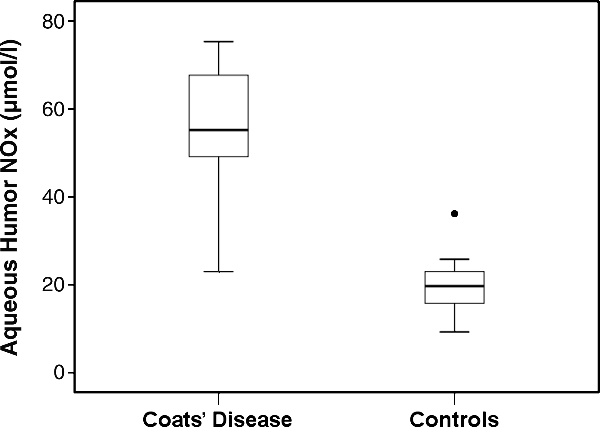
Box plot of the aqueous humor levels of nitrite and nitrate (NOx) in the Coats' disease and control groups. The box for each group represents the interquartile range (25–75th percentile), and the line within this box represents the median value. The bottom and top bars of the whisker indicate the minimum and maximum values, respectively. Outlier values are indicated (circles).
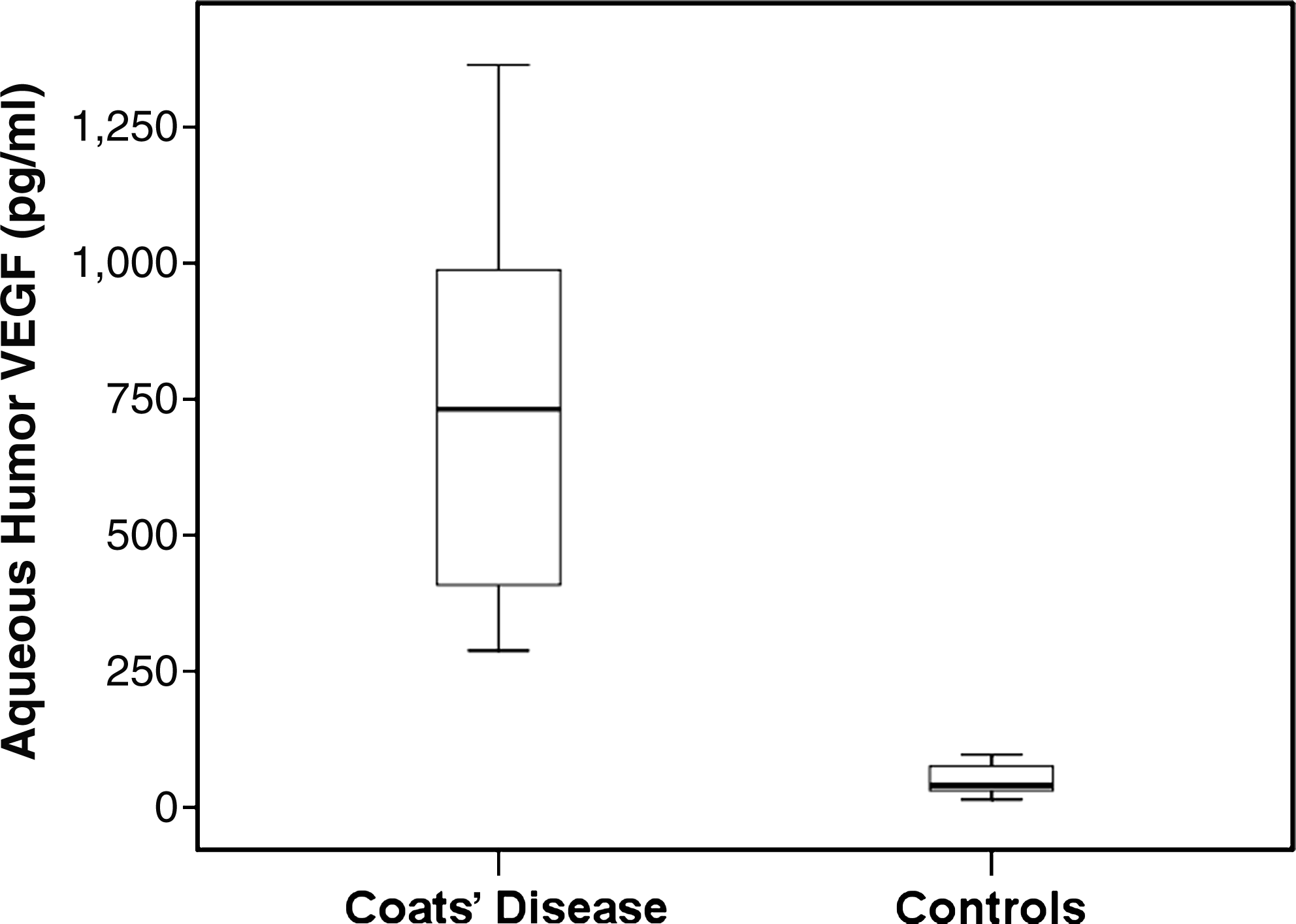
Box plot of the aqueous humor vascular endothelial growth factor (VEGF) levels in the Coats' disease and control groups. The box for each group represents the interquartile range (25–75th percentile), and the line within this box represents the median value. The bottom and top bars of the whisker indicate the minimum and maximum values, respectively.
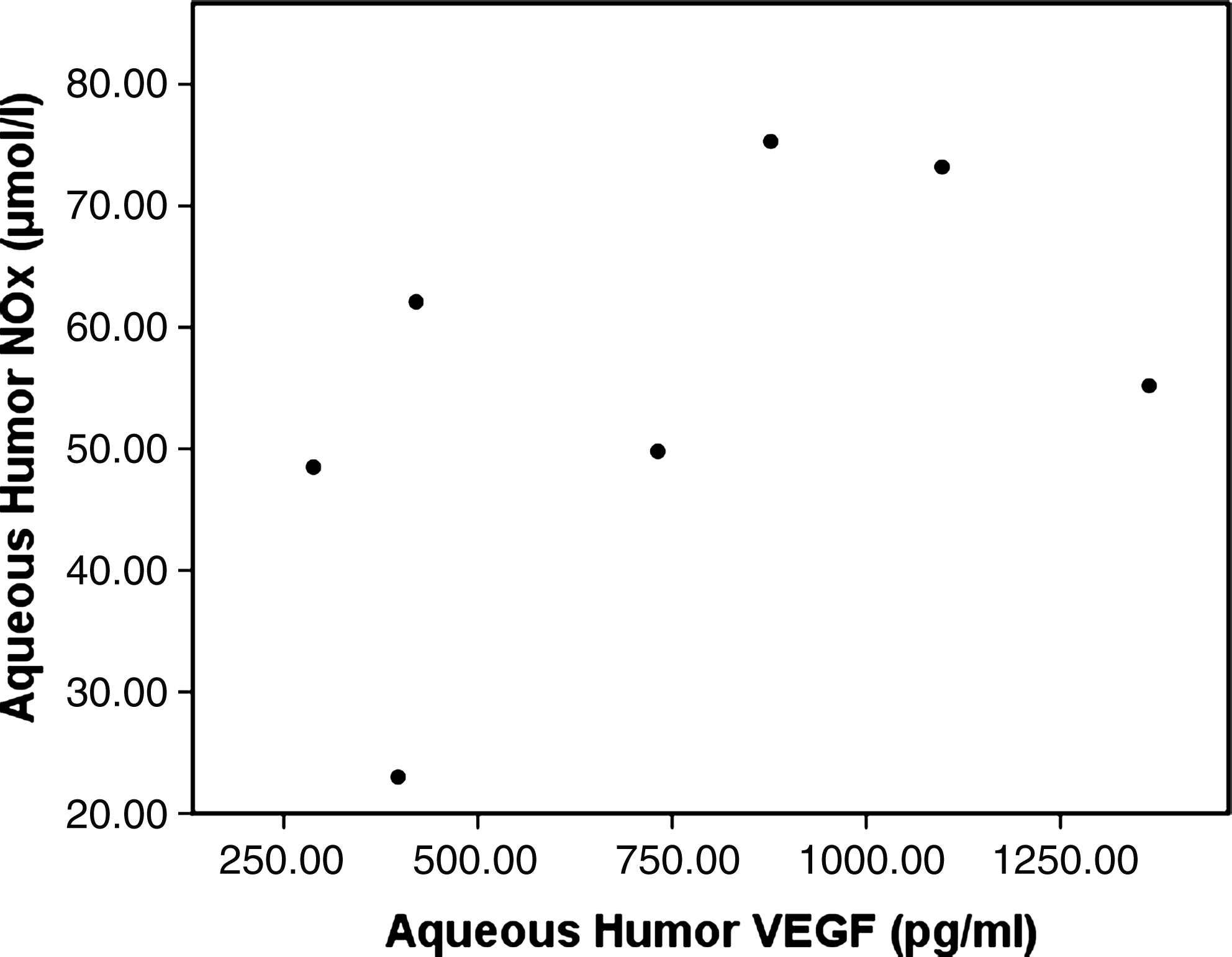
Lack of relationship between VEGF and NOx in the aqueous humor of patients with Coats' disease.
Discussion
The pathogenesis of Coats' disease is uncertain. Telangiectatic blood vessels experience a loss of endothelial cells and pericytes with subsequent mural disorganization and a breakdown of the blood–retinal barrier. 17 These blood vessels leak lipoproteins into the retina, which accumulate there. Hence, lipoproteins break through the external limiting membrane of the retina causing non-rhegmatogenous exudative retinal detachment.2,20 The microvascular changes in Coats' disease may be associated with the exacerbation of retinal vascular abnormalities as a result of impaired oxygenation of the outer retina and secondary expression of increased VEGF. 21 VEGF is considered a key molecule in the regulation of angiogenesis, which not only causes pathologic angiogenesis but also induces vascular leakage and exudation in many ocular disease states. 22 A recent report indicated that the VEGF level was elevated in Norrie disease pseudoglioma (NDP) homologue knockout mice, 23 and mutation of the NDP gene has been postulated to play a role in the pathogenesis of Coats' disease. 24 VEGF levels in ocular samples with Coats' disease have been studied previously. A single case report showed an elevated VEGF level in the vitreous of a patient with stage 4 Coats' disease, and this level significantly decreased after intravitreal injection of an anti-VEGF agent. 5 Another study, including 4 patients with Coats' disease, showed that the intraocular VEGF level is >100 times higher in diseased than control eyes. 6 Several case reports also showed that bevacizumab, a recombinant humanized monoclonal antibody against all VEGF isoforms, may be a valuable adjunctive therapy for Coats' disease.7–9 In this study, elevated VEGF levels were noted in the aqueous humor of the eyes of patients with Coats' disease, and VEGF levels were significantly higher in patients with Coats' disease than the control group. Our results confirm those of previous reports and provide further evidence to support the role of VEGF in Coats' disease.
NO is widely recognized to be an important intercellular messenger in the cardiovascular and nervous systems and immunological reactions, including those in the eye. Generally, this molecule, which is formed by constitutive NOS, eNOS, and nNOS, contributes to the physiological regulation of ocular hemodynamics and cell viability and protects vascular endothelial cells and nerve cells or fibers against pathogenic factors associated with glaucoma, ischemia, and diabetes mellitus. On the other hand, NO formed by iNOS expressed under influences of inflammatory mediators evokes neurodegeneration and cell apoptosis, leading to serious ocular diseases. One of the well-characterized functions of NO is acting as a mediator of vascular dilation and permeability. 10 Takeda et al. found a higher expression of nNOS and eNOS in the retinas of diabetic rats than in those of control rats, indicating that increases in constitutive NOS could be associated with retinal vascular permeability. 25 Further, VEGF produces a dose-dependent upregulation of NO generation in human endothelial cells, 26 and it has been shown that NO is involved in signaling VEGF's permeability-enhancing effects.27,28 In addition, research has indicated that NOS inhibition blocked VEGF-induced vascular hyperpermeability in all ocular tissues. 29 As one of the fundamental abnormalities in Coats' disease is increased retinal vascular permeability, we hypothesize that NO is a contributing factor in the pathogenesis of Coats' disease and have evaluated the aqueous humor NO levels in patients with this disease. The results show that patients with Coats' disease have elevated NOx levels in the aqueous humor. To our knowledge, this is the first investigation of NO levels in patients with Coats' disease, and our results suggest that inhibiting NOS may benefit patients with this disease.
Although we presume that elevated NOx levels in Coats' disease are mainly correlated with increased retinal vascular permeability, other pathologic processes such as retinal ischemia and inflammation may also be involved. It is well known that NO has a role in ischemic processes. 10 Retinal ischemia induced in rats by bilateral common carotid artery occlusion resulted in the expression of nNOS and iNOS in Müller and retinal ganglion cells. 30 These phenomena may be involved in ischemic damage of the retina. Further, clinical research showed that aqueous humor NO levels increased in patients with retinal artery and ischemic retinal vein occlusion.13,31 In Coats' disease, retinal ischemia is usually found in areas of telangiectasia.2,3,21 According to FA, peripheral retinal capillary nonperfusion was notable in all of our cases, indicating that retinal ischemia may also have contributed to the elevated aqueous humor NOx levels in the present study. On the other hand, several studies suggest that NO is involved as a proinflammatory mediator in immunological reactions or inflammation in the eye.10,14 A previous study reported elevated aqueous humor NO levels in patients with uveitis. 32 In the eyes of patients with Coats' disease, several histopathological studies have shown that lipid-laden macrophages are present in the sensory retina and subretinal fluid,2,17,20 and inflammatory macrophages are considered as a source of NO. 33 These findings indicate that immunological reactions or inflammation and NO may be involved in the pathogenesis of Coats' disease.
Although the aqueous humor concentrations of NOx and VEGF were higher in our patients with Coats' disease than our control subjects, no correlation was observed between NOx and VEGF. As the angiogenic effects of VEGF can be mediated by eNOS-generated NO and NO has been suggested as a downstream imperative of VEGF,26–28 this was an unexpected result. One possible explanation may be that the aqueous humor NOx level may be affected by serum diffusion. In a similar study on PDR, NOx and VEGF were increased but not related in the vitreous fluid of patients with PDR. 34 The authors believed that serum diffusion could play a significant role in the intravitreous enhancement of NOx, but not the increase of VEGF. In Coats' disease, the disruption of the blood retinal barrier could favor the passage of NO from systemic circulation into the eye and affect the aqueous humor concentration of NOx. Another possible explanation may be that the aqueous humor NOx level may not be influenced by VEGF alone but multifactorially such as certain cytokines related to ischemic and inflammatory processes. Further, there have been conflicting assertions regarding the interaction between VEGF and NO. Although VEGF upregulates the expression of endothelial eNOS and elicits the subsequent production and release of NO from endothelial cells, NO has also been shown to serve as an endogenous inhibitor of VEGF expression in vivo. 35 It is possible that a reciprocal regulation between VEGF and NO exists in the retina. If this is applicable in the pathogenesis of Coats' disease, it is not surprising that no correlation was observed between aqueous humor NOx and VEGF in the patients in the present study.
Our study showed that patients with Coats' disease had higher NOx and VEGF levels in their aqueous humor than controls, but no correlation was observed between NOx and VEGF. While these results suggest that VEGF and NO may play important roles in the pathogenesis of Coats' disease, further studies are needed to clearly elucidate the relationship between VEGF and NO and other cytokines in Coats' disease. A better understanding of the pathogenesis of Coats' disease will be key in developing new treatment. Based on our results, anti-VEGF therapies or treatments based on the inhibition of NOS would be likely to provide promising approaches for patients with Coats' disease.
Footnotes
Author Disclosure Statement
The authors reported no conflicts of interest and are solely responsible for the content and writing of this article.