
Abstract
Abstract
Purpose:
Fluconazole is a bis-triazole antifungal agent with a low molecular weight (306 Da). It is hydrophilic in nature and has low protein binding. It is available as eye drops for the treatment of ocular mycoses, the second most common cause of blindness in developing countries. However, its administration often results in poor patient compliance and limited use due to its short half-life (15–30 min) and a low log P (0.25). Therefore, fluconazole was incorporated into a novel sorbitan (spans) based elastic (spanlastic) vesicular system with intent to achieve a prolonged and better effect. Spanlastics are to niosomes what Transfersomes® are to liposomes.
Methods:
Developed spanlastics consisted of spans and an edge activator prepared by the ether injection method. Developed vesicles were characterized for size, shape, and the number of vesicles/ml by optical microscopy. Entrapment efficiency was determined by the dialysis method, and the ex vivo corneal permeability study was performed using porcine cornea. A 3-tier safety of the novel formulation was established by the Ames test, the 3-(4, 5-dimethylthiazol-2-yl)-2, 5-diphenyltetrazolium bromide (MTT) assay, and in rabbits according to the OECD guidelines 404 and 405.
Results:
Spanlastics were smaller in size (3 times) and showed a better permeation in comparison to a corresponding niosomal formulation. The system showed an increase (3-fold) in the apparent permeability coefficient compared to the marketed formulation Zocon® (0.3% w/v solution of fluconazole) due to its elastic nature. The developed system was found to be stable for 2 months under refrigerated conditions and under extreme storage conditions. Safety was established in terms of genotoxicity (Ames test), cytotoxicity (MTT assay; mouse peritoneal macrophages), acute dermal/eye irritation/corrosion, and chronic eye irritation/corrosion tests (OECD guidelines).
Conclusion:
The developed system is novel and provides an effective and safe formulation of fluconazole.
Introduction
Due to physiological and anatomical constraints,7–9 only a small fraction of the administered drug, effectively 1% or even less of the instilled dose, is ocularly absorbed.10,11 This forces the clinician to recommend a frequent dosing at an extremely high concentration, resulting in a pulsed bioavailability profile. Antifungal treatment furthermore involves aggressive and chronic therapy. 12
Fluconazole (FLZ) is a stable, water-soluble, bis-triazole antifungal with low molecular weight (306 Da), low protein binding (∼11%), and low toxicity. It is available as eye drops for the treatment of ocular mycoses. Important considerations in reference to its effectiveness are its (a) short ocular half-life, of almost 15 min in the debrided eye and 30 min in the nondebrided eye 13 and (b) low partition coefficient (log P 0.25). 14 The latter indicates poor permeability across the ocular epithelial membrane, ultimately leading to poor bioavailability and effectiveness.
The aim of the present study was to develop an effective ocular carrier system for fluconazole and similar agents, so as to result in effectual permeation across the corneal epithelium coupled with a prolonged effect. Antifungal agents need to be administered very frequently and for long periods to cure the infection. Hence, it was proposed to incorporate fluconazole into a suitable elastic vesicular system that will attain this target.
In 1992, Cevc and Blume 15 introduced the first generation of highly deformable, elastic liposomes, referred to as Transfersomes®. A second generation of elastic vesicles (EVs), mainly consisting of nonionic surfactants (NISs), was introduced in 1999 by Van den Bergh. 16 In 1997, Touitou et al. 17 developed ethosomes, soft vesicular carriers mainly consisting of phospholipids and ethanol. These elastic vesicles have been found to deeply and easily penetrate through the skin.18–20
Shen and Tu 21 developed ganciclovir elastic liposomes that showed significant transocular absorption as compared to the drug solution. The results were attributed to the particle size (200 nm) and the elasticity of liposomes. They proposed that deformable elastic liposomes can enter the corneal structure that is similar to the stratum corneum. A recently published article from our laboratory 12 advocates the advantage of elastic vesicles over niosomes. In that, the former can reach even the posterior eye segment due to its elasticity and nanosize. This elasticity is attributed to the use of edge activators (EAs). EAs are the surfactant molecules that provide flexibility to the lipid bilayer membranes of these vesicles. These surfactant molecules destabilize the lipid bilayers and, in turn, increase the deformability of the vesicles. 22 The surfactant present in these vesicles causes them to induce pores in lipid structures, such as membranes, and also provokes a solubilization (lysis) in the higher concentration range.
Methods
Materials
Fluconazole was gifted by Sreenivasa Pharma. Pvt. Ltd. Other materials used in the study are glutathione, oxidized, (Hi Media Laboratories), sodium cholate (Gift sample from New Zealand Pharmaceutical Ltd.), sodium deoxycholate (Gift sample from Panacea Biotec Ltd.), sodium taurocholate (Gift sample from New Zealand Pharmaceutical Ltd.), cholesterol (Sigma-Aldrich Chemie GmbH) and Zocon® (Fluconazole eye drop 0.3%w/v; Ahlcon Parenterals).
Preparation of elastic vesicles
Ether injection method
The NISs used in the study included sorbitans such as Span 40 (S40), Span 60 (S60), and Span 80 (S80). Various EAs, namely, sodium taurocholate (EA1), sodium cholate (EA2), sodium deoxycholate (EA3), and tween 80 (EA4), were used, and their efficiency to result in a most suitable system was also evaluated. NISs were used in varying combinations to produce different formulations. NIS dissolved in ether was injected into the continuously stirred aqueous phase consisting of EAs maintained at 60°C. The ethereal solution was injected using an 18-gauge needle at the rate of ∼0.25 mL/min. 23
As ether is regarded as a safe solvent by USP and also Spans are soluble in it, ether was used for the development of formulations. 24
Characterization of elastic vesicles
Vesicles were evaluated for shape, size, lamellarity, and abundance (no. of vesicles/mm3). Preoptimization was done by evaluating aggregration/irregularity in shape upon storage, for 1 month in the refrigerator (8°C). Formulations showing no or minimal aggregation upon storage were selected for further studies. The morphological characters (viz. shape uniformity and lamellarity) of prepared vesicles were monitored employing optical microscopy (eclipse i80; Nikon). Vesicles were suitably diluted with water, and the no. of vesicles per cubic millimeter was counted by optical microscopy using a hemocytometer. The vesicles in 80 small squares were counted, and the no. of vesicles/mm3 was calculated using the following formula
25
:
To determine the total drug content, 1 mL of the formulation was disrupted using isopropyl alcohol (IPA), and the solution was analyzed spectrophotometrically at 262 nm. IPA was chosen as a suitable solvent for disrupting the prepared vesicles. 12
The entrapment of vesicles was determined using the dialysis bag method,
26
and the % entrapment was calculated as follows:
Selected vesicles were observed under an optical microscope (Nikon eclipsed i80; magnification 40×) and a transmission electron microscope (TEM; at magnification 20,000× and 200,000×) for structural attributes such as lamellarity, uniformity of size, shape, and physical stability characteristics, that is, aggregation and/or irregularity. The size and size distribution of vesicles were determined by a dynamic light-scattering method, using a computerized inspection system (Malvern Zetamaster, ZEM 5002; Malvern).
pH of the formulations was measured using Cyberscan, Eutech pH 510, at 25°C. Zeta potential of the vesicular dispersions was measured using the Malvern's zetasizer. The measurements were done at 25°C, and the electric field strength was around 23.2 V/cm. The zetasizer measures the zeta potential based on the Smoluchowski equation (ζ=UE η/ɛ; where ζ is zeta potential, UE is electrophoretic mobility, η is viscosity of the medium, and ɛ is dielectric constant). 27 Comparative measurement of the elasticity of prepared vesicles and the corresponding niosomes was carried out by extrusion measurement. 28 The vesicles were passed (extruded) through a polycarbonate membrane with a pore size 1.0 μ at constant pressure.
Ex vivo corneal permeability studies
Ex vivo corneal permeability studies were performed using the membrane diffusion technique. The studies were conducted within a jacketed cell and maintained at a constant temperature (37°C±0.2°C), under constant stirring using a magnetic stirrer. This study was performed as reported earlier. 29 The cell used was a 2-limbed reservoir; on 1 limb of which cornea was mounted, and the other limb was used as the sampling port (volume=20 mL). The preparation (0.3 mL) to be studied was placed on the cornea. Porcine cornea was used for the study, and the cornea was mounted within half an hour of sacrifice of the animals. The diffusion medium used was freshly prepared glutathione bicarbonate ringer, pH 7.4. Aliquots of the medium were withdrawn after a fixed time interval from the sampling port and were replaced with equal quantity of fresh media to maintain a constant volume. Sink conditions were maintained throughout the study. Samples were analyzed spectrophotometrically at 262 nm.
The apparent corneal permeability coefficient (Papp) of different formulations was determined according to the following equation:
where
Stability of formulation
The formulations were evaluated for stability in terms of drug leakiness and aggregation behavior. The formulations were kept in 15-mL sealed glass ampoules and stored under different conditions (refrigerator, ie, 2°C–8°C and room temperature, ie, 35°C±2.0°C). Aliquot samples from the formulations were withdrawn at definite time intervals (0, 1, and 2 months) and analyzed spectrophotometrically for any change in the extent of entrapment with respect to the initial sample (0 month; 100% entrapment). The samples were also evaluated microscopically to observe the extent of aggregation.
Freeze–thaw cycle
Formulations were subjected to a series of 4 repeated freeze–thaw cycles 30 at −20°C overnight.
Stability upon sterilization
Final vesicular formulations were sterilized by autoclaving at 121°C (15-psi pressure) for 20 min. Any change in vesicles in terms of both change in size and abundance and also change in % entrapment (leakiness) was observed poststerilization.
Differential scanning calorimetry
Differential scanning calorimetry (DSC) thermograms were obtained on DSC (Metler Toledo, Switzerland). The calorimeter was calibrated for temperature and heat flow accuracy using the melting of pure indium (mp 156.6°C and ΔH of 25.45 J g−1). The temperature range was from 0°C to 400°C with a heating rate of 5°C per minute. The gas used was nitrogen with a purging rate of 50 mL/min.
Safety studies
Since the vesicular systems developed and reported by us were novel systems and composed of surfactants only (spans and EAs), it was necessary to evaluate them for safety. We employed a 3-tier system of evaluation that included in vitro genotoxicity, in vitro cytotoxicity, and in vivo ocular irritation (OECD guidelines) study in rabbits. Details of these tests are given below.
Determination of mutagenic activity (Ames test)
The test was carried out as per details given by Maron and Ames. 31
MTT assay: in vitro cytotoxicity in mouse peritoneal macrophages (normal cells)
The cytotoxicity of drug loaded elastic vesicles and plain drug solution was evaluated in triplicate using the mouse peritoneal macrophage cell line. 32 Mouse peritoneal macrophage cell lines were cultured under standard conditions (5% CO2, 98% humidity, 37°C) in a medium supplemented with 10% heat-inactivated fetal bovine serum, penicillin (100 units/mL), streptomycin (100 μg/mL), L-glutamine (0.3 mg/mL), pyruvic acid (0.11 mg/mL), 0.37% NaHCO3, and 50 μM of 2-mercaptoethanol. The cells were plated in 96-well plates at a density of 0.6×10⁶ in 200 μL of medium per well and incubated with different concentrations of the test material (the vesicular dispersion or solution containing 0.3% w/v of FLZ was diluted with cell suspension to achieve 1, 5, and 10% v/v) for 48 h. The medium was refreshed with a fresh medium containing 100 μg/mL of 3-(4, 5-dimethylthiazol-2-yl)-2, 5-diphenyltetrazolium bromide (MTT) for 3 h. Plates were centrifuged at 200 g, and the supernatant was aspirated, and violet color MTT-formazan crystals were dissolved in 100 μL dimethyl sulfoxide. Thereafter, plates were stirred for 20 min, and optical density (OD) was measured at λ 570 nm (reference wavelength, λ 620 nm) on an ELISA plate reader (Thermo Labs). Cell growth was calculated by comparing the absorbance of treated versus untreated cells. The OD of violet color formazan crystals was directly proportional to the cell viability.22,23 The plot was drawn between % cell viability and the different concentrations of tested materials.
In vivo safety study
All the animal study protocols were approved by the Institutional Animals Ethics Committee, Panjab University, Chandigarh, India.
Acute dermal irritation/corrosion test as per the OECD guideline 404
According to the OECD guideline 405, before considering the in vivo eye irritation/corrosion test, preferably a study of the in vivo dermal effects of the substance should be conducted and evaluated in accordance with the OECD Testing Guideline 404. 33
The substance to be tested was applied in a single dose on the skin of a rabbit; untreated skin areas of the test animal served as the control. The degree of irritation/corrosion was observed and scored as per the details described. 33
Acute eye irritation/corrosion test as per OECD guideline 405
Acute dermal irritation/corrosion study was followed by an acute eye irritation/corrosion study in accordance with the OECD guideline 405. 34 The degree of eye irritation/corrosion was evaluated by scoring lesions of conjunctiva, cornea, and iris, at specific intervals. The test indicates that the animals should also be observed for other effects in the eye.
Chronic repeat instillation irritation/corrosion test
This study was planned to further evaluate whether the vesicular systems were safe for a long-term therapy or not. Same scale and testing parameters were followed as were used for conducting the acute study. This study was a repeated dose study in which 5 repetitive doses of the vesicular dispersion were instilled in the conjunctival sac at an interval of 5 min, for a period of 1 week. Animals were evaluated for ocular irritation/corrosion based on the prescribed scale. 34
Challenge test
The test consists of challenging the preparation with a prescribed inoculum of suitable microorganisms, storing the inoculated preparation at a prescribed temperature, withdrawing samples from the container at specified interval of time, and counting the organisms in the samples so removed. 35
The preservative properties of the preparation are adequate if, in the condition of test, there is a significant fall in the number of microorganisms in the inoculated preparation after the times and at the temperatures prescribed. The preservative used in the preparation was 0.03% w/v sodium perborate. 36
Statistical analysis
The raw data obtained from in vitro studies were analyzed by applying the correction factor for volume and drug losses during sampling. All results are expressed as the mean±standard deviation. The results were analyzed for statistical significance by a 1-way analysis of variance (ANOVA) test followed by the Tukey's test or the Students pair t-test, whichever applicable.
Results
In the present study, we prepared elastic vesicles (spanlastics) using NISs and EAs in suitable ratios so that they exhibit flexibility at all levels of combination.
Characterization of elastic vesicles
Various parameters related to morphology of the vesicles were taken into account for selecting a suitable NIS and EA and their respective NIS:EA ratio (Table 1). Based on the results, final combination ratios were selected. It may be noted that 25 combinations for each NIS were evaluated with respect to the vesicle size, shape, abundance (Table 1), and lamellarity. Higher cell count and stability were observed in case of vesicles prepared from Span 60 in comparison to Span 40 and Span 80.
Very few, 1,000–10,000; few, 10,000–15,000; fairly good, 15,000–20,000; good, 20,000–30,000; abundant, 30,000–40,000; very abundant, 40,000-above; S40, Span 40; S60, Span 60; S80, Span 80; Chol, cholesterol; EA1, sodium taurocholate; EA2, sodium cholate; EA3, sodium deoxycholate; EA4,tween 80; AG, aggregation; IR, irregularities.
Various combinations of span and EAs were tried (Table 1). Based upon the preliminary evaluation, suitable ratios of Span 60 and the corresponding EAs were selected (Table 2). The selection criterion was based on the highest cell count and stability of vesicles upon storage for a period of 1 month, especially in terms of aggregation/irregularity and cell disruption. The niosomal formulation (f1) was taken as control, and developed formulations were compared with it (80:20 ratio was selected based upon the highest cell count observed at this combination).
The selected ratios were developed into ocular formulations containing 0.3% w/v of FLZ (unentrapped drug was not removed).
Entrapment efficiency of the vesicles
The formulations f3 and f5 showed a significantly high entrapment, that is, 65.73% and 62.69%, respectively (Table 2), which was 39% and 32%, respectively, more than the f1 (47.21%) and thus were opted as the formulations of choice. The cell count/mm3 was also higher for f3 and f5. In addition to being large in size f1 was also invariably uni- or bilamellar in nature. Moreover, they were less abundant than all other types (Tables 1 and 2).
Drug content
The actual amount of FLZ added for all practical purposes was 3.0 mg/mL, and the drug content of all the formulations was not found to be significantly different from the added amount (Table 2).
Vesicles shape and type
Microscopy
f3 and f5 were examined microscopically (Nikon eclipsed i80; magnification 40×). Observation under an optical microscope indicated the vesicles to be small in size, round in shape, and bilamellar/multilamellar (Fig. 1c, d) showing no aggregation or irregularities. The particles were uniformly distributed as shown in Fig. 1a and b.
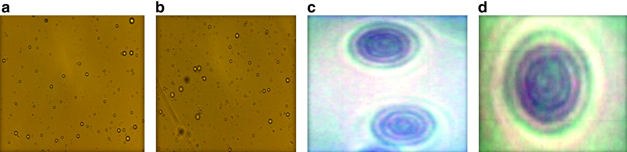
Optical photomicrographs (40× magnification) of f3
Transmission electron microscopy
f3 and f5 were evaluated under TEM, and were confirmed to be small, round, and bilamellar/multilamellar (Fig. 2a and b) and no aggregation/irregularity was observed.
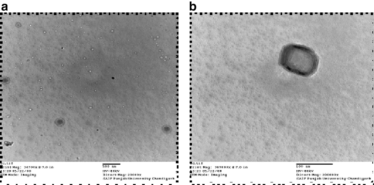
Transmission electron microscopy images of f3
Vesicle size and size distribution
The average particle size of the developed vesicular system is shown in Table 3. It also depicts the effect of sonication on the entrapment efficiency of the vesicles. With the decrease in the particle size upon sonication, a considerable decrease in the entrapment capacity of the vesicular system was observed. While the decrease was 25%–30% in case of f3 and f5, it was even more (37%) for f1. Hence, formulations were not subjected to any sonication, considering that the particle size achieved otherwise is acceptable for ocular administration. However, it may be observed that nanovesicles of size <200 nm could be achieved with ultrasonication of 3 min at a 98% amplitude. Further optimization of the ultrasonication method and/or of the formula may result in suitable nanovesicles with minimum compromise of entrapment. However, the deformation to an ultrathin shape can serve the purpose of the nanoparticle, even if the initial dimensions are of 2–3 μm.
pH of the formulations
Unbuffered solutions with pH 4.0–8.0 are, however, usually acceptable. 37 The pH in both cases was found to be 7.0. Almost no change in the pH of these dispersions was observed (for up to 2 months) when stored under refrigerated conditions.
Zeta potential
Zeta potential of the formulations f3 and f5 was measured using Malvern's zetasizer at 25°C. The observed zeta potential of the optimized formulations f3 and f5 was found to be −25.2 and −13.7, respectively. The negative value of the zeta potential obtained for both f3 and f5 indicates a low scope of coalescence of vesicular system as confirmed from optical microscopy (Fig. 1c, d).
Degree of deformation
Developed EVs showed a much smaller change (4.2% for f5 and 11.0% for f3) in average particle size (Table 4) in comparison to niosomes showing a large decrease in size of 64.6% after passage through a 1-μm polycarbonated membrane.
Preparation of formulation f3 and f5 was scaled up (10 times) to produce bigger batches.
Ex vivo corneal permeability studies
Ex vivo corneal permeability data of the various formulations are summarized in Table 5. A 1-way ANOVA followed by the Tukey's test was applied on the data, and significance was considered at P<0.05.
All the values were significantly different from one another, except for those marked similarly as superscript letters a, b, and c.
Statistical analysis of the data indicates that all the formulations, that is, f1, f3, and f5, were significantly better than the marketed product (Zocon®, fluconazole eye drop 0.3%w/v by Ahlcon Parenterals) at all time intervals, as shown in Table 5. All 3 formulations, f1, f3, and f5, showed an improvement in the intrinsic apparent permeation coefficient (Papp) of the FLZ solution from 5.3×10⁻⁶ to 1.3×10⁻⁵ cm/sec.
The % amount permeated was highest in case of f3 (29.0%) and lowest in case of Zocon® (11.4%). An overall slow (initial phase of very slow permeation up to 1.0 h) and low permeation of FLZ from Zocon® was expected because of the hydrophilic nature of the drug. f5 results (28.6%) were almost similar to that of f3, with the value being significantly higher than f1 (26.9%) and Zocon®. 38 However, the apparent permeability coefficient of f1 and f5 was not significantly different while f3 showed a significantly higher Papp.. Fig. 3 shows % permeated at various time intervals of all formulations through the porcine cornea.
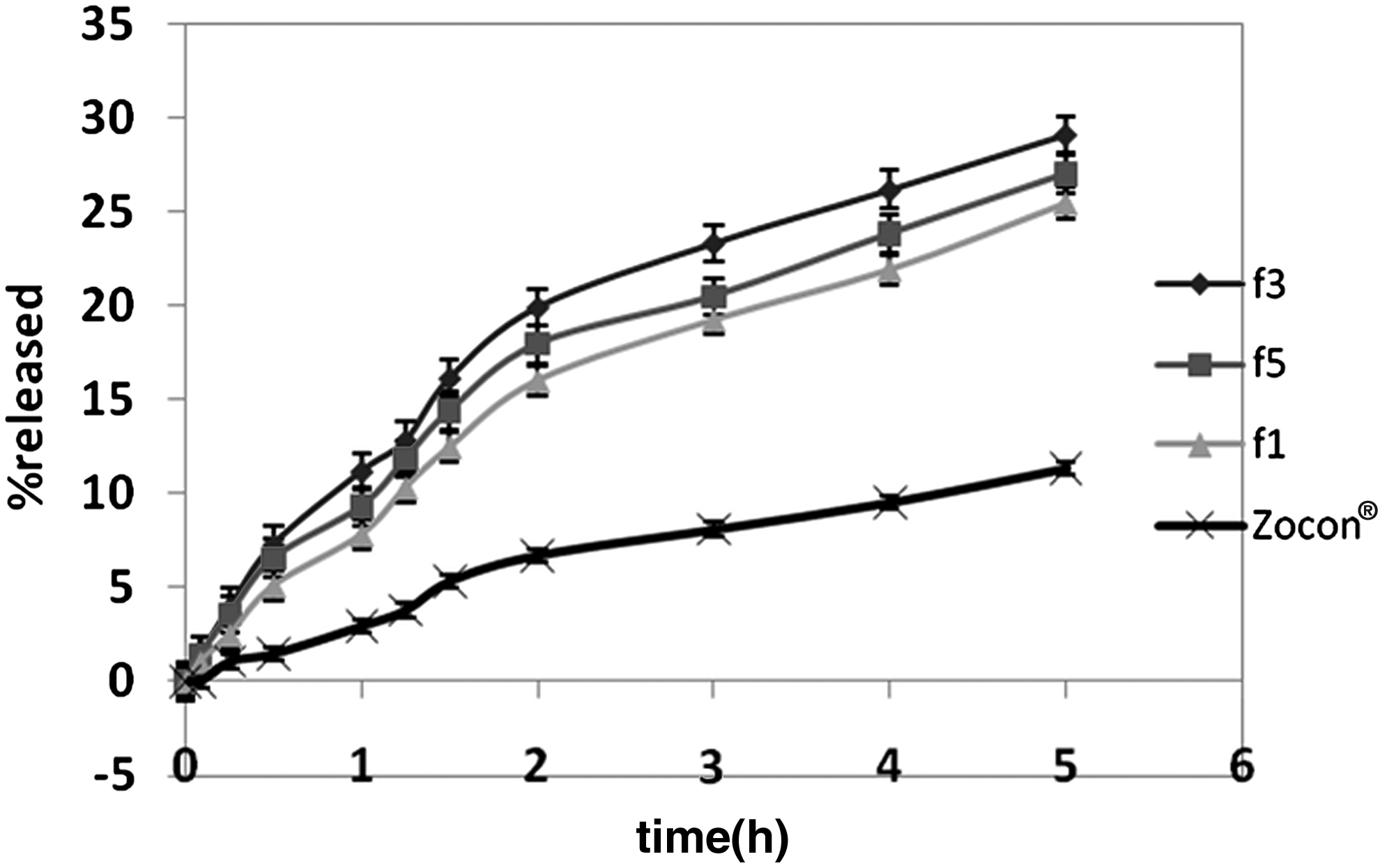
Percent permeated through porcine cornea-versus-time (min) data of f1, f3, f5, and marketed formulation.
Stability studies
The stability studies were conducted under refrigerated conditions of storage (4°C–8°C) and at ambient room temperature (35°C±2.0°C). Stability was measured in terms of % leakiness and aggregation/irregularity of vesicles over a period of 1 and 2 months of storage. There was a little change in the entrapment (almost 5%) under refrigerated conditions of storage, while there was an appreciable change in the entrapment (almost 15%) for formulations f3 and f5, at ambient room temperature. There was mild aggregation under refrigerated conditions and severe aggregation at room temperature for both formulations f3 and f5. The increase in drug leakiness and degree of aggregation at elevated temperatures may be related to an increase in the kinetic energy of the vesicle, resulting in a higher rate of collision between these vesicles and their subsequent rupture.
However, when dispersions were exposed to repeated freeze-thaw cycles, the results clearly showed that both the systems were extremely stable under a stress condition with a percent drug loss of not >2% (Table 6). There was not much change in the vesicle count values as well.
Stability upon sterilization
Autoclaving cannot be described as a suitable method of choice for sterilization of these vesicular systems. Because particle size was found to reduce nearly to half the original size, with percent leakiness of almost 6%.
Differential scanning calorimetry
Fluconazole gave 2 peaks at temperatures 139.71°C and 101.76°C (Fig. 4a), which are characteristic peaks of the drug. A degradation peak at 338.87°C was also observed. The constituent surfactants, namely, Span 60 (Fig. 4b), sodium cholate (Fig. 4c), and tween 80, showed endothermic peaks at 50.65°C, 121.12°C, and −14°C, 39 respectively. The formulations f3 and f5 showed broad peaks at 120.20°C (Fig. 4d) and 210.54°C (Fig. 4e), respectively, and no peak corresponding to fluconazole was observed, indicating its entrapment within the vesicles.

Differential scanning calorimetry thermograms of drug
Further, the physical mixture showed individual peaks for Span 60 and sodium cholate (Fig. 4f), but not of drug, indicating the interaction of the drug with span (which melts at 50.65°C) and tween 80 (Fig. 4g) (the exothermic peak for tween 80 was not observed in the DSC, because instrument did not run below 0°C). Broad and single DSC thermograms observed both for the blank (Fig. 4h, i) and drug-loaded EV dispersions confirm significant interaction of components and point towards their amorphousness.
Safety studies
Determination of mutagenic activity (Ames test)
Since the revertant count (Table 7) produced by spanlastics was not more than the negative control, it was concluded that the formulations were nongenotoxic.
MTT assay: in vitro cytotoxicity in mouse peritoneal macrophages (normal cells)
The toxicity of the developed formulations (f3 and f5) and the corresponding empty vesicles (b3 and b5) was tested for the MTT cell viability assay in mouse peritoneal macrophages. Both the f3 and f5 and their corresponding empty vesicles b3 and b5 did not show any toxic effect on cell proliferation of mouse peritoneal macrophages when applied at a concentration of up to 10% v/v in the cell culture suspension medium. It may, however, be noted that a plain drug solution of FLZ (0.3% w/v) when applied as 1% v/v in the cell culture suspension showed a 50% cell growth inhibition. The extent of cytotoxicity increased dose dependently at 5% and 10% v/v concentrations (Fig. 5). Hence, it may be concluded that not only were the developed vesicles noncytotoxic but also the entrapment of FLZ within these vesicles reduced its inherent cytotoxic effect.

Cell viability versus concentration.
In vivo safety studies
Acute dermal irritation/corrosion test as per the OECD guideline 404
The interpretation of obtained scores (0/80) clearly indicated a nonirritant/corrosive nature of developed vesicles when applied to dermal tissues; hence, they may be considered safe for dermal application. The study was conducted in accordance with the OECD guideline 404. 33
Acute eye irritation/corrosion test as per the OECD guideline 405
Single-instillation study
The scores (0/195) for developed formulations again established that developed vesicular systems are safe for ocular use. There were no signs of irritation/corrosion in any of ocular tissues. The study was conducted in accordance with the OECD guideline 405. 34
Repeated-instillation study
This study is a modification or extension of the previous study and was performed based on the fact that it is usually recommended to frequently repeat the instillation of antifungal preparations for an effective control of the infection. Similar studies have been reported by another group also. 40 The scores obtained from this study also proved the safety of developed systems for ocular use.
A tear film disruption was, however, observed at 1- and 24-h intervals in this study. This may be caused by the adherence of these vesicles to the corneal/conjunctival surface such that they disturb the integrity of the tear film. The surfactant nature of these vesicles may also be responsible for such action. The observation, however, indirectly indicates that the systems were probably present in the eye even after 1 and 24 h of instillation.
The score for f5 was 0/195, whereas a score of 2/195 was observed for f3, which is also too small to be of any serious concern. Hence, the developed vesicles may be considered safe for ocular use. Moreover, the redness of conjunctiva, which was observed at 24 and 48 h (in one of the rabbits), was reversible at 72 h, and as per OECD guidelines, a reversible change indicates the noncorrosive nature of the test material.
Chronic repeat-instillation irritation/corrosion test
As already discussed in earlier sections, the treatment of ocular fungal infection involves long-term therapy. So, we evaluated the developed formulations for chronic repeat instillation (5 times at 5-min intervals) also. The score of both f3 and f5 for eye irritation/corrosion study was 0/312. The interpretation of scores clearly depicted that both vesicular systems were nonirritant/non-corrosive upon the chronic use to all ocular tissues. Considering the low score of the study, both the vesicular systems may be considered safe for a chronic ocular use.
Challenge test
Sodium perborate (0.03% w/v) was incorporated into f3, f5, and a plain drug solution, that is, 0.3% w/v of FLZ, and the challenge test was performed according to British Pharmacopoeia. 35 Suitable positive and negative controls were included in the test.
At 6 h, f3, f5, and the plain drug solution showed almost 70%–84% and 70%–75% reduction with respect to the count at 0 time in case of Staphylococcus aureus and Pseudomonas aeruginosa, respectively, whereas around 100% reduction was observed in case of Candida albicans and Aspergillus niger. No viable microorganisms were observed in both vesicular systems and the plain drug solution after 24 h.
It was clear from the results that vesicles do not interfere with the activity of sodium perborate.
A 0.03% solution of sodium perborate diluted in the same manner as the preservative-containing formulation was also evaluated. The log reduction in the number of organisms at 6 h was >3 for all the strains. This value conformed to the pharmacopoeial requirements (wherein a log 2 reduction is suggested at 6 h). At 24 h, a complete inhibition of microorganisms was observed, with the log reduction lying between 5.66 and 3.98. According to the pharmacopoeia, the limit allowed is log 3 reduction. Hence, it may be concluded that in the present formulation, the use of 0.03% sodium perborate served as an effective preservative.
Discussion
First-generation elastic vesicles, also referred to as Transfersomes, were designed by Cevc et al.15,41,42 These deformable vesicles are a novel type of liquid-state vesicles composed of phosphatidylcholine in combination with the EAs (sodium cholate). The vesicle elasticity was obtained by combining stabilizing and destabilizing molecules within 1 lipid membrane. Subsequent studies have demonstrated that Transfersomes are more effective than standard liposomes and achieve an enhanced drug absorption across mouse and human skin.43,44
A new series of elastic and rigid vesicles consisting solely of surfactants have been reported in the last decade. 45 These surfactant-based elastic and rigid vesicles consisted of the bilayer-forming surfactant L-595 (sucrose–laurate ester) alone or in combination with the micelle-forming surfactant PEG-8-L (octaoxyethylene–laurate ester),46,47 respectively.
For our study, suitable ratios of Span 60 (Span 40 and Span 80 did not show suitable results) and various EAs were selected instead of Span 40 and Span 80 combinations with EAs. Since span was the basic constituent of these EVs, they are referred to as spanlastics by us. Spanlastics prepared and reported by us may be considered to be a modification of niosomes in the same way as Transfersomes were for liposomes. It has been reported that niosomes prepared by Span 80 show a high degree of disruption and aggregation, which may be the reason behind the instability of vesicles prepared with Span 80. 48 Presence of saturated alkyl chains in Span 60 probably bestows more sustainability to the developed vesicles. Span 40 though reported to produce stable vesicles 49 produced comparatively less-stable vesicles in the present study. Results indicated that as the concentration of the surfactant or EAs increased, the no. of vesicles/milliliter initially increased, but beyond a particular concentration of EAs, the abundance decreased. This observation may be ascribed to the fact that an increase in the concentration of EAs up to a certain level favors the formation of vesicles; thereafter, however, it causes lysis of the span bilayers. In other words, beyond a certain threshold concentration, a further increase in the amount of surfactant probably dissolves the lipid bilayer, leading to the breakdown of vesicular structures. This supposition is the extension of the concept that maximum micelle formation takes place at critical micelle concentration.
Particle size is an important parameter in in-process control and, particularly, in quality assurance, because the physical stability of vesicular dispersions depends on particle size and the particle size distribution. Particle size of the ophthalmic preparation should be <10 μm to avoid eye irritation. 50 The developed formulations are well within this range and hence are appropriate for ocular use. EAs tend to form vesicles that are more spherical and hence have a smaller particle size. 51 The small particle size of the vesicles as a whole may thus be attributed to the presence of EAs that is attributed to a low aggregation number of the EAs in combination with spans (viz. a viz. presence of cholesterol in case of niosome). The reason for higher entrapment in case of f3 and f5 may be attributed to (i) bigger particle size of f3 and f5 than that of f2 and f4 and (ii) the multilamellar property of both f3 and f5.
The ideal pH for maximum comfort when an ophthalmic preparation is instilled in the eye is indicated to be of the order of 7.2±0.2. 52 Instillation of a solution with a pH different from tears is sometimes irritating and may cause a painful sensation and also initiate lacrimation.
Since the vesicular systems are prepared using ether injection and also are surfactant-based, safety was an important issue. Hence, a 3-tier safety evaluation (genotoxicity, cytotoxicity, and acute and chronic toxicity studies as per OECD guidelines) was done that showed the developed system to be safe for topical use.
The extremely high flexibility of the membrane of EVs permits them to squeeze through pores much smaller than their own diameters. This deformability characteristic permits EVs to penetrate through the biological membranes, spontaneously minimizing the risk of complete vesicle rupture while squeezing through. The flexibility of f3 and f5 may be attributed to the presence of EAs. EAs are often single-chain surfactants that destabilize the vesicles and increase the deformability of the bilayer by lowering the interfacial tension. Niosomes consist of cholesterol, which is known to confer rigidity onto the niosomal structure; this may explain the lower degree of deformation associated with these f1 vesicles.
A better permeation was achieved by the developed systems can be explained based upon 2 mechanisms of transportation. First, the elastic vesicles interact with the epithelial cell membrane and act as penetration enhancers, and subsequently modify the intercellular lipid lamellae (mechanism 1). Second, the elastic vesicles can act as drug-carrier systems, whereby intact vesicles carrying the drug pass through the intercellular spaces and reach across the biological membrane (mechanism 2). This is an extension of the mechanism proposed for other EVs, to act as suitable drug-carrier systems that can cross the membranes intact. 41 It is believed that the successful passage of such carriers is based on 2 important factors: the highly stress-dependent elasticity of the vesicle bilayers and the existence of an osmotic gradient. A significant high permeability achieved with f1 could also be because of the penetration-enhancing effect of Span 60. 53
Conclusions
Sorbitan (spans)-based elastic (spanlastic vesicles) novel vesicular systems were developed in this proposal to act as carrier systems for fluconazole, which is a hydrophilic drug with a short ocular elimination half-life and low log P of 0.25.
The developed spanlastic systems were found to be safe in terms of genotoxicity, cytotoxicity, acute dermal/eye irritation/corrosion, and chronic eye irritation/corrosion test.
It may thus be concluded that the developed systems showed great promise, with their novelty being an added advantage. The established safety added to the confidence for use in ocular, topical, and even oral routes. Use of a combination of surfactants is expected to considerably increase their permeability. The low cost of ingredients of these vesicular systems is also of great significance. Moreover, the method of preparation is highly spontaneous, and hence it will be easy to scale-up and commercialize these vesicles.
Footnotes
Author Disclosure Statement
No competing financial interests exist.