




Abstract
Abstract
Purpose:
To evaluate the activity of lowering intraocular pressure (IOP) by Cassiae seed in the DBA/2J mouse glaucoma model.
Methods:
Young male (mean age: 3 months) inherited glaucoma mice (BDA/2J) were enrolled in this study. To evaluate the potential of Cassiae seed in the treatment of glaucoma, all subjects were divided into 6 groups. There were 1 sham group, positive control identified as group 2 topical brimonidine and group 3 oral acetazolamide, and groups 4–6 Cassiae seed extract (CSE) groups. The lactate dehydrogenase (LDH) level in the anterior aqueous humor and the changes of IOP were investigated. Contents of total polyphenol glycosides in the CSE were measured using a high-performance liquid chromatography (HPLC) method. Chromatographic separation was performed on a Cosmosil 5C18-MS reverse-phase HPLC column (4.6×250-mm i.d., 5 μm) with methanol/0.1% H3PO4 as the mobile phases in a gradient elution mode at a flow rate of 1.0 mL/min and an injection volume of 10 μL. The wavelength of UV detector was set at 254 nm.
Results:
The LDH level in the anterior aqueous humor and IOP significantly decreased after treatment with CSE. The IOP-lowering effect of CSE was comparable to those of oral acetazolamide and brimonidine instillation. There were no abnormal findings in the external appearance, and body weight change after treatment with CSE for 5 weeks. Chrysophanol and physcion were measured by an HPLC method to obtain total polyphenol glycosides of the CSE, and were involved in the IOP-lowering function.
Conclusion:
Cassiae seed may be safe and beneficial for treating glaucoma due to its significant IOP- and LDH-lowering activities.
Introduction
Cassia tora L. (of the family Leguminosae) is a well-known traditional Chinese medicinal plant. Seeds of the plant called Jue Ming Zi in Chinese (the so-called Cassiae Seed) were used to treat eye diseases and improve the eyesight in ancient China. Several studies reported that Cassiae seed possesses various pharmaceutical activities, including antioxidant activity, 5 antihepatotoxic activity, 6 strong growth-promoting activity toward Bifidobacterium bifidum in the gastrointestinal tract, 7 hypolipidemic effects, 8 inhibitory activity against platelet aggregation, 9 and estrogenic and antiestrogenic activity. 10 Nowadays, it is also applied as one of the components in many herbal drinks in Taiwan and China for health purposes. However, relatively little scientific information is available on the role of Cassiae seed in curing ocular diseases.
Several types of experimental glaucoma were first developed in the 1940s and 1950s, primarily in rabbit and monkey species, to provide animals for special pharmacologic and pathophysiologic investigations. Most methods for inducing glaucoma involve partial destruction of the aqueous humor outflow to abruptly raise the IOP. Recent studies exposed animals to steroids for a long time and at high dosage levels (via eye drops, an injection under the cornea, or drug administration). The mechanism is to increase outflow resistance, which increases IOP. 11 Laser-induced glaucoma was also reported that make use of the photocoagulation of the trabecular meshwork of the iridocorneal angle of animals to induce elevation of the IOP. 12 While the above models certainly increase the IOP of animals, there are some disadvantages such as being laborious, expensive, time consuming, vulnerable to infection, and inducing undesired IOP levels and durations, and an abrupt elevation in the IOP. In fact, glaucoma is typically a progressive disease with varying IOP insults and chronic asynchronous retinal ganglion cell (RGC) death. The ideal glaucoma model in rats should be convenient, inexpensive, reproducible, accurate, and as similar to human glaucoma in its effects as possible. The IOP elevation and glaucoma in BDA/2J mice are natural, age-related variables, progressive, and asynchronous. 13 In addition, unlike artificially triggered high IOP by a laser or steroid injection, it does not cause animal infection or death, phthisis bulbi, or unexpected higher IOPs. The aim of the study was to evaluate the activity of lowering IOP by Cassiae seed in the DBA/2J mouse glaucoma model.
Methods
Materials
Cassiae seed (C. tora seed) was purchased from a local herbal store in Pingtung, Taiwan, and was identified botanically by Mr. Huang at the Medicinal Plant Research Laboratory, Department of Pharmacy, Tajen University (Pingtung, Taiwan). A voucher specimen was deposited at the herbarium (TU-P-99001) of the Department of Pharmacy, Tajen University. Physcion and chrysophanol were purchased from Tauto Biotechnique Company. LC-grade methanol was obtained from Tedia. Orthophosphoric acid was purchased from Merck. The Cosmosil 5C18-MS reverse-phase high-performance liquid chromatographic (HPLC) column (4.6×250-mm i.d., 5 μm) was obtained from Nakalai Tesque. All other chemicals were of analytical reagent grade.
Preparation of the Cassiae seed extract and determination of total polyphenol glycosides in the Cassiae seed extract
Six hundred grams of Cassiae seed was added to 3 L of distilled water and then refluxed for 2 h in a reflux extraction apparatus (Angu). Subsequently, the aqueous extract solution was filtered through a filter paper and a filter funnel. The filtered extract was dried and lyophilized (Panchun) and then stored in an electronic dry cabinet (Komry) for the following study. Contents of total polyphenol glycosides in the Cassiae seed extract (CSE) were measured using an HPLC method based on the difference in the amounts of aglycones (chrysophanol and physcion) in the CSE before and after acid hydrolysis. 14 A Hitachi L-2130 HPLC pump system equipped with a diode array detector (L-2450) and an L-2200 autosampler were used to analyze the aglycones, chrysophanol, and physcion, on a Cosmosil 5C18-MS reverse-phase HPLC column (4.6×250-mm i.d., 5 μm) at 254 nm. The mobile phase was methanol (A) and 0.1% H3PO4 (B). The conditions of the solvent gradient elution were 57% (A) at 0–3 min, 57%–90% (A) at 3–20 min, 90% (A) at 20–35 min, 90%–57% (A) at 35–40 min, and 57% (A) at 40–50 min at a flow rate of 1.0 mL/min and an injection volume of 10 μL. The CSE at 150 mg was dissolved in 10 mL of a 1.2 N HCl solution to produce a CSE sample with a concentration of 15 mg/mL. Samples were protected from light with aluminum foil and then incubated at 80°C in a water bath for 1 h. An aliquot of the solution (1 mL) was filtered (0.45 μm) and then subjected to the HPLC assay.
Test animals
A lot of male DBA/2J (inherited glaucoma) mice were from the Taipei Animal Laboratory Center. The animals, with a mean age of 3 months, were maintained under standard laboratory conditions (a 12-h light/dark cycle and a temperature of 22°C±2°C). Standard chow (contents with >25% crude protein, >4.5% crude fat, <12% water, and <9% ash; Fwusow Industry Co. Ltd) and sterilized water were available ad libitum. Mice were procured 1 week before the experiments to allow them to acclimatize to the laboratory environment. This study was approved by the appropriate animal care and use committee of Tajen University with approval No. IACUC-99-27.
Effect of CSE on the IOP in the DBA/2J mouse glaucoma model
In accordance with the various treatments, all the male DBA/2J mice (total 33 mice) were divided into 6 groups (n≥5). Group 1 (sham group) received placebo therapy. Groups 2–6 also consisted of mice with inherited glaucoma (DBA/2J mice with gray hair and body lengths of about 20 cm). Group 2 received topical 0.15% brimonidine tartrate (Alphagan P®; Allergan) instillation twice a day. Group 3 was given acetazolamide (78.13 mg/kg/day by gastric gavage; Taiwan Veterans Pharmaceutical). In addition, groups 4–6 were given 50, 100, and 250 mg/kg/day CSE, respectively, by gastric gavage. Checking the IOP and anterior chamber tapping (so-called paracentesis) were assessed with an anesthetic regime of 50 mg/kg pentobarbital sodium via an intraperitoneal injection. All procedures in the experiment were performed in a very short time when the mice were semiunconscious and cooperative. In week 1, the IOP of the left eye of the mice was measured with a Tono-pen XL hand-held applanation tonometer (Reichert). The tip of the tonometer was allowed to touch the center of the cornea of the mice. This procedure was carried out under light microscopy to avoid an inaccurate central corneal thickness (CCT) reading. The results were read automatically, and the mean IOP was acquired after 5 repeated measurements, to diminish the bias from the CCT and corneal opacity. In addition, 0.1 mL anterior aqueous humor fluid was taken by paracentesis from the anterior chamber of each right eye with a 1-mL needle under light microscopy. The anterior aqueous humor fluid was immediately used for the lactate dehydrogenase (LDH) analysis. Because there was no suture of the tapping wound, antibiotic, 0.5% moxifloxcin hydrochloride (Vigamox®; Alcon Laboratories), was immediately applied to prevent the occurrence of endophthalmitis. From weeks 2 to 4 during the experiment, the IOP of the left eye of each mouse was checked. No further tapping of the aqueous humor was necessary. In addition to checking the IOP, anterior chamber tapping for the LDH test was carried out at the end of week 5. During all of the experiments, the external appearance and daily body weight gain of each mouse were carefully recorded.
LDH measurement
Analysis of LDH levels used an UV-kinetic method in the laboratory of the Department of Pathology, Kaohsiung Armed Forces General Hospital (Kaohsiung, Taiwan). The aspirated anterior aqueous humor was centrifuged to measure light absorbability at 340 nm using a Roche Cobas C501 analyzer (Roche Diagnostics). We analyzed LDH levels before and after various treatments (Nikon).
Statistical analysis
All values are shown as the mean±standard deviation. Changes in the LDH level, IOP, and body weight before and after treatment were analyzed by an analysis of variance (ANOVA) test. To compare the percentage change of IOP from baseline in every group during each week after 5 weeks of various treatments by ANOVA, generalized estimating equation and Scheffe's post hoc method were used. If the measured IOP was <18 mmHg, we suggested that the period time for treatment had reached the target IOP, and success was thus defined. A P value of<0.05 was considered significant.
Results
To obtain the contents of total polyphenol glycosides in the CSE, aglycones (chrysophanol and physcion) in the CSE before and after acid hydrolysis were measured using an HPLC method. The HPLC chromatograms of authentic chrysophanol and physcion standards are shown in Fig. 1A. The retention times of chrysophanol and physcion were 24.627 and 27.373 min, respectively. The calibration curve was constructed by plotting chrysophanol and physcion response areas versus concentrations, which ranged 0.625–10.000 and 3.125–50.000 μg/mL, respectively. The correlation coefficient (r2) of the linear regression analysis was >0.9995. Because chrysophanol and physcion in the CSE exist solely in glycoside form, these 2 aglycones were thus undetected in the CSE. The HPLC chromatogram of the CSE hydrolysate after acid hydrolysis is shown in Fig. 1B. Chrysophanol and physcion contents were, respectively, determined by standard curve calculations to be 0.67±0.01 and 1.09±0.03 μmoL/g. The contents of total polyphenol glycosides were calculated by subtracting the amounts of aglycones in the CSE from those in hydrolysate to be 1.76 μmoL/g.
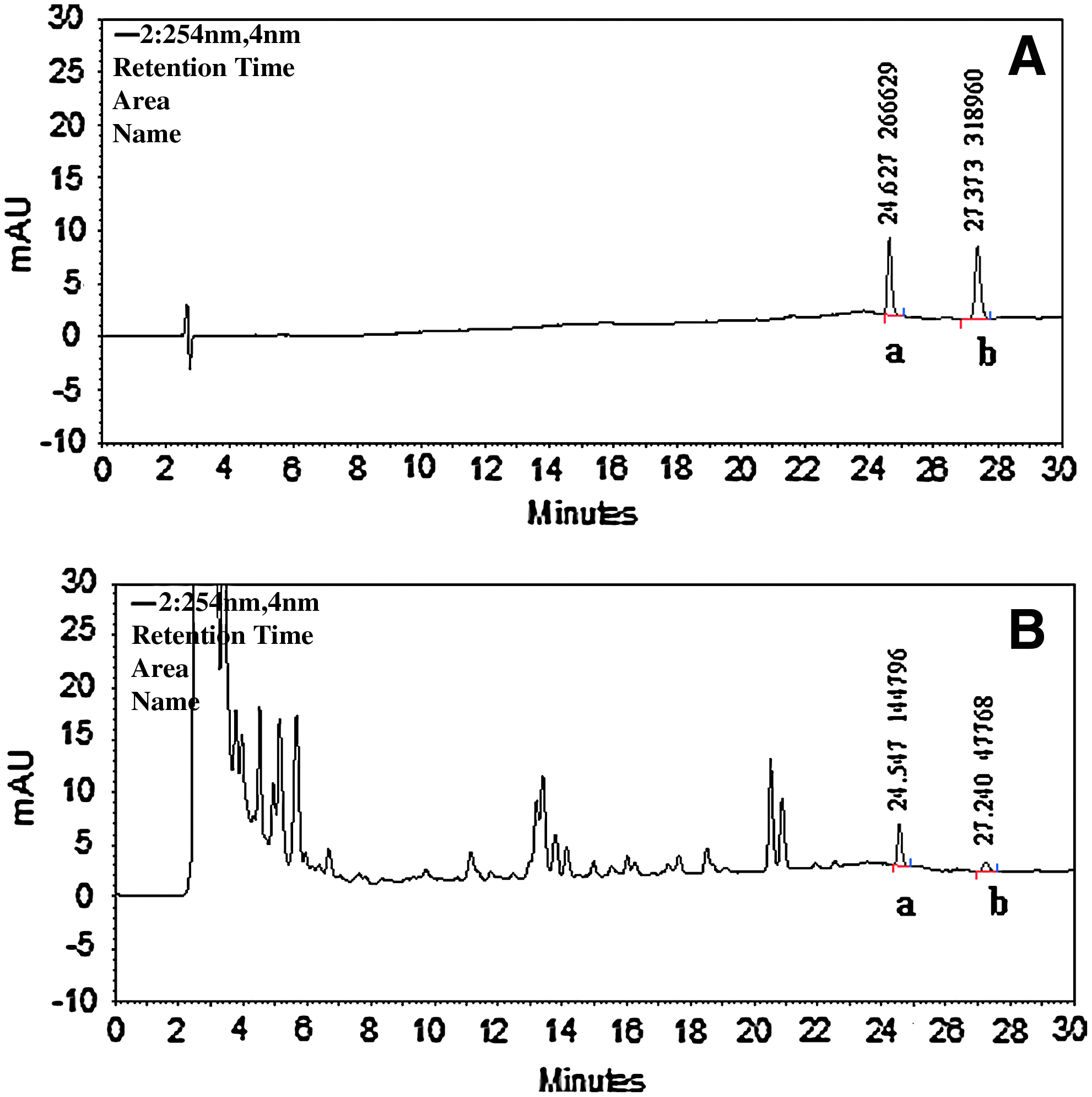
High-performance liquid chromatography chromatograms of chrysophanol and physcion authentic standards
Body weight changes of the 6 groups before and after treatment for 5 weeks were evaluated, and the results are shown in Fig. 2. In terms of the body weight change after 5 weeks of treatment, mice in group 3 (oral acetazolamide) had significantly retarded growth and actual body weight loss (P<0.05), while the other groups experienced normal weight gain. In addition, in all DBA/2J mice that were given oral acetazolamide, the appearance showed a poorer hair quality; the daily exercise performance was poor, and the animals even had an unstable gait. The results indicated that mice with oral acetazolamide treatment showed obvious adverse effects on body weight and appearance.
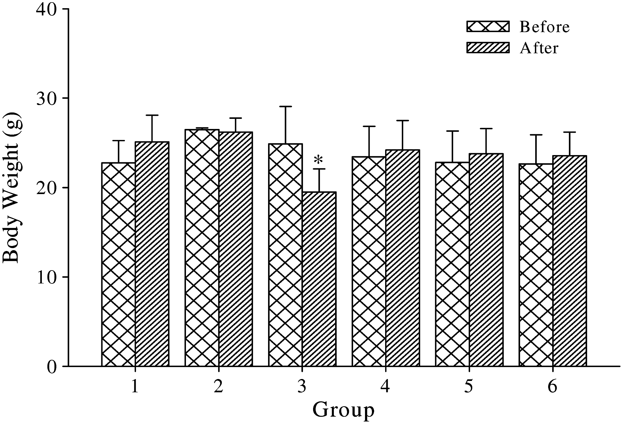
Variation in body weight before and after various treatments for 5 weeks (n≥5). *P<0.05 significantly differed before and after treatment for 5 weeks by analysis of variance (ANOVA).
The IOP of glaucomatous mice in all groups before and after 5 weeks of treatment showed statistically significant differences according to the ANOVA test (Fig. 3). At first, the mean IOP in group 1 was elevated after 5 weeks without treatment. In addition, it showed that DBA/2J mice treated for 5 weeks with topical brimonidine, oral acetazolamide, and any dose of CSE, all achieved success (the measured IOP<18 mmHg). In the meantime, we compared the time to achieved success point in every group, and results showed that group 3 had achieved it by the 3rd week (with oral acetazolamide), group 6 by the 4th week (with high dose of CSE), and groups 2, 4, and 5 by the 5th week (with topical brimonidine and low and medium oral doses of CSE), but not group 1 (Fig. 4). This means that the rate of IOP change by a high dose of CSE was even better than that of brimonidine. We also evaluated the percentage of IOP change at the same time. The definition of the percent change from the baseline is the percentage change of measured IOP in the week from week 1 onward, in each group. Therefore, from the week 2 to week 5, the lower and lower IOP were found (except group 1). First, we found that the percentage change of IOP in group 3 (oral acetazolamide) and group 6 (high dose of CSE) showed a remarkable difference (P<0.05) in week 3. In week 4, groups 3–6 revealed a remarkable change (P<0.05) (except for brimonidine use in group 2). In week 5, groups 2–6 all showed the significant percentage change of IOP (P<0.05). In addition, it was surprising to find that the percentage change of IOP in groups 3, 4, 5, and 6 had already been reduced to about 40% by the 5th week (Table 1). From these results, we concluded 2 important findings. First, the effects of lowering the IOP with the CSE were as good as those of brimonidine and acetazolamide. The high dose of CSE even showed strong and lasting activity in obviously reducing the IOP. Second, the IOP-lowering effects of CSE showed a dose-dependent relationship. Higher doses of CSE reduced the IOP more quickly. In addition, any dose of CSE could achieve the IOP (<18 mmHg) after 5 weeks of treatment (Table 1). The ability to reduce the IOP for the medium and high doses of CSE was comparable to groups treated with brimonidine and acetazolamide.
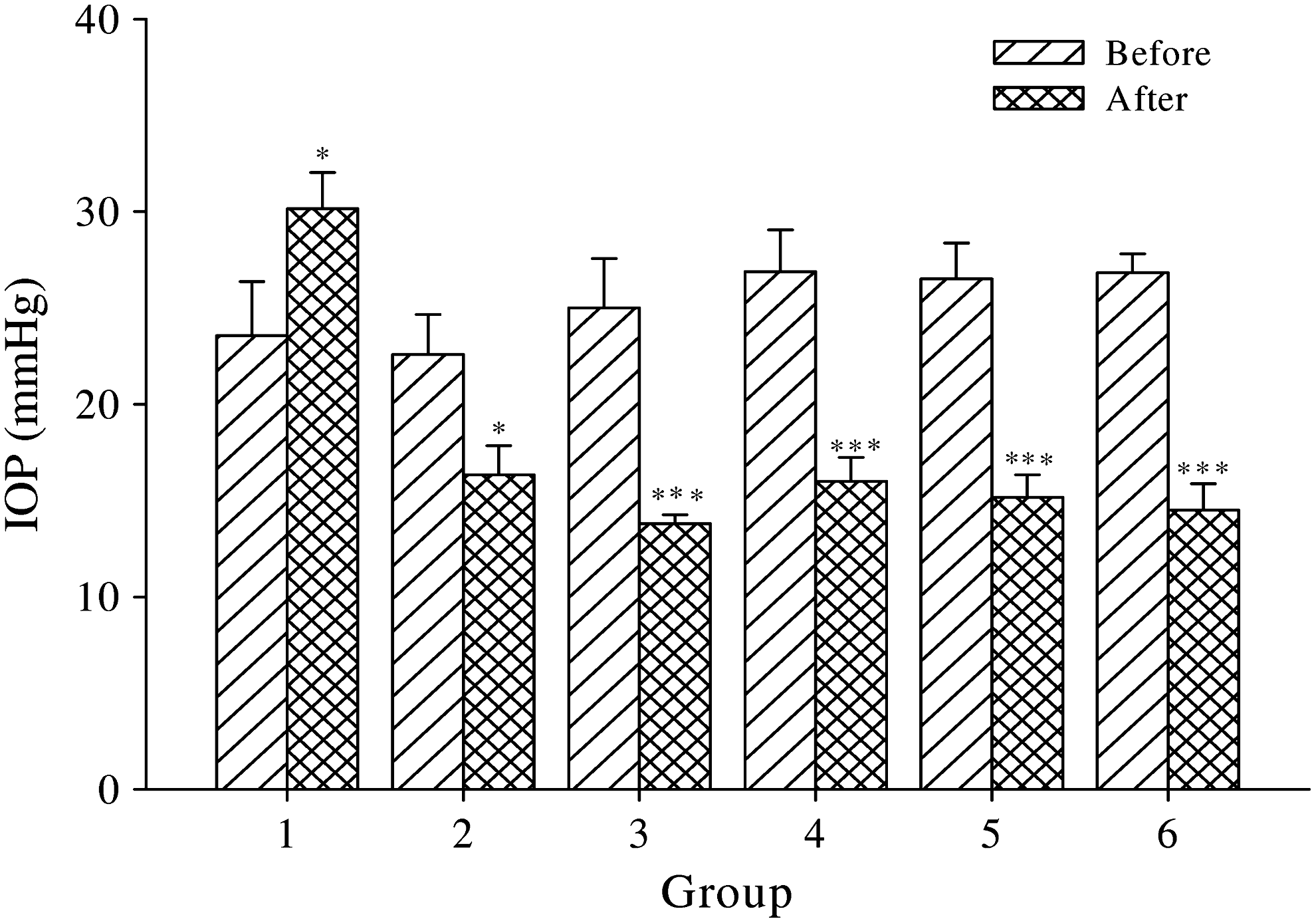
Variation in intraocular pressure (IOP) before and after various treatments for 5 weeks (n≥5). *P<0.05 and ***P<0.001 significantly differed before and after treatment for 5 weeks by ANOVA.

Different IOPs of each group within each week (n≥5). *P<0.05 significantly differed from the IOP baseline (week 1) by ANOVA.
P<0.05, significantly differed from the IOP baseline (week 1) in each group by analysis of variance.
IOP, intraocular pressure.
As we know, LDH is released by injured and dead cells. LDH levels in the anterior aqueous humor before and after treatment for 5 weeks are shown in Fig. 5. LDH levels in group 1 (sham group) revealed elevation apparently for 5 weeks (P<0.05). Before treatment in group 2, the average LDH was 236.6±9.0 U/L. After treatment with topical brimonidine instillation, it was reduced to 186.8±7.1 U/L (P<0.05). In group 3, the level of LDH revealed no remarkable change after oral acetazolamide. In group 4, the LDH level showed no apparent change after a low dose of CSE. However, LDH levels before and after medium and high doses of CSE in groups 5 and 6 showed significant differences (P<0.05). This indicates that treatment of glaucomatous mice with topical brimonidine instillation, and medium and high doses of CSE, showed less cellular dysfunction in the anterior aqueous humor.

Variation in lactate dehydrogenase (LDH) levels before and after various treatments for 5 weeks (n≥5). *P<0.05 significantly differed before and after treatment for 5 weeks by ANOVA.
Discussion
Glaucoma is a disease characterized by a specific pattern of optic head and visual field damage. Higher IOPs are an important risk factor for developing glaucomatous optic nerve damage. If one fails to effectively control the IOP, the progression of glaucoma may further cause the death of RGCs, resulting in loss of vision. Thus, many researchers devoted themselves to resolving this troublesome disease. In our study, the modern model of DBA/2J mice was selected for the investigations. The mean age of these mice was 3–4 months in our experiment according to Fan et al. 15 We found that the average IOP varied between 10 and 30 mmHg according to a Tono-pen tonometer. After 5 weeks of therapy, the results showed that topical use of brimonidine, oral acetazolamide, and any doses of CSE all exhibited significant IOP-lowering activity and all achieved the target IOP (of <18 mmHg). The IOP-lowering effectiveness at high doses of CSE was even better than that of brimonidine based on the time to reach the target IOP during treatment. According to ancient medical documentation and experience, Cassiae seed is a herbal medicine used to treat ocular diseases and improve vision. This article provides the first scientific evidence of the IOP-lowering activity of Cassiae seed, which is beneficial in treating glaucoma.
Oxidative stress induced by reactive oxygen species was implicated in the pathophysiology of glaucoma. 16 Extensive oxidative stress may result in reduced trabecular meshwork function, which leads to cell loss, compromised integrity of the trabecular meshwork, and pathologic consequences. 17 Damaged structures caused by oxidative stress will decrease the drainage of the aqueous humor and then cause IOP elevation in patients with glaucoma. 18 It was reported that the LDH activity in the anterior aqueous humor of the glaucoma group was higher than that of the normal group in rabbits and people. 19 Recently, the LDH level in the anterior aqueous humor was used as an indicator to determine the level of cellular dysfunction in glaucoma. A literature review did not reveal LDH levels of the anterior aqueous humor of DBA/2J mice. In our study, we found that the LDH in the anterior aqueous humor of DBA/2J mice aged 3 months in group 1 experienced almost double the baseline after 5 weeks of placebo therapy, signifying accumulated cellular dysfunction in the anterior chamber in glaucoma mice. In addition, DBA/2J mice with topical brimonidine, and medium and high oral dosages of CSE for 5 weeks, were found to have significantly reduced LDH levels. It was reported that the effect of pretreatment with an antioxidant, prostaglandin (PG) analogs, β-adrenergic receptor antagonists, and local carbonic anhydrase inhibitors markedly reduced oxidative stress. 12 They concluded that the impact of oxidative stress on the trabecular meshwork can be minimized by the use of antioxidants and IOP-lowering agents. These results are partly comparable to ours. Similarly, our results indicated that brimonidine and the CSE can significantly reduce cellular dysfunction after oxidative stress, with the exception of oral carbonic anhydrase inhibitors (acetazolamide). In Japan, brimonidine was ever reported to have antioxidant and neuroprotective effects.20,21 Excellent antioxidative activity was reported for Cassiae seed in China. 22 In experiments, they confirmed that the pharmaceutical mechanism of Cassiae seed is to increase the antioxidative ability of ocular tissues through analyzing LDH and superoxide dismutase levels in the periocular blood. In our investigation, CSE with LDH-lowering activity in the anterior aqueous humor was associated with its lowering the occurrence of cellular dysfunction in the trabecular meshwork.
Our study is the first report to prove the effectiveness of Cassiae seed in reducing the IOP. It would be interesting to understand the mechanism of Cassiae seed of in lowering the IOP. Recently, Memarzadeh and coworkers identified that higher systolic and mean arterial blood pressures were associated with a higher prevalence of open-angle glaucoma. They came to the conclusion that lowering the blood pressure may have the benefit of reducing the development of glaucomatous damage. 23 Strikingly, a number of studies have demonstrated the ability of Cassiae seed to decrease blood pressure. 24 Thus, Cassiae seed possesses significant IOP-lowering activity that could be associated with its systemic before hypotensive function. However, acute phenomenon-persistent ocular hypotensive activity may be another mechanism of action.
In addition, diuretic activity is another factor involved in the IOP-lowering function. For example, acetazolamide, a weak diuretic and carbonic anhydrase inhibitor, is widely used to control IOP in acute glaucoma. The mechanism involves decreasing the production of the aqueous humor and enhancing the function of carbonic anhydrase in the renal proximal tubular epithelium. Thus, in addition to enhancing diuresis, it can also be used to reduce the IOP in various types of glaucoma, especially in juvenile open-angle glaucoma. A literature review indicated that some compounds extracted from Cassiae seed have diuretic effects. For example, chrysophanol has diuretic effects, but the mechanism remains unknown. Physcion can inhibit the function of 15-hydroxyprostaglandin dehydrogenase and retard the metabolism of PGs. In fact, PGs can facilitate vasodilation of the smooth muscle of afferent arterioles of glomeruli, and hence increase renal blood flow, the glomerular filtration rate, and the function of diuresis. According to animal studies, PGs play an important role in diuresis.25,26 In our study, 2 phenolic compounds of chrysophanol and physcion with diuretic activity found in the CSE could be involved in lowering the IOP.
As for adverse effects, a poor external appearance, retarded growth, and weight gain were obviously observed in the group treated with acetazolamide for 5 weeks. Oral acetazolamide treatment was reported to cause poor weight gain in a small subset of children. 27 Due to carbonic anhydrase inhibition of acetazolamide, the loss of excessive HCO3− in the urine results in renal tubular acidosis, so-called metabolic acidosis. Metabolic acidosis exerts an antianabolic effect in bone growth centers, which is partly related to a state of resistance to growth factor (GF) and insulin-like growth factor 1. It also interferes with the hepatocellular action of GF and may be a mediating factor for growth failure. This phenomenon could explain the retarded growth and abnormal weight gain of DBA/2J mice treated with oral acetazolamide. We also found less-shiny hair and an unstable gait in this group. As far as we know, carbonic anhydrases are distributed not only in the ciliary body and renal tubular lumen but also in endothelial cells of capillary vessels. Therefore, oral acetazolamide will cause the loss of potassium ions, resulting in hypokalemia. Hypokalemic patients treated with acetazolamide may exhibit muscle weakness in the clinic. Thus, acetazolamide may reduce the exercise capacity associated with increased perception of leg fatigue and an unstable gait. However, Cassiae seed may be a safe herbal medicine because of the normal external appearance and body weight gain in DBA/2J mice treated with CSE for 5 weeks.
Conclusion
We demonstrated that the CSE possesses a significant IOP-lowering activity, which was comparable to those of brimonidine and acetazolamide. In addition, the CSE may significantly reduce the cellular dysfunction in glaucoma in the anterior aqueous humor based on lower LDH levels. There were no abnormal findings in external appearance and body weight change after treatment with the CSE for 5 weeks in DBA/2J mice. In conclusion, Cassiae seed may be a safe and beneficial herbal medicine for treating glaucoma due to its significant IOP- and LDH-lowering activities.
Footnotes
Author Disclosure Statement
The authors have no financial or proprietary interest in any product mentioned in this article. There were no financial supports and/or grants for this study. No competing financial interests exit.