
Abstract
Abstract
Purpose:
To investigate the varied effects of sperminated pullulans (SP) with different amino residues on cornea permeability and its local toxicity.
Methods:
Three groups of rabbits were used: control, low-amino residue content SP (SP-L), and high-amino residue content SP (SP-H). The in vitro and in vivo spreading assays were combined with high performance liquid chromatography (HPLC) to measure the concentration of puerarin in the external medium or aqueous humor when 0% SP, 0.2% SP-L, and 0.2% SP-H were included. The toxicity of SP was determined by corneal hydration values, Draize score, aqueous humor protein concentration, corneal endothelial evaluation, as well as light microscopy and electron microscopy.
Results:
The application of 0.2% SP-L and 0.2% SP-H to the cornea in vitro increased puerarin apparent permeability coefficient by 1.96-fold (P<0.05) and 2.95-fold (P<0.01), respectively. SP-H showed stronger effect than SP-L (P<0.05). For the in vivo assay, those were 1.81-fold (P<0.05) and 3.71-fold (P<0.01), respectively. With the SP application, the corneal hydration values were <83% and Draize scores were <4, with no apparent changes in histological observations.
Conclusion:
SP is one potential adjuvant promoting puerarin permeability to the cornea, and the high-content amino residue SP showed stronger effect, without ocular toxicity.
Introduction
Cationic polymer and the complex are newly developed adjuvants to enhance the drug permeability locally, showing ideal biocompatibility and mucosal adhesion.3,4 Sperminated pullulans (SP) (Fig. 1) is a type of cationic polymer, and has been shown to promote the insulin absorption by bronchial epithelial cells, as well as the FD-4 penetration across the Calu-3 cell monolayer. 5 The penetration-enhancing effect is positively correlated to the content of amino residues of the SP. However, whether SP could be used as potential adjuvants for puerarin application and whether SP is toxic for cornea application are still unknown. The present study set out to examine the effects of SP on cross-cornea transportation of puerarin, and whether this enhancement on penetration is dependent on the amount of amino residues of SP. The toxicity of SP application was also evaluated.

The chemical structures of sperminated pullulans (SP).
Methods
Animal
One hundred twenty New Zealand white rabbits (male or female, 2.0–3.0 kg) from the animal facility of the First Clinical College of Harbin Medical University with a slit-lamp examination of the eyes were used for the present experiment. The procedures performed on these animals were in accordance with the guidelines of the Association for Research in Vision and Ophthalmology and have been approved by the local ethics committee of animal research in the First Clinical College of Harbin Medical University.
For the in vitro diffusion experiment, the rabbits were sacrificed, and the cornea with 2-mm sclera was isolated, followed by a wash with the glutathione-bicarbonate-Ringer (GBR) solution. Then, the cornea was placed between the supply pool (superior surface of the cornea) and the acceptance pool of the Franz diffusion pool at 37°C. The supply pool was filled with 1 mL GBR solution and the acceptance pool with 9 mL GBR solution. After the system was balanced for 10 min, the solution in the supply pool was changed into 1 mL GBR solution with 0.3% puerarin, or with 0.2% SP-L/SPH-H. Then, at 30, 60, 90, 120, 150, 180, 210, and 240 min, 100 μL solution was taken from the acceptance pool (same amount of GBR solution was put back) to measure the puerarin concentration with high performance liquid chromatography (HPLC).
For the in vivo experiment, 60 rabbits were randomly divided into 3 groups: the control group, the low-amino residue content SP (SP-L) group, and the high-amino residue content SP (SP-H) group. The animals were kept away from food overnight before the experiment. For the diffusion test, after the animals were sedated, 50 μL 0.3% puerarin in phosphate-buffered saline (PBS), or the solution prepared with 0.2% SP-L/SPH-H were dropped into the conjunctiva of the bilateral eyes. Then, the eyes were closed for 30 s. The drug administration procedure was repeated for 3 times with a 5-min interval each. The animals were set free afterward. With local anesthesia, 200 μL aqueous humor was withdrawn from the 6 o'clock direction of the cornea at 0.5, 1, 2, 3, 4, 6, 8, 10, 16, and 24 h after drug administration.6,7 The aqueous humor was centrifuged at 12,000 r/min for 20 min, and the supernatant was used for the HPLC analysis.
HPLC analysis
The LC-6A HPLC instrument and the supersorb ODS C18 (4.6×250 mm, 5 μm) column were used. The examination wavelength was 249 nm; the flow speed was 0.7 mL/min; and the temperature of the column was 40°C; the flow phase was methanol:water:0.1 M PB, pH 7.4=11:79:10. The washing sequence was as follows: mobile phase, puerarin dissolved in mobile phase, blank aqueous humor, puerarin dissolved in aqueous humor, aqueous humor sample, and aqueous humor sample and puerarin.
Data analysis
For Q, which was the cumulative permeability per unit area,
Cn was the drug concentration at time t, Ci was before time t, V0 was the solution volume in the acceptance pool, and V was the volume of sample obtained.
For the apparent permeability parameters Papp (cm/s):
C0 was the initial drug concentration in the supply pool, A was the effective area for transportation and ΔQ/Δt was calculated by the slope of the Q–t curve.
For the increase in the infiltration rate ER:
For the pharmacokinetics in the in vivo experiment, DAS2.0 software was used to simulate the changes of drug concentration following the time.
Toxicity assessment
The corneal hydration value measurement: wet cornea was weighted as Wb, and then the tissue was dried at 70°C for 12 h, with the weight Wa. The corneal hydration value=(Wb−Wa)/Wb×100%.
For the Draize test, 4 rabbits in each group (control, SP-L, and SP-H) were used. 0.05 mL SP solution was dropped into the left eye conjunctiva, while the right eye was treated with 0.05 mL PBS (pH 7.4). The treatment lasted for 14 days with 4 times per day. The reactions of cornea, iris, and conjunctiva were scored at 1, 2, 4, 12, 24, 48, and 72 h after drug administration, as well as the 6, 24, 48, 72 h, and 7 days after the last drug administration. 0–3.9 was considered as nonirritating; 4–8.9 as mild irritation; 9–12.9 as modest irritation; and 13–16.9 as severe irritation.
For the sodium fluorescein staining, the drug administration lasted continuously for 14 days. 0.2% sodium fluorescein was dropped into the eye conjunctiva at 24 h, 7 days, 14 days from the first drug administration and 7 days after the last drug administration.
For the aqueous humor protein concentration, the drug administration lasted continuously for 14 days. Seven days after the last drug administration, the animal was sacrificed and 0.2 mL aqueous humor was obtained. 0.05 mL aqueous humor, 0.05 mL 1g/L standard solution, or 0.05 mL distilled water was added into a sample tube, a standard tube, and a blank tube, respectively, followed with 3.0 mL CBBG250 visualization reagent. The solution was mixed and left for 10 min before measuring the light absorption at 595 nm. The protein content (g/L)=standard tube absorbance×(sample tube absorbance−blank tube absorbance)/(standard tube absorbance−blank tube absorbance).
For endothelial cell count, the endothelial cell of the cornea was counted at 7 days after drug administration, with the noncontact corneal endothelial cell counter. For the trypan blue and alizarin red staining, 7 days after drug administration, 5 cornea samples from each group were stained in 0.25% trypan blue for 90 s, followed with a saline wash before 1% alizarin red staining for 4 min. Upon microscopic examination, 5 random views were selected to count the total number of endothelial cells and trypan blue-stained cells. The cornea endothelial cell viability rate=the total number of trypan blue-stained endothelial cells/total endothelial cells×100%. Also, 3 eye samples from each group were prepared for HE staining or EM observation.
Statistical analysis
SPSS16.0 software was used for a statistical analysis and the data were represented as x±s. Multiple groups were compared using the one-way ANOVA analysis and between 2 groups were using the Turkey test. P<0.05 was determined as significant difference.
Results
The in vitro permeation results
The effects of 0.2% SP-L and 0.2% SP-H on puerarin permeation are shown in Fig. 2. By 4 h, 0.2% SP-L increased the accumulation of puerarin from 9.96±0.85 μg/mL to 19.88±5.78 μg/mL, while 0.2% SP-H increased the accumulation of puerarin to 27.91±4.22 μg/mL. Compared to the control group, SP-L and SP-H increased puerarin permeation by 1.96-fold (P<0.05) and 2.95-fold (P<0.05), respectively. The increased amount of amino residues in SP-H showed stronger effect than SP-L (P<0.05). The corresponding steady-state flow, apparent permeability coefficient, and the ratio of infiltration enhancement are shown in Table 1.
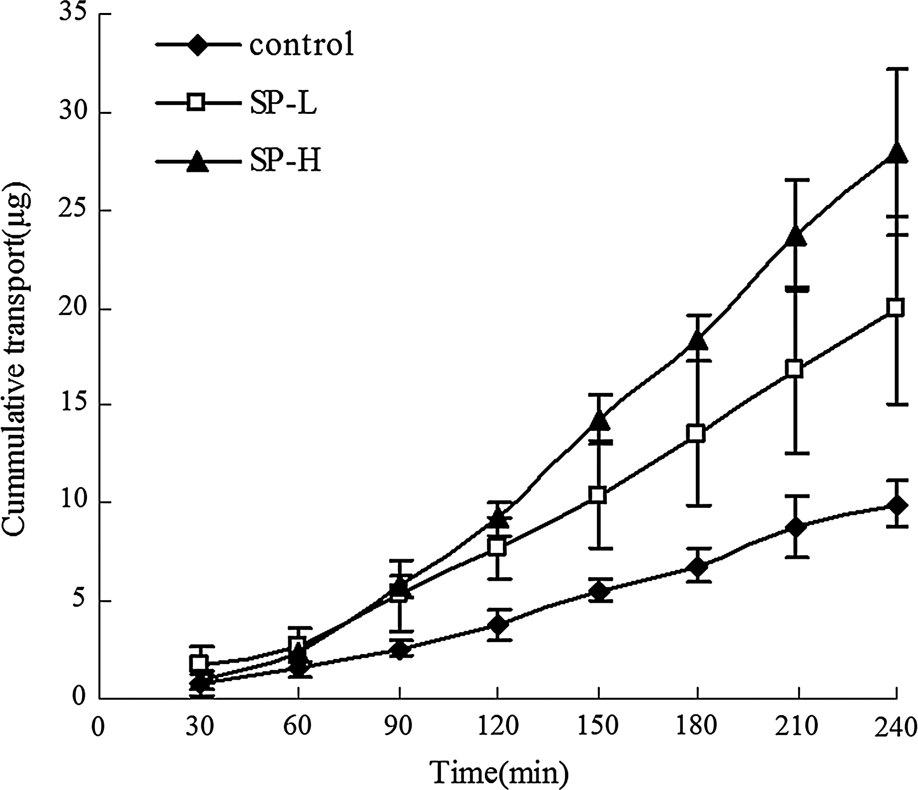
Effect of SP on puerarin permeation through excised rabbit cornea.
P<0.05 versus control group.
P<0.01 versus control group.
P<0.05 versus SP-L group.
SP-H, high-amino residue content sperminated pullulans; SP-L, low-amino residue content SP.
The in vivo permeation experiments
The results of puerarin concentration in the aqueous humor following 0.2% SP-L and 0.2% SP-H application are shown in Fig. 3. The pharmacokinetics is presented in Table 2. The uses of 0.2% SP-L and 0.2% SP-H significantly increased the peak value of puerarin concentration in the aqueous humor, with the bioavailability increased by 1.81-fold (P<0.05) and 3.71-fold (P<0.01), respectively. The increased amount of amino residues in SP-H also showed stronger effect than SP-L (P<0.05).
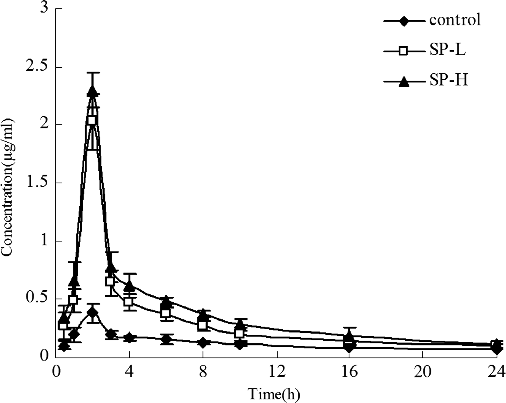
Effect of SP on puerarin concentration in aqueous humor in vivo.
P<0.05 versus control group.
P<0.01 versus control group.
P<0.05 versus SP-L group.
Eye toxicity assessment
The values of corneal hydration in the control group, the SP-L group, and the SP-H group were 78.12±1.56, 78.76±1.35, and 79.48±0.61, respectively (P>0.05). All values were in the normal range, suggesting that there was no damage of 0.2% SP to isolated cornea tissue.
For the Draize test, 0.2% SP solution did not increase the eye-blink frequency and tear secretion. The slit-lamp examination revealed no conjunctival edema, clear cornea, and normal iris. The Draize scores in the control group, the SP-L group, and the SP-H group were 0, 0.75±0.96, and 0.5±0.58, respectively, suggesting that there is no irritation of SP solution to the eye tissue.
For the sodium fluorescein staining, in all observed time points the cornea epithelial cells were not stained with sodium fluorescein, indicating the integrity of the cornea epithelium after SP solution application.
The aqueous humor protein concentration is shown in Table 3. There were no significant differences between the SP-L/SP-H and the control group (P>0.05), or between the SP-L and SP-H groups (P>0.05).
The counting of cornea epithelial cells is shown in Table 3. There were no changes in the viability of cornea epithelial cells between the experiment and the control groups (P<0.05). For the trypan blue and alizarin red staining, Fig. 4 showed that no cells were stained as blue, and the cells showed an arranged sequence.
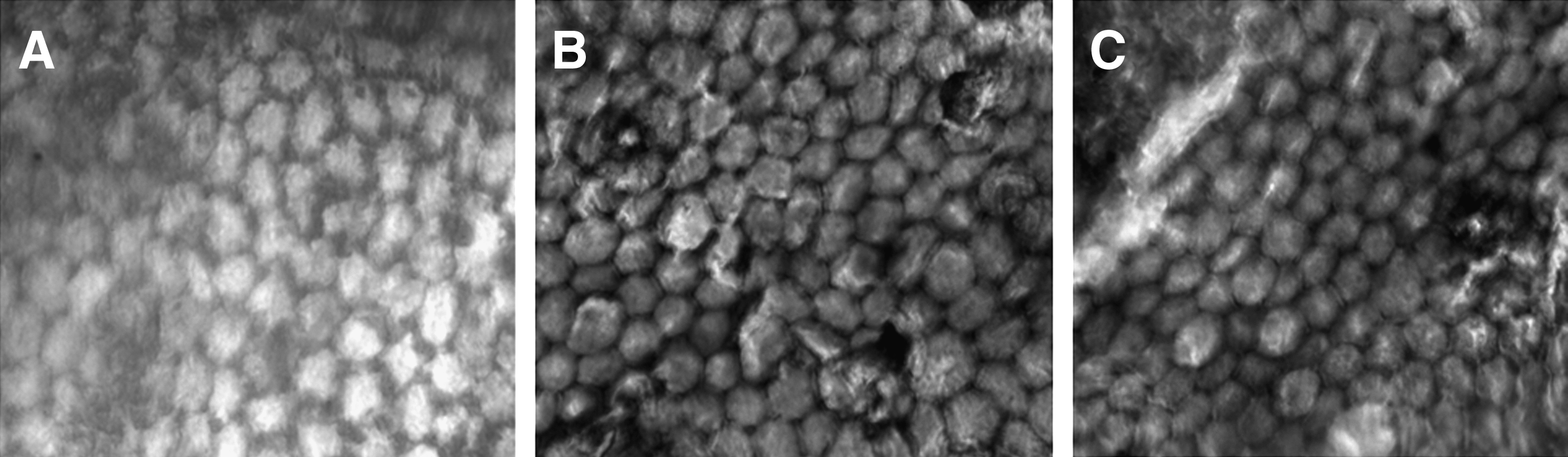
The morphology of corneal endothelium by staining with alizarin red and trypan blue.
Finally, the EM study showed no ultrastructure changes in the cornea, iris, retina, and optic nerve obtained from SP-solution-treated groups (Fig. 5). The euchromatin in the epithelial cell showed an even distribution, without nucleus shrinkage. The basement layer was composed by fibroblasts, with arranged collagen fibers; the endothelial cells showed an integrated membrane, and clear structures of the nucleus.
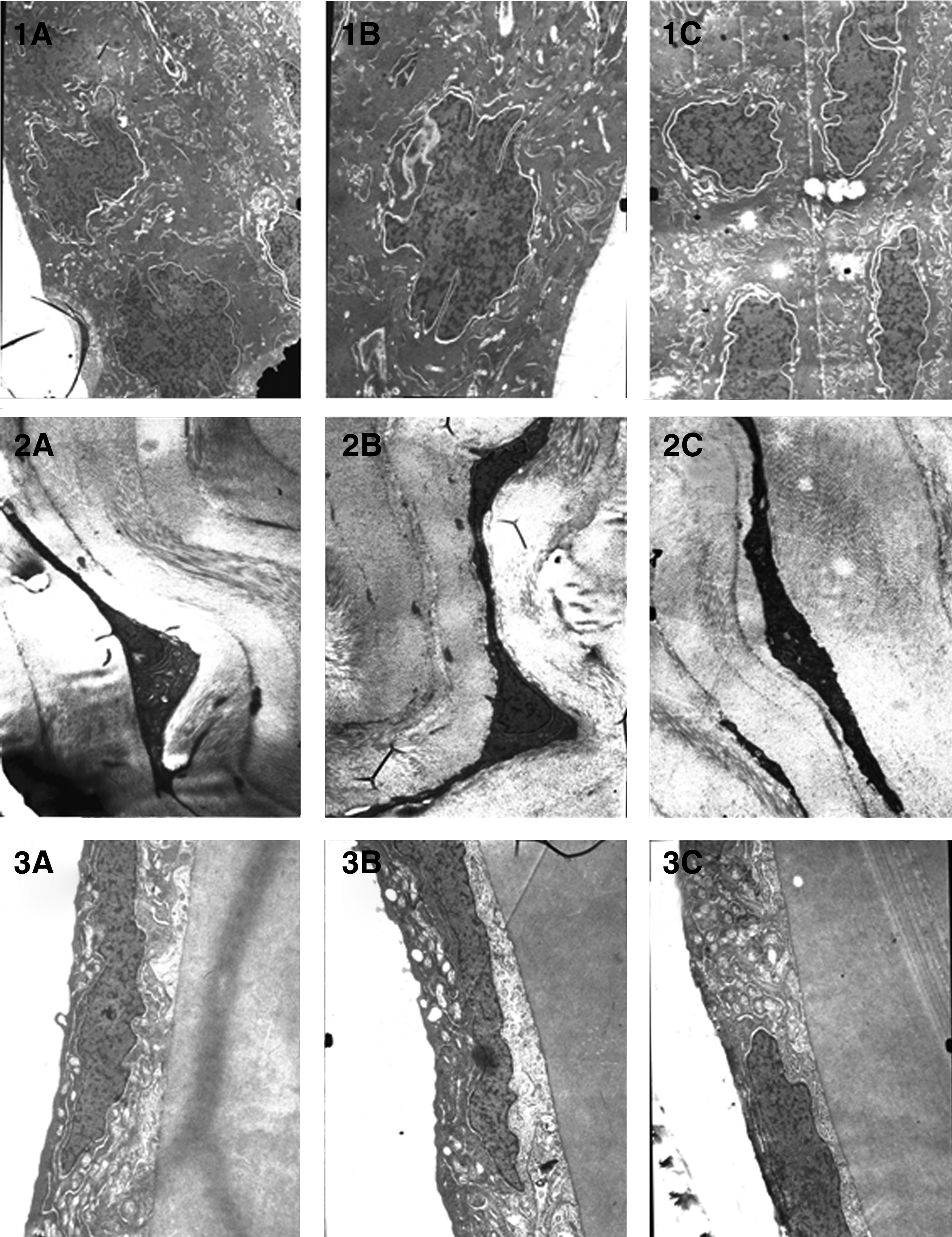
Photographs of TEM of cornea.
Discussion
Puerarin could improve the local microcirculation and decrease the IOP at the same time,2,8 which is ideal for glaucoma therapy: some current antiglaucoma agents reduce the IOP, and decrease the circulation at the same time, leading to the malnutrition of the eye. Moreover, puerarin could antagonize the glutamate excitotoxicity in the nervous system. However, the low solubility restricts the use of puerarin and argues for the use of other permeability enhancers upon application.9–11 The present study investigated the effects of coapplication of SP on puerarin penetration of the cornea, and demonstrated SP as a potential enhancer, the efficiency of which depends on the amount of amino residues. The increased amino residues could lead to higher density of charges of the SP molecules, therefore promoting the interaction with the cell membrane. This also suggested for new directions in screening for other compounds as permeation enhancers in the future.
In the in vivo experiments, the drops of drugs after administration could be diluted by tears and decrease the effective concentration of the drug outside of the cornea. Also, the aqueous humor undergoes circulation and removes the penetrated drug molecules inside. This explains the peak and washout phase during the in vivo experiment. In the present study, SP not only increased the peak value of puerarin concentration but also extended the washout phase, indicating a longer period with effective drug concentration. One potential explanation is that the adherence of SP to cornea prolonged the transportation of puerarin.
Very interestingly, the toxicity assessment with multiple criteria indicated that SP is rather safe for cornea application. The cornea showed no hydration, and the Draize score suggested for lack of irritation. Also, the epithelium showed no apparent damage with sodium fluorescein and trypan blue staining, and EM observation indicated normal ultrastructure. This is consistent with other systems, SP showed low toxicity in vivo as well. 12
In conclusion, the present study demonstrated the puerarin permeation-enhancing effects of SP on cornea application, both in vitro and in vivo. The effect is positively correlated to the amount of amino residues on the SP. Also, the use of SP causes no toxic effects to the cornea tissue.
Footnotes
Author Disclosure Statement
No competing financial interests exist.