
Abstract
Abstract
Purpose:
We previously documented that ocular surface epithelial cells could regulate ocular surface inflammation and suggested that, while Toll-like receptor 3 upregulates, EP3, one of the prostaglandin E2 receptors, downregulates ocular surface inflammation. Others reported that rebamipide, a gastroprotective drug, could not only increase the gastric mucus production, but also suppressed gastric mucosal inflammation and that it was dominantly distributed in mucosal tissues. The eyedrop form of rebamipide, approved in Japan for use in the treatment of dry eye diseases, upregulates mucin secretion and production, thereby suppressing superficial punctate keratopathy on the ocular surface of patients with this disease. In the current study, we investigated whether rebamipide has anti- inflammatory effects on the ocular surface.
Methods:
To examine the effects of rebamipide on polyI:C-induced cytokine expression by primary human conjunctival epithelial cells, we used enzyme-linked immunosorbent assay and quantitative reverse transcription-polymerase chain reaction assay. We studied the effects of rebamipide on ocular surface inflammation in our murine experimental allergic conjunctivitis (EAC) model.
Results:
Rebamipide could suppress polyI:C-induced cytokine production and the expression of mRNAs for CXCL10, CXCL11, RANTES, MCP-1, and IL-6 in human conjunctival epithelial cells. In our EAC model, the topical administration of rebamipide suppressed conjunctival allergic eosinophil infiltration.
Conclusions:
The topical application of rebamipide on the ocular surface might suppress ocular surface inflammation by suppressing the production of cytokines by ocular surface epithelial cells.
Introduction
Rebamipide, a gastroprotective drug, has been prescribed for the treatment of gastric ulcers and gastritis. The drug has been reported to increase gastric mucus production8,9 and to suppress gastric mucosal inflammation.10,11 According to Naito et al. (1996), rebamipide was dominantly distributed in mucosal tissues; its mean mucosal concentration after ingestion was more than 100 times its mean serum concentration. 12
In Japan, rebamipide in an eyedrop form has been approved for use in the treatment of dry eye disease. As the drug upregulates the production and secretion of mucin, it helps to suppress superficial punctate keratopathy on the ocular surface in patients with this disease.
In the current study, we investigated whether rebamipide could suppress cytokine production in human conjunctival epithelial cells, and whether it could suppress ocular surface inflammation in our murine EAC model.
Materials and Methods
Human conjunctival epithelial cells
Our study was approved by the institutional review board of Kyoto Prefectural University of Medicine, Kyoto, Japan. All experimental procedures were conducted in accordance with the tenets of the Declaration of Helsinki. Written informed consent was obtained from all patients after they were given a detailed explanation of the purpose of the research and the experimental protocols.
For enzyme-linked immunosorbent assay (ELISA) and quantitative reverse transcription polymerase chain reaction (RT-PCR), primary human conjunctival epithelial cells (PHCjECs) harvested from the conjunctival tissue obtained at the time of conjunctivochalasis surgery were cultured using a previously described method. 1 Briefly, conjunctival tissues were washed and immersed for 1 h at 37°C in 1.2 U mL−1 purified dispase (Roche Diagnostic Ltd., Basel, Switzerland). Then, the epithelial cells were detached, collected, and cultured in a low-calcium defined keratinocyte-SFM medium with defined growth-promoting additives (Invitrogen, Carlsbad, CA), including insulin, the epidermal growth factor, fibroblast growth factor, and a 1% antibiotic–antimycotic solution. Cell colonies usually became visible within 3 to 4 days. After reaching 80% confluence in 7–10 days, the cultured PHCjECs were used in subsequent procedures.
Enzyme-linked immunosorbent assay
We performed ELISA to confirm protein production. The amount of CXCL10, CXCL11, RANTES, MCP-1, and IL-6 released into the culture supernatant was determined by ELISA using the human CXCL10, CXCL11, RANTES DuoSet (R&D Systems, Inc., Minneapolis, MN) or the OptEIA™ MCP-1, IL-6 set (BD Pharmingen, San Diego, CA), respectively, in accordance with the manufacturer's instructions. 1
Quantitative RT-PCR
Total RNA was isolated from PHCjECs using the RNeasy Mini kit (QIAGEN, Valencia, CA) according to the manufacturer's instructions. For the RT reaction, we used the SuperScript™ Preamplification kit (Invitrogen). Quantitative RT-PCR was on an ABI-prism 7700 (Applied Biosystems, Foster City, CA) instrument using a previously described protocol 1 and the manufacturer's instructions. The primers and probes for CXCL10, CXCL11, RANTES, MCP-1, IL-6, and human GAPDH were purchased from Applied Biosystems. To amplify cDNA, PCR was performed in a 25-μL total volume that contained a 1 μL cDNA template in 2×TaqMan universal PCR master mix (Applied Biosystems) at 50°C for 2 min and 95°C for 10 min, followed by 40 cycles at 95°C for 15 s and 60°C for 1 min. The results were analyzed with sequence detection software (Applied Biosystems). The quantification data were normalized to the expression of the housekeeping gene GAPDH.
Murine EAC
Balb/c mice were purchased from CLEA (Tokyo, Japan) and sensitized at 6–12 weeks of age. They were maintained on a 12-h light/12-h dark cycle under specific pathogen-free conditions. All experimental procedures were approved by the Committee on Animal Research of Kyoto Prefectural University of Medicine. All studies were performed in accordance with the ARVO Statement for the Use of Animals in Ophthalmic and Vision Research.
Using our murine EAC model 6 on day 0, the mice were immunized with a subcutaneous injection into their left hind footpads of ragweed (RW) adsorbed on alum (200 μg RW and 2.6 mg alum in a total volume of 200 μL). On day 7, they received an intraperitoneal injection of RW adsorbed on alum and on day 18, their eyes were challenged with RW in PBS (500 μg in 5 μL per eye) or PBS alone (controls, 5 μL per eye). We administered rebamipide eyedrops (0.6%, 2%, or 6% rebamipide) 1 h before and 2-, 5-, and 8 h after short RW pollen challenge on the day of antigen challenge. Eyes were collected 24 h postchallenge for a histologic study.
Histological analysis
Using our previously described method, 6 we dissected whole eyeballs together with the eyelids and conjunctiva. This was followed by fixation in 10% neutral buffered formalin and embedding in paraffin blocks. Vertical 6-μm-thick sections were affixed to microscope slides, deparaffinized, and stained with the Luna's method. Stained eosinophils were counted under a light microscope. The number of eosinophils infiltrating the lamina propria mucosae of the tarsal of the conjunctiva in the entire section was recorded. We used sections from the central portion of the eye; they included the pupil and optic nerve head. Since the cell number varied according to the area counted, cell-count data are expressed as the number of infiltrating eosinophils divided by the area of the count (mm2) measured by Scion Image software (Scion Corp., Frederick, MD). Data are presented as the mean±SEM of all examined mice.
Compounds and reagents
Rebamipide was supplied by Otsuka Pharmaceutical Co., Ltd. (Tokyo, Japan). RW, RW extract, and aluminum hydroxide (alum) were purchased from Polysciences, Inc. (Warrington, PA), LSL Co., Ltd. (Tokyo, Japan), and Sigma (St. Louis, MO), respectively.
Data analysis
Data were expressed as the mean±SEM and evaluated by Student's t-test using the Microsoft Excel software program.
Results
Rebamipide downregulated the production of cytokines induced by polyI:C stimulation
As many transcripts in PHCjECs are significantly upregulated upon polyI:C stimulation, 1 we first examined whether rebamipide downregulated the polyI:C-induced production of cytokines in these cells. We performed ELISA to assess the effects of rebamipide on the polyI:C-induced production of CXCL10, CXCL11, RANTES, MCP-1, and IL-6. To determine the optimal dose, we administered rebamipide at doses of 2-, 0.2-, and 0.02 mM. As polyI:C-induced cytokine production was suppressed dose dependently (data not shown), we concluded that the optimal dose was 2 mM. PHCjECs were therefore exposed to 10 μg/mL polyI:C and 2 mM rebamipide for 24 h. We found that rebamipide significantly attenuated the production of CXCL10, CXCL11, RANTES, MCP-1, and IL-6 (Fig. 1). We also examined the polyI:C-induced production of IL-8, however, it could not be downregulated by rebamipide (data not shown).
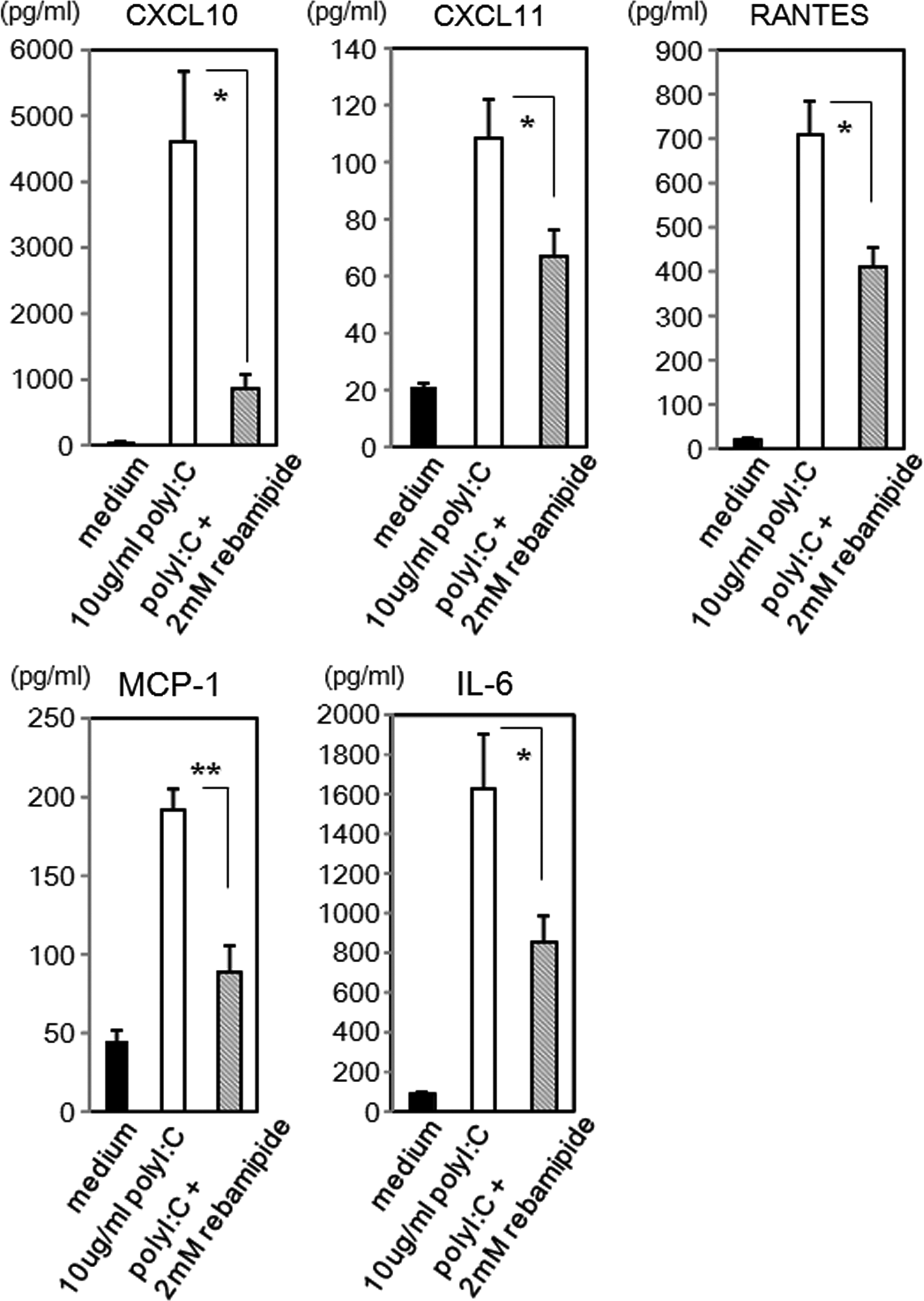
Effect of rebamipide on polyI:C-induced cytokine production. PHCjEC were exposed to 10 μg/mL polyI:C and 2 mM rebamipide for 24 h. Data are representative of 3 separate experiments and are given as the mean±SEM from one experiment carried out in 6 wells per group. (*P<0.05, **P<0.005.)
Rebamipide downregulated the mRNA level of cytokines induced by polyI:C stimulation
We performed quantitative RT-PCR to examine the effects of rebamipide on the polyI:C-induced expression of mRNAs for CXCL10, CXCL11, RANTES, MCP-1, and IL-6 in PHCjECs. After exposing the cells to 10 μg/mL polyI:C and 2 mM rebamipide for 6 h, the expression of mRNAs for these cytokines was significantly attenuated (Fig. 2).
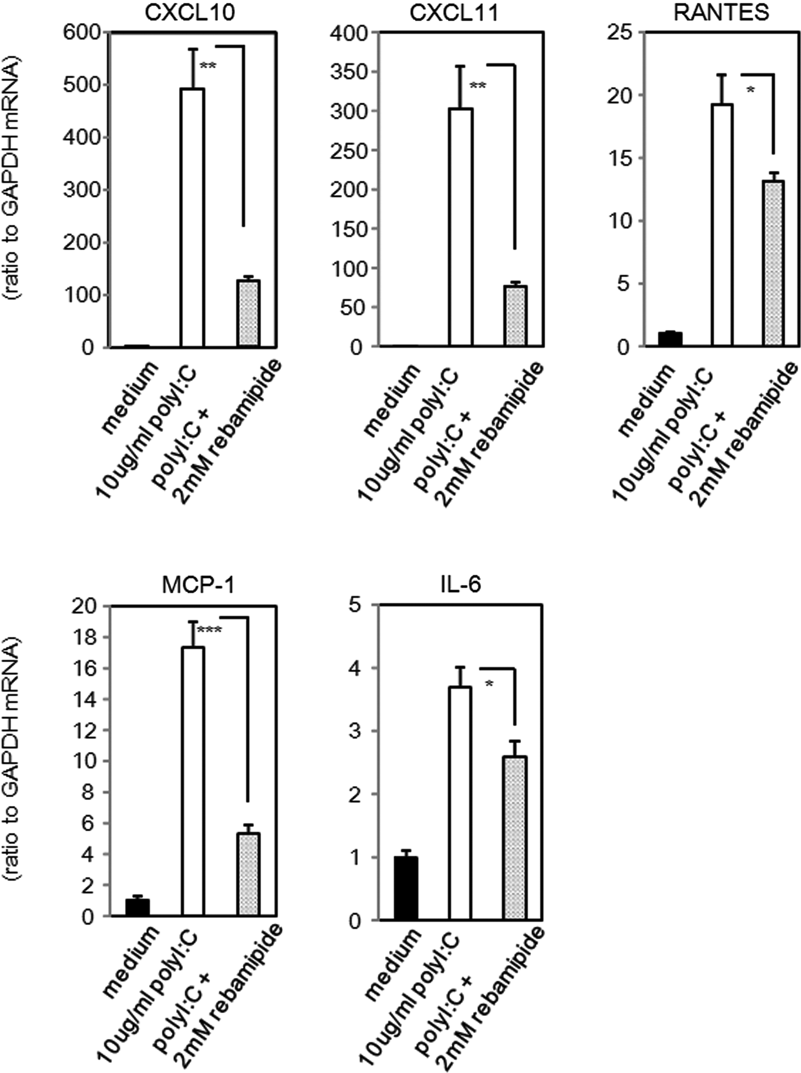
Effect of rebamipide on the polyI:C-induced mRNA expression of cytokines. PHCjEC were exposed to 10 μg/mL polyI:C and 2 mM rebamipide for 6 h. The quantification data were normalized to the expression of the housekeeping gene GAPDH. The y axis shows the increase in specific mRNA over unstimulated samples. Data are representative of 3 separate experiments and are given as the mean±SEM from one experiment carried out in 9 wells per group. (*P<0.05, **P<0.005, ***P<0.0005.)
Effect of rebamipide eye drops
We already knew that TLR3, a receptor of polyI:C, positively regulated allergic eosinophilic inflammation 5 and that rebamipide suppressed the polyI:C-induced production of cytokines, including allergy-related proteins such as RANTES in conjunctival epithelial cells in vitro. Therefore, we investigated whether the allergic eosinophilic inflammation in EAC could be suppressed by rebamipide eyedrops. On the day of challenge, we administered rebamipide eyedrops (0.6%, 2%, or 6% rebamipide per 5 μL) at 4 different time points to the eyes of RW-sensitized wild-type mice. We found that the infiltration of eosinophils was suppressed in a dose-dependent manner. At concentrations of 2% and 6%, the antigen-induced infiltration of eosinophils was significantly inhibited compared to the vehicle-treated controls (Fig. 3 and Supplementary Fig. S1; Supplementary Data are available online at www.liebertpub.com/jop).
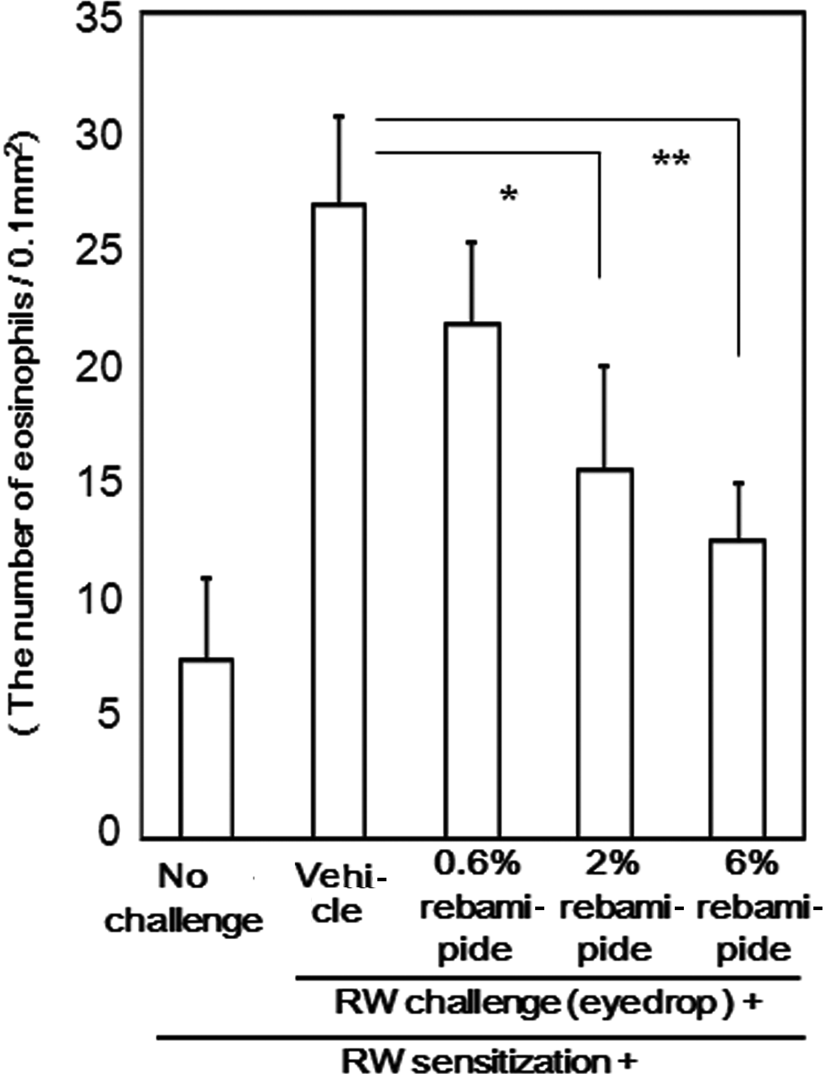
Effect of rebamipide on conjunctival allergic eosinophilic infiltration in murine experimental allergic conjunctivitis. The number of eosinophils in the lamina propria mucosae of the tarsal conjunctiva was quantified. The data are shown as the mean±SEM of samples from all examined mice (vehicle: n=16, 0.6% rebamipide: n=16, 2% rebamipide: n=21, 6% rebamipide: n=14).
Discussion
Elsewhere we showed that ocular surface epithelial cells could regulate ocular surface inflammation1–4,13–20 and posited that TLR3 upregulates, while EP3, a PGE2 receptor, downregulates ocular surface inflammation.5–7
Here we demonstrate that in human conjunctival epithelial cells, rebamipide suppressed the polyI:C-induced production and expression of mRNAs for cytokines, and that its topical administration suppressed allergic eosinophil infiltration in our EAC model. These findings suggest that the topical delivery of rebamipide to the ocular surface might suppress ocular surface inflammation.
Rebamipide, a gastroprotective drug, has been used to treat gastric ulcers and gastritis. In rats, the drug accelerated the healing of acetic acid-induced gastric ulcers. 21 Although the precise mechanisms of action remain unknown, its beneficial effect on the gastric mucosa has been variously attributed to increased gastric mucus production,8,9 suppression of gastric mucosal inflammation,10,11 inhibition of neutrophil activation,22,23 and hydroxylradical scavenging.22,24
Naito et al. administered rebamipide tablets (100 mg) to 32 patients with chronic gastritis. They were then subjected to gastroscopy and the rebamipide level in the gastric mucosa and in serum from venous blood was assayed. Between 30- and 120-min postingestion, the mean mucosal concentration of rebamipide was 60.0±109.8 μg/g tissue, higher than 0.1 mM (37 μg/mL), and its mean serum concentration was 0.25±0.23 μg/mL, lower than 1.0 μM (0.37 μg/mL). Their findings indicate that the concentration of rebamipide in the gastric mucosa was attributable to local penetration, suggesting that its blood level and systemic distribution play only a minor role in its antioxidative and antineutrophilic activities and that rebamipide acts directly on the gastric mucosa. 12
In Japan, rebamipide eyedrops are used to treat dry eyes, because it could increase the level of mucin-like substances and improve the corneal and conjunctival epithelial damage. Urashima et al. 25 investigated the effects of rebamipide on the number of periodic acid Schiff (PAS)-positive cells in the conjunctiva, the mucin content in the cornea and conjunctiva of normal rabbits, and the effects of rebamipide on the desiccation-induced corneal damage. They found that it increased the number of PAS-positive cells in the conjunctiva and increased the amount of mucin-like substances in the conjunctiva and cornea. It also lowered the rose bengal scores of the cornea in their desiccation-induced corneal damage model. They proposed rebamipide as a candidate drug for the treatment of human corneal and conjunctival epithelial damage because it acts to increase the level of mucin-like substances.
Based on findings suggesting that rebamipide affects the mucosa directly, we considered that it may also have effects on ocular surface epithelial cells and that it may suppress ocular surface inflammation. We found that rebamipide did suppress the polyI:C-induced production of cytokines in conjunctival epithelial cells and that its topical administration suppressed allergic eosinophil infiltration in our EAC model. Therefore, we suspect that the topical application of rebamipide to the human ocular surface may suppress ocular surface inflammation.
Our observations suggest that the use of rebamipide may open new strategies for treating human ocular surface inflammation, including allergic conjunctivitis and dry eye diseases by modifying epithelial cell functions. However, the mechanisms underlying the anti-inflammatory and antiallergic effects of rebamipide remain to be elucidated because the receptor for rebamipide remains unknown. It was reported that rebamipide increased PG levels, including PGE2 on gastric tissue. 26 We reported that PGE2 could also downregulate the production of polyI:C-induced cytokines in human ocular surface epithelial cells. 3 Intriguingly, IL-8 production induced with polyI:C could not be downregulated by rebamipide as same as by PGE2, although the suppression effect by rebamipide of the 5 cytokine production (CXCL10, CXCL11, RANTES, MCP-1, and IL-6) might be smaller than by PGE2. Therefore, it is possible that rebamipide might exert the anti-inflammatory effects through PGE2. On the other hand, it is also possible that rebamipide might suppress the inflammation induced by not only TLR3, but also other mechanisms.
Investigations are underway in our laboratory to identify the precise molecular mechanisms of its anti-inflammatory and antiallergic effects because their identification may help to define the mechanisms of regulation of inflammation involving epithelial cells.
Footnotes
Acknowledgments
We thank Chikako Endo and Maki Kobori for technical assistance. This work was supported, in part, by grants-in-aid for scientific research from the Japanese Ministry of Health, Labour and Welfare, the Japanese Ministry of Education, Culture, Sports, Science and Technology, the Kyoto Foundation for the Promotion of Medical Science, the National Institute of Biomedical Innovation of Japan, and the Intramural Research Fund of Kyoto Prefectural University of Medicine.
Financial Relationship with Manufacturer
We declare that the work described in the present article was carried out in collaboration with Otsuka Pharmaceutical Co., Ltd., who supplied rebamipide used in this study.
Authors' Contributions
Material contributions to the research: Mayumi Ueta, Chie Sotosono, Norihiko Yokoi, and Shigeru Kinoshita; writing and review contributions to the manuscript: Mayumi Ueta.
Author Disclosure Statement
No competing financial interests exist.
References
Supplementary Material
Please find the following supplemental material available below.
For Open Access articles published under a Creative Commons License, all supplemental material carries the same license as the article it is associated with.
For non-Open Access articles published, all supplemental material carries a non-exclusive license, and permission requests for re-use of supplemental material or any part of supplemental material shall be sent directly to the copyright owner as specified in the copyright notice associated with the article.