
Abstract
Abstract
Purpose:
To report the clinical course of a highly myopic woman treated by a single intravitreal injection of bevacizumab during the first trimester of pregnancy.
Methods:
Observational case report. A 35-year-old woman affected by pathologic myopia complained of blurred vision in her left eye in the fourth week of pregnancy. A subfoveal myopic choroidal neovascularization (CNV) was diagnosed on the basis of slit-lamp fundus biomicroscopy and fluorescein angiography. After discussing the treatment-related risks, she was administered an intravitreal injection of bevacizumab in her seventh gestational week. During pregnancy, fetal ultrasound and ophthalmic examination were performed monthly. After delivery, the mother and infant were followed quarterly for 12 months.
Results:
The patient had an uneventful prenatal course and delivered a healthy full-term infant. Significant visual improvement with no documented adverse events related to treatment was obtained.
Conclusions:
In our experience, a single intravitreal bevacizumab injection administered during the first trimester of pregnancy did not provoke any complications, and was effective in myopic CNV treatment. Further studies are warranted to provide more detailed information about this treatment and the related risks in pregnant women.
Introduction
Case Report
A 35-year-old woman was referred to our retinal service for recent-onset visual decrease with blurred vision in her left eye. Her medical history was unremarkable. Best-corrected visual acuity (BCVA), with a spherical equivalent refraction error of -13 diopters, was 20/50 in her left eye, with no metamorphopsia. Fundoscopic findings revealed a myopic appearance of the fundus, with a papillary temporal crescent in both eyes. In the left eye, mild foveal edema along a lacquer crack, with a small hemorrhage on its upper edge, was visible. Before undergoing fluorescein angiography (FA), the patient referred the possibility that she might be pregnant. Nonetheless, FA was performed, and subfoveal dye leakage confirmed the presence of an active CNV (Fig. 1B). Spectral-Domain coherence tomography (SD-OCT) revealed a blurred hyper-reflective area with fuzzy borders in correspondence with the CNV (Fig. 1C). After CNV diagnosis, an obstetrical consultation revealed that she was in the fourth week of pregnancy. The patient was informed of her macular disease by a collegial advisory council that included an obstetrician and a teratology information service consultant. The disease's natural course and treatment options were examined, particularly the risk of treating her disease with anti-VEGF during early pregnancy. Visual acuity further decreased to 20/50, and the patient decided to undergo an intravitreal bevacizumab injection. Informed consent was obtained, and the patient was treated in her seventh week of pregnancy. After treatment, the pregnancy was closely monitored; she underwent monthly fetal ultrasound examination, along with Doppler assessment of uteroplacental circulation (the latter from the 24th week of pregnancy). SD-OCT evaluation and a complete ophthalmic examination were also performed monthly. After delivery, the mother and infant were followed quarterly for a period of 12 months by both ophthalmogists and pediatricians.
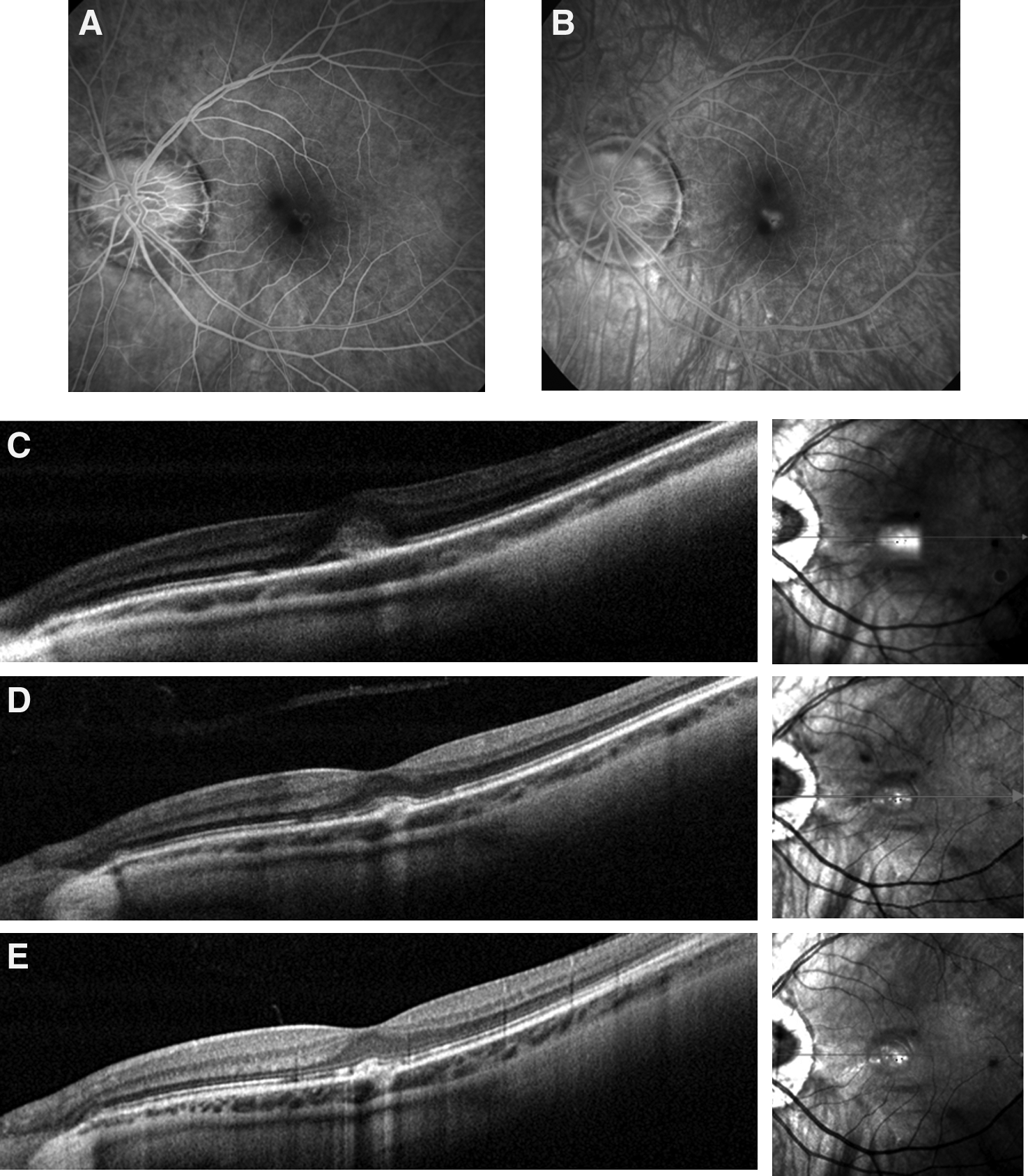
Fluorescein angiography shows hyper-fluorescence confined to the early phase of the angiogram
Results
No fetal, systemic, or ocular injection-related complications were reported. A monthly fetal ultrasound showed normal growth, and monthly umbilical, middle cerebral, and uterine artery Doppler ultrasound flowmetry always showed normal findings. The woman delivered a healthy male infant vaginally at term, without pregnancy-related complications such as hypertension, proteinuria, and threatening preterm delivery.
The baby showed a normal Apgar score (10/10), a normal birth weight (3.75 kg), and a normal visual behavior with no congenital anomalies, such as pulmonary or cardiovascular problems. He had a normal growth, reaching all developmental steps appropriately up to 12 months of age.
The patient's baseline BCVA improved from 20/50 to 20/32 at month 1 and increased to 20/25 at month 12 after the intravitreal injection. Monthly SD-OCT scans showed progressive reduction of the hyper-reflective dense area in correspondence with the CNV that almost disappeared 2 months after the injection (Fig. 1D). At month 6, only a slight thickening of the retinal pigment epithelium (RPE) line was detectable, and both the photoreceptor inner/outer segment (IS/OS) and the external limiting membrane lines were perfectly delineated (Fig. 1E). No further injections were required during follow-up.
Discussion
The management and optimal treatment of subfoveal CNV in pregnancy is still uncertain. Bevacizumab is a full-length humanized monoclonal VEGF antibody used as first-line therapy in PM-related CNV treatment, intravitreally administered off-label. 3 The use of intravitreal bevacizumab during pregnancy is controversial: Although a small amount of the drug (1.25 mg/0.05 mL) is delivered intravitreally, it can have systemic diffusion, 4 possibly leading to placental vascular injury. Moreover, the inhibition of VEGF can play a role in serious maternofetal complications, such as preeclampsia. 5 Our knowledge regarding fetus exposure to bevacizumab is based on preclinical studies and limited clinical experience.6–9 Preclinical studies in rabbits showed teratogenic effects and an increased number of miscarriages after being intravenously administered bevacizumab. 10 In our case, the patient was administered an intravitreal bevacizumab injection during the first trimester of pregnancy, more specifically at 7 weeks and 3 days of gestation. It is known that the first trimester of pregnancy is crucial for fetal development, and to the best of our knowledge, only a few reports exist with regard to the exposure to intravitreal bevacizumab during this period. One of these reports described 2 cases of early-term miscarriage at 4 weeks and 5 weeks of gestation, 7 days and 10 days after intravitreal bevacizumab, respectively, in 2 women unaware of their pregnancy; the short period between the treatment and miscarriages did not lead to a clear correlation between these 2 events. 7 The other reports showed that patients delivered healthy full-term infants with no documented adverse events related to treatment, and no cases of pregnancy complication in 2 cases of CNV of an origin other than age-related macular degeneration, and in one case of PM-related CNV, respectively.8,9
Our experience confirms the preliminary results of the previous case reports, reporting an uneventful pre- and post-natal course in a highly myopic woman and her baby. After treatment, visual acuity had favorably improved, with long-term stabilization.
The patient's decision was paramount in the collegial decision for treatment; she was fully aware of the benefits and risks of anti-VEGF treatment, and she thought the natural course of disease outweighed the risk of fetal damage due to drug exposure. The opportunity to wait until the end of the first trimester was rejected owing to the progressive impairment of visual function that worried the patient. The alternative treatment considered was verteporfin photodynamic therapy (PDT), which was widely used for subfoveal myopic CNV treatment until a few years ago. Verteporfin, similar to bevacizumab, showed teratogenicity in preclinical studies, but adequate studies conducted on humans are lacking (Source: verteporfin product insert). We ruled out verteporfin, as the intravenous route of administration might lead to a higher concentration in the utero-placental circulation. Moreover, PDT failed to reveal statistically significant benefits for subfoveal myopic CNV at 2-years follow-up. 11 Alternative anti-VEGF treatment, such as Ranibizumab, was considered. Since both bevacizumab and ranibizumab are currently “off label” for the treatment of myopic CNV, and considering the cost difference (ranibizumab costs ∼$2,000 per dose, while bevacizumab costs ∼$50), 12 the assignment was based on patient choice.
In summary, at present, there is no “on label” treatment for subfoveal myopic CNV in pregnancy. A multidisciplinary approach and extensive discussion with the patient appears fundamental when deciding treatment. In our experience, a single intravitreal bevacizumab injection administered during the first trimester of pregnancy did not show any complications, and appeared effective in myopic CNV treatment. However, despite the encouraging results reported to date, further studies are warranted that provide more detailed information on this treatment and its related risks during pregnancy.
Footnotes
Acknowledgment
The authors wish to thank Michael John of the Vita-Salute San Raffaele University for the English language editing of this article.
Author Disclosure Statement
F.B. is an advisory board member for Allergan, Novartis, Farmila-Thea, Bayer, Pfizer, Alcon, Bausch & Lomb, Genentech, Alimera Sciences, Sanofi Aventis, and Thrombogenics.
No competing financial interests exist for the other authors.