
Abstract
Abstract
Nanoparticles can be used for the treatment of various retinal diseases. Due to small sizes, they can improve bioavailability of therapeutic agents and pass through biological barriers of the eye, such as the cornea, conjunctiva, sclera, and even more, blood–retinal barriers. Another important characteristic of nanoparticles is the ability to be fabricated based on the researchers' design through chemical processes. In this regard, they can act as a novel drug delivery system, enabling targeted therapies for angiogenesis-related blindness (ARB). With these possibilities, many researchers have utilized nanoparticles as novel therapeutic options for the treatment of exudative age-related macular degeneration and diabetic retinopathy, both of which are characterized by pathologic neovascularization. In this review, we summarize various attempts and rationales in using nanoparticles to treat ARB. After that, we discuss the toxicity of nanoparticles on the retina. We expect this review to be a stepping stone for novel therapeutics for ARB, resulting in improvement in visual outcomes.
Nanoparticles: Novel Therapeutic Possibilities for Retinal Diseases
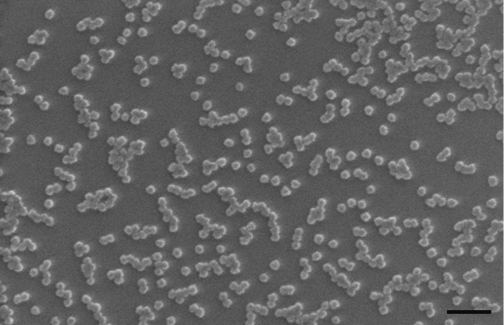
Scanning electron microscope image of 20-nm-sized silicate nanoparticles. These nanoparticles are examples of bare metal nanoparticles. Scale bar, 100 nm.
As for retinal diseases, there are 2 clinical trials currently registered in ClinicalTrials.gov (www.clinicaltrials.gov; Search date: 27 May, 2012; Search word: nanoparticle, eye, retina). One trial is a phase 2 trial studying the efficacy of intravenously infused albumin-stabilized nanoparticles containing paclitaxel in treating patients with metastatic melanoma of the eye (ClinicalTrials.gov Identifier: NCT00738361). The other phase 2/3 trial aims to examine the utility of cyclodextrin nanoparticle eye drops containing dexamethasone for the treatment of diabetic macular edema (ClinicalTrials.gov Identifier: NCT01523314). These studies show that there are researchers who see potential of nanoparticles in the treatment of retinal diseases by optimizing and stabilizing the delivery of therapeutic agents. In this review, we discuss the results of preclinical studies investigating possible application of nanoparticles in angiogenesis-related blindness (ARB). For the sake of precise discussion, we first define pathologic conditions that can be called ARB. Then, various attempts using nanoparticles in the treatment of pathologic neovascularization are summarized. In this section, we can realize that almost all forms of currently available nanoparticles are utilized in this field: nanocapsules (nanocarrier), nanoconjugates, and nanoparticles themselves. After that, we discuss the underlying rationales in the use of nanotechnology in overall retinal diseases and deal with the issue of nanoparticle-associated toxicity that is an unavoidable part of discussion regarding medical application of nanoparticles.
Treatment Modalities for ARB
ARB indicates vision-threatening retinal diseases, which are characterized by pathologic angiogenesis, such as age-related macular degeneration (AMD), diabetic retinopathy (DR), and retinopathy of prematurity (ROP). 2 All these 3 diseases affect substantial population of specific age groups: the elderly, the middle-aged, and premature infants, respectively. Clinical manifestations of diseases differ from each other in some degree. In patients with AMD, subretinal fluid or hemorrhage ensues after choroidal neovascularization (CNV), 3 whereas in diabetes mellitus (DM) patients, macular edema (ME) and extensive vitreous hemorrhage (VH) are more common clinical findings. 4 In ROP, the neovascularization process occurs at the junction between the vascularized and avascular retina. 5 However, commonly, in the process of diseases, neovascularization occurs which results in complications, including fluid collection between retinal layers, retinal detachment, and VH.
Therefore, the mainstay of treatment modalities for ARB is effective control of pathologic neovascularization by modulating pro- and antiangiogenic factors. Focal treatment modalities, such as panretinal photocoagulation for DR and laser photocoagulation for ROP, suppress the secretion of angiogenic factors, including the vascular endothelial growth factor (VEGF) by destruction of the peripheral retina, resulting in regression of pathologic angiogenesis.5,6 A more direct approach regarding angiogenic factors is the use of anti-VEGF antibodies or fusion proteins bound to VEGF. Currently, FDA-approved anti-VEGF agents are pegatanib sodium (Macugen®; Eytech Inc., Cedar Knolls, NJ), ranibizumab (Lucentis®; Genetech Inc., South San Francisco, CA), and a recently approved fusion protein aflibercept (EYLEA; Regeneron Pharmaceuticals, Inc., Tarrytown, NY). Bevacizumab (Avastin®; Genetech Inc.), a widely used anti-VEGF antibody for the treatment of ARB, has been used as an off-label drug.
These drugs have shown a positive effect on visual function in patients with ARB. With the treatment of ranibizumab and bevacizumab, many large, randomized trials demonstrated improvements in visual acuity of patients with AMD. 7 Furthermore, significant gains in visual acuity by aflibercept at 6 months were maintained at 1 year in patients with diabetic ME. 8
However, currently utilized anti-VEGF agents have limitations in common. First, in some pathologic conditions, there are concerns that anti-VEGF agents cannot show sustained effect on neovascularization. Despite bevacizumab's efficacy in the treatment of stage 3 ROP in a prospective, controlled, randomized, multicenter trial, 9 the effect of the drug might be only transient.10,11 Similarly, although VEGF inhibitors demonstrated efficacy as a short-term treatment option for certain patients with diabetic ME, long-term improvement in visual acuity and general long-term efficacy are unknown. 12 Second, repeated intravitreal injections are inevitable to get a sustained effect, resulting in increased risk of injection-related infection and retinal injury. 13 Third, direct inhibition of VEGF is expected to influence neuronal function of the retina, because VEGF is not only a growth factor for endothelial cells, but also a protective factor for neuronal cells.14,15 Recently, we also demonstrated that bevacizumab would suppress the differentiation of retinoblastoma cells even at concentrations that do not affect cellular viability, suggesting the negative effect of anti-VEGF therapies on the function of photoreceptors in the mature retina. 16 In this regard, although anti-VEGF agents have revolutionized the treatment of ARB to improve visual outcomes, there is still a need to develop novel therapeutics.
Nanotechnology: a Tool to Make Our Therapeutics for ARB More Powerful
Nanoparticles have received attention from researchers for possibilities to suppress pathologic angiogenesis in in vitro and in vivo studies. Interestingly, currently available techniques in manufacturing nanoparticles for medical uses are widely utilized in studies investigating the efficacy of nanoparticles on ARB (Fig. 2). The first form of antiangiogenic nanoparticles is the one with expression plasmid for specific genetic materials. Zhang et al. devised polylactic-co-glycolic acid (PLGA) nanoparticles containing the plasmid DNA expressing hypoxia inducible factor (HIF)-1α short hairpin RNA and a green fluorescent protein. 17 These nanoparticles lasted for 4 weeks in the retinal pigment epithelium (RPE) layer after they were administered via the intravitreal route and successfully suppressed CNV induced by laser in mice. Nanoparticles of Park and colleagues are based on PLGA:Chitosan and contain the expression plasmid for plasminogen kringle 5, a domain with antiangiogenic property. 18 They showed that intravitreal injection of nanoparticles induced expression of plasminogen kringle 5 in the retina and suppressed retinal neovascularization in the oxygen-induced retinopathy rat model. In this study, the expression of kingle 5 was maintained for 4 weeks, and overexpression of VEGF and intracellular adhesion molecule-1 was attenuated for the same study period. The same group also investigated the antiangiogenic effect of the nanoparticles on CNV, demonstrating substantial efficacy. 19 Interestingly, intravitreally injected PLGA:Chitosan nanoparticles resulted in expression of kringle 5 even at deep retinal layers near a experimentally induced laser lesion. Liu et al. investigated liposome-protamine-hyaluronic acid nanoparticles with small interfering RNA (siRNA) targeting the human VEGF receptor 1 mRNA. 20 In this study, the effect of nanoparticles containing siRNA enhanced the effect of naked siRNA, suppressing CNV lesion effectively.
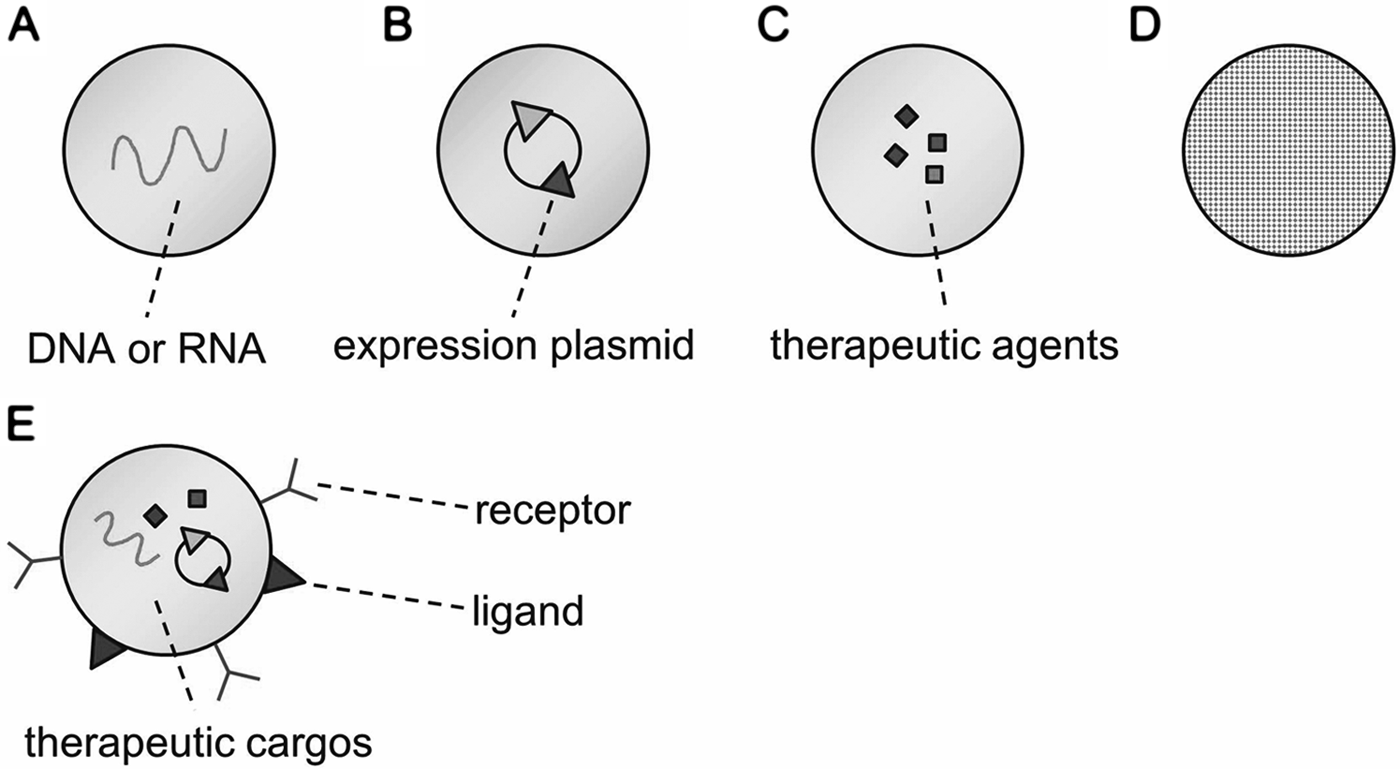
Various types of nanoparticles are suggested for the treatment of angiogenesis-related blindness.
Packing antiangiogenic molecules into nanoparticles is another example of antiangiogenic nanoparticles. Based on the idea that blocking specific types of integrins reduced CNV, Kim and Csaky formulated nanoparticles of polylactic acid (PLA)/PLA-polyethylene oxide (PEO) encapsulating the water-soluble integrin-antagonist peptide, C16Y. 21 In this study, nanoparticles released C16Y peptide over 6 weeks in the water and intravitreally administered nanoparticles are demonstrated to be endocytosed by the RPE cells. This study demonstrated the ability of nanoparticles to transport therapeutic agents to the retina with sustained effects. Without nanoparticles, intravitreally injected peptides would have very short half-lives, as the authors of the study suggested.
With the advancement of nanotechnology, fabrication of nanoparticles with specific surface molecules has been possible. Singh et al. intentionally manufactured nanoparticles in which the surface was coated with a linear arginine-glycine-aspartic acid (RGD) peptide, transferrin, or a combination of both. 22 These 2 molecules were selected to improve targeted delivery of anti-VEGF intraceptor plasmid to the neovascular lesion of the eye. According to the study, functionalized nanoparticles enhanced the efficacy of gene delivery, resulting in significant reduction in CNV lesions in the animal model.
The last interesting part of nanoparticles in the treatment of ARB is that bare nanoparticles demonstrate antiangiogenic properties by themselves. Nanoceria is known to scavenge reactive oxygen species and can inhibit the progression of pathologic retinal neovascularization by this mechanism. 23 Metal nanoparticles also inhibit proliferative vasculopathy. Kalishwaralal et al. demonstrated antiangiogenic action of silver nanoparticles on VEGF-induced proliferation and migration of endothelial cells in in vitro assays, 24 and the same group reported that silver nanoparticles could suppress the vascular permeability induced by advanced glycation end-products, suggesting possible application of them in the treatment of retinal complications of diabetes. 25 We also investigated antiangiogenic effect of gold and silicate nanoparticles.26,27 The exact mechanism of antiangiogenic action of bare nanoparticles is yet to be elucidated. However, repeated evidences from separate research groups increase the validity of the results of antiangiogenic effect of nanoparticles themselves. We summarize the results of studies on antiangiogenic effect of nanoparticles in Table 1.
CNV, choroidal neovascularization; HIF-1α shRNA, hypoxia inducible factor-1α short hairpin RNA; OIR, oxygen-induced retinopathy; PEG-LPH, PEGylated liposome-protamine-hyaluronic acid; PLA, polylactic acid; PEO, polyethylene oxide; PLGA, polylactic-co-glycolic acid; RGD, arginine-glycine-aspartic acid; VEGFR1, vascular endothelial growth factor receptor 1.
Rationales in the Use of Nanotechnology for Retinal Diseases
As we can see examples in the treatment of ARB, nanoparticles enable us to utilize novel therapeutics or currently available therapeutic agents more efficiently. The following sections discuss underlying rationales in the use of nanotechnology for retinal diseases, including ARB, retinal degeneration, and uveitis, the most actively studied fields in the eye in relation with nanotechnology.
Rationale 1: improved bioavailability
Nanoparticles can enhance the concentration of therapeutic agents in the retina via different routes of administration. Koirala and colleagues investigated the relative expression of RPE-specific vector when the plasmids compacted into polyethylene glycol nanoparticles and the naked plasmids were administered by subretinal injection. 28 Interestingly, nanoparticles increased the expression of enhanced green fluorescent protein effectively, and the increase was maintained over 1 month. In the study with solid lipid nanoparticles loaded with myriocin, an inhibitor of serin palmitoyl-CoA transferase, Strettoi et al. examined the effect of topically administered nanoparticles. 29 The authors proved that by topical administration, solid lipid nanoparticles reached the outer nuclear layer and between photoreceptors and the RPE. As intravitreally injected myriocin reduced the loss of photoreceptor cells, topically administered myriocin-laden solid lipid nanoparticles evidenced a protective effect on the number of photoreceptor rows in the outer nuclear layer in a mouse model of retinitis pigmentosa. Nanoparticles can improve the penetration of therapeutic agents, enabling topical administration of them to affect deep retinal layers.
Nanoparticles can improve bioavailability of intravenously administered drugs. The study of Sakai and colleagues was interesting in that they demonstrated the presence of nanoparticles in the retinal layers and more efficacies of betamethasone-laden PLA nanoparticles than betamethasone alone. 30 Systemically administered PLA nanoparticles loaded with betamethasone effectively inhibit the inflammation of the retina in a rat model of experimental autoimmune uveitis (EAU). Furthermore, bioavailability of therapeutic agents injected into the vitreous cavity was also enhanced by the aid of nanoparticles. Tamoxifen-loaded PEG nanoparticles resulted in significant suppression of inflammatory reaction in rats with EAU, whereas free tamoxifen did not induce positive effects. 31 These studies show that nanoparticles improve the bioavailability of therapeutic genes or drugs significantly.
Rationale 2: as a novel drug delivery system
Most studies regarding the action of nanoparticles in the treatment ARB have investigated the possible application of nanoparticles as a novel drug delivery system (DDS). The most noticeable field using nanoparticles as DDS is the gene therapy. As previously mentioned, genetic materials can be packed into nanoparticles.17–20,22,28 Furthermore, therapeutic agents are also safely delivered in nanoparticles to enhance the efficacy.21,29–31 Although wide use of anti-VEGF agents has lessened psychological burdens of intravitreal injection, maintanence of effective concentration at the retinal layers is still the problem of drug development for ARB. 32 A more attractive aspect of nanoparticles is that they can prolong the effective period of therapeutic agents. Recurrent injections even at 7 to 8 times a year are inevitable drawbacks of currently available anti-VEGF agents. Detailed pharmacokinetic information about nanoparticles in the eye is not published, but Koirala et al. suggested positive possibility of prolonged effectiveness of nanoparticles in their study showing that the expression of the target gene was maintained over a month. 28 Overall, insertion of therapeutic materials into nanoparticles is a good option for a novel DDS.
Rationale 3: increased surface molecules
Another rationale for the powerful effects of nanoparticles might be due to their physical properties. Dividing certain materials into tiny pieces definitely increases the surface area for the constant volume (Fig. 3B). Simply, if the diameter of a ball is divided in half, the volume decreases to one-eighth (

Dividing certain molecules into tiny pieces definitely increase the surface molecules, enhancing the interaction between therapeutic and target molecules.
Rationale 4: overcoming biological barriers
Penetration or diversion of biological barriers in the eye is also a hot issue in the development of therapeutics for retinal diseases. As we discussed in the previous section regarding the bioavailability, nanoparticles seem to penetrate anatomical barriers of the eye, such as the cornea, conjunctiva, and sclera.28,29 Therefore, they can affect the retinal diseases even when they were administered via topical or intravenous routes. The characteristic of overcoming biological barriers can provide various administration options for therapeutic agents. Furthermore, intravitreally injected nanoparticles can penetrate the retinal layers. In this way, they can be used for the treatment of CNV. In a study with intravitreally injected human serum albumin nanoparticles, Kim et al. demonstrated that nanoparticles were detected in the choroidal space of the rat eyes. 34
Interestingly, it seems that nanoparticles can pass through the blood–retinal barriers (BRB). We previously reported that nanoparticles of which diameter were small enough could penetrate BRB and exist throughout the whole retinal layer. 35 In this study, 20-nm-sized gold nanoparticles were distributed in all retinal layers when they were administered intravenously. Taken together, nanoparticles seem to improve the penetration of therapeutic molecules through biological barriers of the eye.
Rationale 5: targeted therapy
Another important strength of nanoparticles in the treatment of ARB is the possible application as a targeted therapy. As previously mentioned, there is already an attempt using nanoconjugates that have surface molecules abundant in CNV lesion for the treatment of CNV. 22 Chemotherapeutic docetaxel nanoparticles also utilized a ligand targeting prostate-specific membrane antigen. 1 Currently, nanotechnology regarding the manipulation of the surface of nanoparticles is rapidly developing; therefore, conjugation of certain ligands and nanoparticles is possible. This technology enables us to invent therapeutic options to attack specific cell types or lesion sites as an emerging targeted therapy. 36 In suppression of neovascular endothelial cells, specific receptors can be good candidates as recognizable targets for nanoparticles. Salehi-Had et al. utilized αvβ3 integrin ligand coupled nanoparticles to potentiate the targeted delivery of therapeutic genetic material to choroidal neovascular membrane. 37
Toxicity of Nanoparticles on the Retina
Nanotechnology helps to make novel therapeutics possible and currently available drugs more efficient by improving bioavailability and assisting targeted therapy. However, there are still concerns over toxicities of nanoparticles, and these concerns should gain attention. Researchers have studied the toxic effect of nanoparticles on the retina, evidencing conflicting results. Merodio et al. investigated the tolerability of bovine serum albumin nanoparticles. 38 They reported that there was neither inflammatory reaction nor alterations in the tissue architecture in the retina after intravitreal injection of nanoparticles. Furthermore, the treatment with nanoparticles did not change the expression of arrestin and rhodopsin autoantigens. There is another study showing an inflammatory change in the posterior vitreous and the retina 18 to 24 h after intravitreal injection of PLA nanoparticles. 39 However, the intensity of inflammation decreased 48 h of the treatment with PLA nanoparticles. In the study of Ding et al., there was elevation of interleukin-8 mRNA and monocyte chemotactic protein-1 at 1 day, but it was normalized 2 days after injection. 40 Furthermore, other signs of local inflammatory responses were not changed.
Bakri et al. evaluated the retinal toxicity of intravitreal gold nanoparticles at 2 different concentrations, evidencing no definite retinal or optic nerve toxicity at 1 month. 41 Prow et al. examined the toxicity of nanoparticles depending on the core materials of nanoparticles and the routes of administration. 42 In their study, intravenously administered chitosan nanoparticles showed inflammation in most of the treated eyes. However, nanoparticles from other core materials did not induce retinal pathology or inflammation. In addition, subretinal administration of nanoparticles was also nontoxic to the retina. There is a study comparing the effect of PEO tail block length of magnetic nanoparticles on the toxicity of nanoparticles. 43 These studies demonstrate that certain aspects of nanoparticles, such as concentrations, core materials, routes of administration, and surface molecules may contribute the toxicity of nanoparticles. (Table 2)
Time interval between injection of nanoparticles and evaluation of toxic effects.
BSA, bovine serum albumin; ELISA, enzyme-linked immunosorbent assay; H, histology; IHC, immunohistochemistry; MCP-1, monocyte chemotactic protein-1; MPO, myeloperoxidase; PCEP, poly[[(cholesteryl oxocarbonylamido ethyl) methyl bis(ethylene) ammonium iodide] ethyl phosphate]; PEG, polyethylene glycol; PLA, polylactic acid; PMN, polymorphonuclear neutrophil; qRT-PCR, quantitative real-time polymerase chain reaction; TNF-α, tumor-necrosis factor-α; N/A, not available.
Recent studies also have implications in the development of therapeutic agents using nanotechnology. Wielgus et al. investigated phototoxicity and cytotoxicity of hydroxylated fullerene nanoparticles. 44 The eye is the organ for sensation of visual and light stimuli; therefore, phototoxicity of specific drugs is required to be carefully investigated. The authors demonstrated that a combination of light exposure and a high concentration of nanoparticles might lead to retinal damage from in vitro cytotoxicity assays. Sanders et al. evaluated the phototoxicity of titanium dioxide nanoparticles in various concentrations and types of titanium dioxide. 45 In their study, titanium dioxide nanoparticles decreased the viability of human RPE cells when cells were exposed to higher concentrations of nanoparticles with ultraviolet A radiation. Therefore, we should have more emphasis on the concentration and general information of nanoparticles to make sure which aspects of nanoparticles induce the retinal toxicity.
Conclusions
Of course, nanoparticles cannot be a magic bullet for ARB. However, they can be a good bypass as a novel DDS and a platform for new therapeutic concepts. With the aids of nanoparticles, we can improve the bioavailability of currently available therapeutic agents and utilize them in more target-specific ways. Furthermore, there is a possibility that nanoparticles exert therapeutic effect by themselves. By conjugating nanoparticles with surface molecules targeting specific cell types in the eye, cell type and lesion-specific therapy is also possible. To develop novel therapeutic options using nanoparticles, researchers should provide general information about nanoparticles and their basic characteristics, including the size, shape, zeta potential, and treated concentration more openly in publications. These approaches help researchers to share therapeutic concepts and strategies in a more controlled way and to avoid unnecessary criticisms regarding nanoparticles. We hope that this review can be a stepping stone for open discussion and development of novel nanotherapeutics for patients with ARB suffering from visual deterioration.
Footnotes
Acknowlegments
This study was supported by the Bio-Signal Analysis Technology Innovation Program of MEST/NRF, Republic of Korea (2012-0006058), the Mid-Career Researcher Program of MEST/NRF, Republic of Korea (2012-0004931), and the Global Core Research Center (GCRC) grant from NRF/MEST, Republic of Korea (2012-0001187).
Author Disclosure Statement
No competing financial interests exist.