
Abstract
Abstract
Purpose:
To compare the results of subscleral trabeculectomy (SST) augmented with mitomycin-C (MMC) versus Ologen™ implant regarding intraocular pressure (IOP) control and incidence of complications.
Methods:
Sixty eyes of 60 patients, who planned to undergo SST, were divided into 2 groups. Group I eyes (included thirty eyes) were operated upon with SST augmented with intraoperative MMC. Group II eyes (included 30 eyes) were operated upon with SST using an Ologen implant. IOP and bleb status, as well as reporting postoperative complications, were followed up.
Results:
The follow-up period was 12 months. At 12 months postoperatively, the mean IOP was 19.33±3.22 mmHg in group I, and 19.87±4.17 mmHg in group II, with no significant difference between groups. One case in each group had hyphema, and 4 cases in group I and 2 cases in group II had shallow anterior chamber. One case in group I and no cases in group II had blebitis. There was no significant difference regarding the complications between both groups.
Conclusion:
We conclude that the use of the Ologen implant in SST is comparable to the use of MMC with advantage of avoiding the potential dangerous complications related to MMC use in the early (12 months) follow-up period.
Introduction
The healing response in the wound is reported to be the single most important risk factor in determining final intraocular pressure (IOP) after glaucoma-filtering surgery. 7 So, inhibition of scar formation during the process of wound healing should promote greater success.5,8
Mitomycin-C (MMC) applied intraoperatively during glaucoma-filtering surgery has improved the success rate and produced lower IOP, specially in the eyes at the risk of failure. 9 However, complications like cataract formation, avascular filtering blebs, thinning of the conjunctiva, subsequent blebitis, and endophthalmitis have been reported.10–13
Ologen™ is a bioengineered, biodegradable, porous collagen glycosaminoglycan matrix implant. It has been tried in animal models with promising results in wound healing after glaucoma-filtering surgery, with a potential of long-term IOP control.14,15 This study was conducted to compare the results of SST augmented with MMC versus Ologen implant.
Patients and Methods
This is a prospective randomized controlled study, which included 60 eyes of 60 patients who received ophthalmologic service at the Menoufia University Hospital during the period of February 2009 to January 2011. Patients included in the study had POAG, chronic angle-closure glaucoma (CACG), uveitic glaucoma, pseudoexfoliation glaucoma (PEXG), and pseudophakic glaucoma not controlled medically (more than 21 mmHg despite medications) as shown in Table 1. Exclusion criteria were neovascular glaucoma, age <18, and previous ocular surgery or laser procedures.
Independent samples test.
Fisher exact test.
Pearson chi-square test.
POAG, primary open-angle glaucoma; CACG, chronic angle-closure glaucoma; PEXG, pseudoexfoliation glaucoma.
Patients were randomly enrolled in 2 groups. Group I included 30 eyes that underwent SST augmented with 0.2 mg/mL MMC (Bristol-Myers-Squibb) for 2 min. Group II included 30 eyes that underwent SST augmented with Ologen (Ologen; Aeon Astron Group B.V. Leiden, the Netherlands) model 830601 6×2 mm implant (Fig. 1).
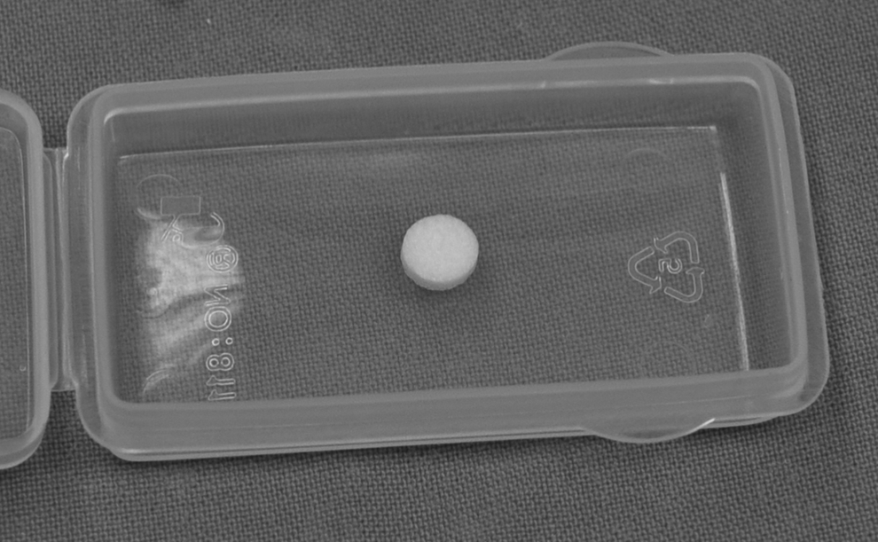
Ologen implant.
A comprehensive ophthalmic examination, including best-corrected visual acuity testing, slit-lamp examination, Goldmann applanation tonometry, fundus examination, and examination of ocular motility, was carried out for all patients. Preoperative data included age, gender, type of glaucoma, IOP, type, and number of antiglaucoma medications. A written informed consent was taken from all patients, and the research was approved by the institutional review board. All measures were in accordance with the tenets of the Declaration of Helsinki.
Surgical technique
All surgeries were performed by 2 surgeons (Hatem M. Marey and Amin F. Ellakwa). All patients were operated under local subtenon anesthesia using 2 mL of a mixture of 2% lidocaine (Sigma Pharmaceuticals, Egypt) and 0.5% bupivacaine (Al-Debeiky Pharma, Egypt) in 1:1 solution.
Group I (SST with MMC)
Fornix-based conjunctival flap was centered on 12 o'clock, with minimal cautery applied as needed. Two sponges 2×2 mm soaked with 0.2 mg/ml freshly prepared solution of MMC was applied on the scleral bed for 2 min, avoiding the conjunctival edge and the corneal epithelium. After removal of the sponges, the application site was thoroughly washed by 200 mL balanced salt solution. Rectangular scleral flap 3×4 mm was created (Fig. 2A), and sclerectomy 1×2 mm was performed, followed by peripheral iridectomy. The scleral flap was closed with two 10/0 nylon sutures at the corners of the flap (Fig. 2B), and the conjunctiva was closed with interrupted 8/0 vicryl sutures (Fig. 2D).

Group II (SST with Ologen)
Surgical steps were the same as group I except for application of MMC. Instead, after suturing the scleral flap with 2 loose 10/0 nylon stitches (Fig. 2B), Ologen 6×2 mm implant was applied on the scleral flap (Fig. 2C), and the conjunctiva was sutured with 8/0 Vicryl-interrupted sutures (Fig. 2D).
The postoperative care in all patients included the administration of topical mixed antibiotic and steroid (tobramycin 0.3% and dexamethasone 0.1%) eye drops 5 times daily for 3 weeks, and cycloplegic drops (cyclopentolate 1%) 3 times daily for 1 week.
In both groups, patients were examined 1 day postoperative and then after 1 week for detection of early postoperative complications. Patients were re-examined at 1, 3, 6, 9, and 12 months postoperatively for evaluation of the IOP and bleb state according to the Moorfield's bleb-grading system. 16 During each visit, complete ophthalmic examination was done. Qualified success was defined as an IOP equal or lower than 21 mmHg with additional topical medication. Absolute success was defined as an IOP equal or lower than 21 mmHg without any additional topical medication.
Statistical analysis was performed using SPSS version 16 (IBM Corporation, Somers, NY) software. Paired t-test was used to detect the difference between pre- and postoperative data in the study groups, and the independent sample test was used to calculate the difference between both groups in numerical variables. Fischer exact and Pearson chi-square tests were used to calculate the difference between groups in categorical variables.
Results
In group I, the mean age was 49.07±5.8 years; 17 cases (56.7%) were men, and 13 cases (43.3%) were women. In group II, the mean age was 50.2±10.2 years; 18 cases (60%) were men, and 12 cases (40%) were women.
Thirteen patients of group I (43.3%) had POAG; 5 cases (16.7%) had CACG; 4 cases (13.3%) had uveitic glaucoma; 4 cases (13.3%) had PEXG; and 4 cases (13.3%) had pseudophakic glaucoma. In group II, 18 patients (60%) had POAG; 4 cases (13.3%) had CACG; 3 cases (10%) had uveitic glaucoma; 2 cases (6.7%) had PEXG; and 3 cases (10%) had pseudophakic glaucoma.
Fourteen cases of group I (46.7%) were receiving 1 antiglaucoma drug; 12 cases (40%) were receiving 2 antiglaucoma drugs; 4 cases (13.3%) were receiving 3 antiglaucoma drugs preoperatively; the mean duration of treatment with topical antiglaucoma drugs was 3.1±1.27 years; and the mean preoperative IOP was 31.46±3.8 mmHg. In group II, 7 cases (23.3%) were receiving 1 antiglaucoma drug; 13 cases (43.3%) were receiving 2 antiglaucoma drugs; 10 cases (33.3%) were receiving 3 antiglaucoma drugs preoperatively; the mean duration of treatment with topical antiglaucoma drugs was 3.03±1.27 years; and the mean preoperative IOP was 29.87±3.44 mmHg. The descriptive and clinical preoperative data of both groups are summarized in Table 1.
In comparing preoperative data for both groups, there was no statistical difference regarding age (P=0.598), gender (P=0.793), type of glaucoma (P=0.76), number of antiglaucoma drugs (P=0.84), and the preoperative IOP (P=0.093) as shown in Table 1.
One month postoperatively, mean IOP was 16.17±2.96 mmHg in group I, and 16.8±5.29 mmHg in group II, with no statistically significant difference between both groups (P=0.57). At 3 months postoperatively, the mean IOP was 16.88±2.52 mmHg in group I, and 17.43±4.86 mmHg in group II, with no statistically significant difference between both groups (P=0.573). At 6 months postoperatively, the mean IOP was 17.57±3.49 mmHg in group I, and 18.5±4.89 mmHg in group II, with no statistically significant difference between both groups (P=0.398). At 12 months postoperatively, the mean IOP was 19.33±3.22 mmHg in group I, and 19.87±4.17 mmHg in group II, with no significant difference between groups (P=0.581); these data are illustrated in Table 2.
Independent samples test.
Pearson chi-square test.
IOP, intraocular pressure.
Regarding complications, 3 cases (10%) in group I and 2 cases (6.7%) in group II had anterior chamber reaction that resolved in the 1st week by conventional treatment. One case (3.3%) in each group had hyphema that resolved by the end of 1st week. Four cases (13.3%) in group I and 2 cases (6.7%) in group II had shallow anterior chamber that was reformed spontaneously by the end of the 1st week. One case (3.3%) in group I and no cases in group II had positive Seidle test that needed additional conjunctival suturing by topical anesthesia. One case (3.3%) in group I and no cases in group II had blebitis that resolved after 2 weeks by additional use of topical gatifloxacin 0.5% drops hourly, atropine 1% 5 times daily, and fuscidic acid 1% drops twice per day. There was no significant difference regarding the complications between both groups (P=0.678) as shown in Table 2.
Regarding bleb appearance at 12th months postoperatively, the mean bleb area score in group I was 2.27±0.98, and in group II was 1.9±0.8, with no significant difference (P=0.118). The mean bleb height score in group I was 2±0.695, and in group II was 1.76±0.71, with no significant difference (P=0.071). The mean vasularity score in group I was 1.8±0.407, and in group II was 1.4±0.498, with a high significant difference (P=0.001), as shown in Table 2.
Comparing the preoperative and postoperative IOP at 1, 3, 6, and 12 months, there was a high significant difference in both groups (P=0.001 for all measures).
Comparing the absolute and qualified success rate between both groups, there was no significant difference between both groups (P=0.424) as shown in Table 3.
Fisher exact test.
Discussion
Since introduced in 1968 by Cairns, STT remains the most common surgical procedure for treatment of glaucoma. 2 However, episcleral fibrosis and subconjunctival scarring decrease the success rate of such surgery, for which adjunctive antimetabolites were used to increase the success rate.17–20 Various studies demonstrated significant enhancement of success rates and postoperative IOP control through an intraoperative use of MMC. 10
MMC is an antitumor antibiotic isolated from Streptomycin caepitorus. It inhibits DNA-dependent RNA synthesis leading to cell death. 21 Its antiproliferative effect works in a dose- and time-dependent way. 22 MMC applied intraoperatively during glaucoma filtration surgery has improved the success rates and produce low IOP in eyes at risk for failure. 9 However, this was accompanied by an increase in adverse effects such as cataract formation, avascular filtering blebs, thinning of the conjunctiva, subsequent blebitis, and endophthalmitis.10–13 Other agents such as corticosteroids, growth factor inhibition, and amniotic membrane have been applied to enhance the results of antiglaucoma surgery.23–25
Studies on animal models showed that the use of a bioengineered biodegradable, porous collagen–glycosaminoglycan matrix implant offers the potential for a new means of providing controlled resistance between the anterior chamber, and the subcojunctival space in early postoperative period. Moreover, it maintains long-term IOP control by avoiding early scar formation and creating a loosely structured filtering bleb.14,15,26
The Ologen implant (Aeon Astron Europe BV, Leiden, The Netherlands) is a porous implant comprising more than 90% lyophilized porcine collagen and <10% lyophilized glycosaminoglycan with a pore size of 10–300 μm. It is a biodegradable implantable scaffold matrix, inducing a regenerative nonscarring wound-healing process without using antifibrotic agents. The matrix improves the regenerating tissue remodeling and prevents scar formation or further infection. 27
In human subjects, the Ologen implant has been tested for augmentation in deep sclerectomy. It was revealed that deep sclerectomy with Ologen implantation is an effective and well-tolerated method for reduction of IOP. 28 A further pilot study revealed nonsignificant differences in postoperative IOP after trabeculectomy with Ologen and sole trabeculectomy. 29
The current study was designed to compare the results of use of Ologen versus MMC as an adjunctive therapy in SST over the 12-month follow-up period. There was no significant difference regarding age, gender, type of glaucoma, number of antiglaucoma drugs used, and the preoperative IOP nullifying the effect of these factors on surgical outcome in both groups.
In the current study, IOP at follow-up visits was significantly lower than the preoperative level in each group independently. This clarifies that both surgeries were efficient in lowering IOP significantly from the preoperative level. Comparing IOP between both groups in the follow-up visits demonstrated slightly lower IOP level in the MMC group. However, there was no statistically significant difference between both groups at 1, 3, 6, and 12 months.
Regarding the success rate, there was no significant difference between both groups. However, more cases in group II (5 cases), than in group I (2 cases), needed antiglaucoma drugs to control the IOP at 21 mmHg or less at the 12-month follow-up visit. This is in contrast to the results of Rosenteter et al., 27 who reported more significant success in MMC group in comparison to the Ologen group with significantly more number of cases in the Ologen group needed antiglaucoma drug to control the IOP at the 12-month follow-up visit. However, all cases in their study were POAG, and they had only 10 cases in each group; 5 (50%) of them in the Ologen group needed antiglaucoma drug to be controlled, which was highly significant.
The success rate in group II (83.3%) is almost similar to that of Nilforushan et al., who had a success rate 80%, but this was for a 24-month follow-up, and their cases were only 10 cases of POAG. 30
There was no significant difference between both groups regarding the complications over the follow-up period. However, there were more cases complicated by shallow anterior chamber in group I (4 cases), than in group II (2 cases). Also, blebitis occurred in one case in group I, and no blebitis occurred in group II, and bleb leak occurred in one case in group I, and no bleb leak occurred in group II. This could be attributed to the use of MMC in group I. There were no complications specific for the use of Ologen in group II over the follow-up period.
Comparing the bleb appearance, there was no significant difference regarding the bleb area or height scores; however, there was significant avascularity in blebs in the MMC group than the Ologen group (P=0.001), this was similar to the results of Rosenteter et al., 27 which is considered as one of the major problems of MMC on blebs.
We conclude that the use of Ologen implant in SST is comparable to the use of MMC with the advantage of avoiding the potential dangerous complications related to MMC use in the early (12-month) follow-up period.
Footnotes
Author Disclosure Statement
No competing financial interests exist.