
Abstract
Abstract
The vitreous humor represents a significant barrier to the penetration of nanoparticulate-based ocular drug delivery systems. The gel structure and biochemical components of the vitreous will impact the rate of nanoparticle movement through the tissue to reach the retinal tissue. Also the structure of the vitreous, flow systems operating within it, age-related structural changes, and the presence of inflammation will also have a potential effect on movement. To effectively target the posterior retina, the physical properties of the nanoparticle formulation are a key element in the design of a system that will penetrate through the vitreous barrier.
Introduction
The Vitreous
The vitreous body fills the region between the lens and the retina and accounts for ∼80% of the tissue volume of the eye. In the adult, the volume is ∼4 mL with a specific gravity close to 1. The main component of the vitreous is water (98%–99% w/w). The solid components consist of collagen and glycosaminoglycans, which—although present in small amounts—are organized to form and maintain a stable gel (Fig. 1). The collagen fibers are composed of “sticky” type II collagen, which is coated with less-adhesive type IX collagen. The type IX collagen is gradually lost through life, allowing the type II fibers to interact and thicken. 5 The presence of a gel in younger animals will impede free molecular diffusion, particularly of high-molecular-weight compounds or suspended solids.
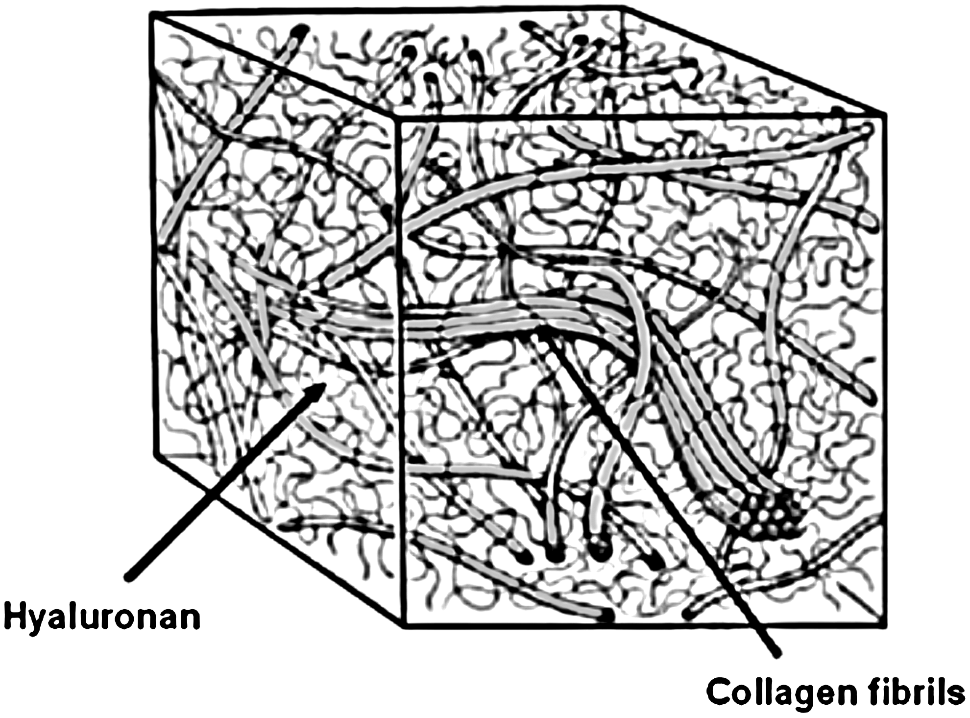
Cross-sectional representation of the structure of the vitreous humor. Collagen fibrils provide structure and strength while maintaining the gel state of the vitreous, whereas the hyaluronan inflates the gel by filling the spaces in between the collagen fibrils [adapted from Le Goff and Bishop (2008) 5 ].
In front of the vitreous body, the lens focuses the image transmitted through the cornea into the posterior chamber, covered by the neural retina. The lens, suspended in the lens capsule by the suspensory ligaments or zonules, obstructs flow from the posterior to the anterior chamber and forward movement has to occur through the zonules. The compartmentalization ensures that a nanoparticulate preparation injected into the vitreous space will provide a partially dispersed reservoir to treat the retina.
Unlike a preparation of large particulates or an implant, the injected volume will remain suspended for a time and not immediately sink to the floor of the vitreous space. Retinal toxicity is associated with settling of large crystals on the cell layer and formulation effects are likely to reduce the severity of the cytotoxicity. 6 Some materials (such as triamcinolone acetonide) stick to the collagen and are used by surgeons to identify the thickened collagenous fibers of the posterior vitreous cortex prior to dissection. 7 Ideally, the dose should remain near the target, often regarded as the posterior pole toward the macular. A short needle will result in more anterior placement, and flow processes in the vitreous will redistribute and ultimately eliminate the dose from the eye from this depot, reducing the potential concentrations at the back of the eye and the effective period of treatment. In addition, the drug may be sequestered by binding to nontarget tissues, including the collagen fibrils, and once through the inner limiting membrane (ILM), by adsorption onto melanin. 8
The Role and Structure of the Vitreous
The role of the vitreous has been debated extensively, and among a previous generation of ophthalmologists, there was a view that the retina might be easily detached, and therefore one role for the vitreous might be to hold the retina in place. Foulds reminded surgeons that in fact the retina is held in place by relatively strong forces and detachment only occurs after a multiplicity of failures. He commented that the true role of the vitreous was to resist rapid changes in size by fluid flows and thus to stabilize the volume. 9 This is supported by the earlier work of Duke-Elder and colleagues who noted that while in vivo the vitreous was capable of withstanding high orders of unbalanced stresses, on removal, small changes in the environment lead to liquefaction. 10 A consequence of this is that the study of the diffusion of nanoparticles in the vitreous body following removal from the globe might yield large artifacts caused by changes in biochemistry and biophysical properties. Lee and colleagues have shown that pars plana vitrectomy performed 2 weeks before intravitreal injection of human vascular endothelial growth factor (VEGF)165 into the vitreous of the rabbit decreased the half-life of the tracer from 2.46 h to 12.5 min with a 4-fold reduction in area under curve (AUC). 11 If a vitrectomy is performed, there appears to be a persistent change in the characteristics of the blood–retinal barrier. Following intravenous administration of fluorescein, this led to increased and faster accumulation of fluorescein in the vitreous. 12 An important role of the vitreous appears to be to decrease the lens exposure to oxygen, and to mop up oxidative radicals by the high levels of ascorbate present in both aqueous and vitreous humor. The consequences of removal of the vitreous are an increased risk of iris neovascularization, following increased oxygen transport, especially if the lens has been removed. Unlike the aqueous humor, which is constantly regenerated, the vitreous humor is a structurally “stagnant” compartment. The impact of surgical techniques on vitreous function has been reviewed by Stefánsson (2009), who has examined effects of vitreous substitution. 13 This has also been the subject of a recent review by Kleinberg and colleagues (2011). 14
The Shell of the Vitreous
In young animals, the firm vitreous can be carefully manipulated from the dissected globe, and removed, with the lens attached to the vitreous humor (VH) by the anterior hyaloid membrane and the lens capsule. The boundary of the featureless, transparent gel is formed by condensed collagen fibrils, known as the vitreous cortex that originates from the vitreous base, straddling and firmly attached to the ora serrata. The vitreous base may be visible in elderly adults as a hyperpigmented structure 4–5 mm in width behind the pars plana. The vitreous is also attached posteriorly strongly to the retina over the retinal blood vessels and at the optic disc, the whole structure joined to the retina at various points within the globe like a tethered barrage balloon. The vitreous is subdivided into 3 regions, with the central vitreous zone forming the bulk of the vitreous body, with the lowest density of fibrils that run anterior to posterior across the globe (Fig. 2). These are anchored in the basal vitreous and posteriorly join the vitreous cortex. At the vitreous base, the fibrils are solidly anchored into the pars plana and anterior retina. 5 In this zone, the fibers are orientated perpendicularly in high density and arc toward the optic nerve head. In some laboratory animals, particularly, the rabbit, the vestigial hyaloid artery originating midway at the posterior lens capsule is clearly visible as a central channel known as the Cloquet's canal. 15 Until the evolution of three-dimensional optical coherence tomography (OCT), the remnants of the structure were not observable in man. 16 This raises the possibility that the more prominent Cloquet's canal in the rabbit may contribute to central fluid flow to a greater degree than in man.
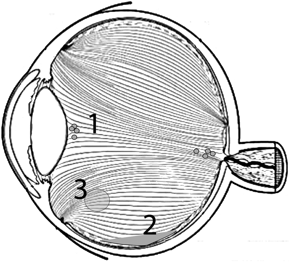
Diagram showing the orientation of collagen fibrils [adapted from Le Goff and Bishop (2008) 5 ] and sites of deposition. A proportion of a dose of microparticles injected into the central vitreous remain associated with the back of the lens or near the optic nerve (1). Heavier, larger particles settle to the floor of the eye (2) and charged nanoparticles may associate with the collagen fibers (3).
Experiments with ink injected into the central vitreous revealed that the central zone was composed of a complex cisternal structure. 17 This can also be revealed by injection of fluorescent beads into vitreous decanted into a cuvette, as described in a previous review. 18
The portion curving forward and inward from the vitreous base is the anterior vitreous cortex and at the front of the eye, the membranes are composed of smooth lamellar sheets and superficial fibers run in parallel to the lens surface. The section coursing posteriorly from the vitreous base is the posterior cortex and has a thickness of 100–110 μm, composed of dense type II collagen fibrils. 19 These are more prominent on the outside of the structure within the cortex, whereas on the inside of the structure, thinner fibrils run from the anterior to posterior position. In the dissected posterior vitreous cortex of older eyes, two holes are noted. The prepapilliary hole is ∼1 mm in diameter. The outer posterior cortex is absent over the optic disc. When the posterior cortex is detached, the cortical fibers protrude through the hole formed at the point of dehiscence and the hole is around 4 times larger than the prepapilliary hole. In infant eyes, Sebag (1989) did not observe a premacular hole but the vitreous blebbed from the prepapilliary hole, which led the author to conclude that it was a true aperture in life. 20
The Inner Limiting Membrane Barrier
The cortex inserts into the basal lamellar layer of the Müller cells and is known as the ILM. The ILM stains with periodic acid Schiff's stain, indicating that a high proportion of proteoglycans are present within the structure. The retinal surface is irregular and in a close arrangement with Müller cells, whereas the outer vitreal surface appears as a smooth, featureless sheet. The strength of attachment varies and is strongest at the vitreous base and around the optic nerve head, forming a ring of thickened tissue (Weiss ring), which is often visible following vitreopapillary separation in posterior vitreous detachment (PVD). 7 The function of the ILM seems to be to support the glial cells during the earliest stages of embryogenesis, 21 as in late embryogenesis, the role is less important and the ILM may be removed; indeed, removal of the ILM has become important in vitreoretinal surgery as peeling is considered as a benefit for most of the traditional retinopathies. 22 Following the introduction of triamcinolone to treat inflammatory disorders, the ILM was found to be accidentally labeled with the insoluble crystalline matter, which became associated with the collagen bundles of the vitreous. Although an association was formed, only a loose binding occurred, as the crystals could be washed from the structures. That being said, this became a useful tool for the surgeon and a review of the use of trimacinolone acetonide as an adjunct to vitreoretinal surgery was conducted by Couch and Bakri (2008). 7 In addition to this, in a previous communication, we mention the early macroscopic evidence from Wolter (1964), 24 suggesting that the ILM of human retina is incomplete, with small breaks in the ILM, which may allow for the migration of phagocytes and neural structures between the vitreous space in the retina. 25 Wolter described the holes to be small and usually associated with blood vessels, with vitreous strands extending through these pores to encircle the retinal blood vessels.
The ILM may pose a significant barrier to intravitreally injected nanoparticles administered for treatment of the retina. Previously, the ILM has been identified as a significant barrier for the penetration of adeno-associated virus (AAV) into the retina, particularly in adults. Dalkara and colleagues conducted experiments examining the contribution of the ILM with 5 fluorescently labeled AAV serotypes. 26 The ILM was digested by treatment with a nonspecific protease. The AAV vectors were seen to traverse a complex pathway and it was shown that for intravitreal transfer, the viral vectors need to bind and accumulate at an intact ILM. In contrast, the viral particles that lacked binding sites for the ILM did not accumulate at the ILM and remained dispersed within the vitreous leading to a failure in gene expression. Heparin sulfate motifs associate with opticin, a small leucine-rich peptide that colocalizes with type XVIII collagen concentrated in the ILM, which may provide association of the AAV vector with the ILM. 27 While this is required for virus transfection, the association of heparin sulfate (HS) with the envelope protein may reduce flux.
The association with the ILM was also described in an early publication by Bourges and colleagues, who studied poly(lactic acid) (PLA) loaded with rhodamine dyes. 28 These were injected into the eyes of rats, and histology and immunohistochemistry were performed at various times after injection. Confocal microscopy of the retina shows accumulation of the particles at the ILM in irregular high-density clusters, which appear to traverse the barrier and thus gain access to the RPE cells. In a related manner, Heiduschka and colleagues showed the penetration of Avastin (bevacizumab) injected intravitreally to cynomolgus monkey (Macaca fascicularis) retina following intravitreal injection. 29 Association with the ILM with residual staining of this layer at 7 and 14 days and strong staining of the outer photoreceptor layer is noted, with the residual antibody remaining associated with the ILM. Thus, following intravitreal administration, tight association with the ILM might be a prerequisite for retinal targeting.
The Impact of Aging
With aging, the collagen elements collapse and thicken, and traction is exerted on the structure resulting in an increase in pigmentation, tears, and retinal detachment. As the eye ages, the gel liquefies and by early middle age, liquid vitreous pockets can be observed in slit-lamp microscopy. This process is known as syneresis and is associated with the dissociation of the collagen hyaluronan complex described by Le Goff and Bishop. 5 The vitreous gel volume decreases, accompanied by collapse of the collagen fibers to form thickened tangled fiber layers. Side illumination with a slit lamp reveals the collapsed structure in autopsy samples. The remnant vitreous pulls away from the retina as a consequence of two processes: the liquefaction of the vitreous gel and the dehiscence of the membranes, associated with a progressive weakening in the adhesion between the ILM and the vitreous cortex. 20 This process is referred to as PVD and can lead to retinal tearing. Sebag (1991) has described the differences in vitreoretinal adhesion on aging using dark-field and electron microscopy in young and old human donor eyes. 30 He comments that in the macula of young subjects, there are strong attachments between the vitreous cortex and the ILM, particularly near the macular hole. The adhesion appears to be provided by vesicular points of attachment configured as a sheet encompassing the macular and the peripapillary posterior pole. Studies in our group have described a model of partial vitreous liquefaction in the rabbit by treatment with ovine testicular hyaluronidase. 31 The degree of vitreous liquefaction generated was similar to that reported in humans between 55 and 60 years of age, with ∼40% gel phase remaining. In partially liquefied vitreous, high-molecular-weight fluorescein isothiocyanate-dextran (average molecular weight 150 kDa) was cleared faster than in controls, suggesting greater convective movement in the liquefied vitreous. Ocular fluorophotometry indicated a temporary forward flux in the partially liquefied vitreous model during the first few hours after injection, which was not observed in normal vitreous. 31 These data suggest that in the elderly patient receiving intravitreal therapy based on a nanoparticle suspension, convective processes for formulation will be important and material placed too far from the posterior pole will fail to be accumulated at the target. The process of PVD is slow, starting as a shallow separation and extending slowly until vitreous separation occurs at the optic disc margin and symptoms are evident. 21 Johnson describes the disorders associated with incomplete PVD as being related to the size and strength of residual adhesion in the perifoveal region.
Flow Processes in the Vitreous
Raised intraocular pressure is a feature of several ophthalmic diseases and induces hydraulic flow in the front of the eye. The effect of increased pressure on vitreal kinetics has been modeled by Missel and it is concluded that, in the normal eye, water flux through the vitreous would be so small that it would only contribute to movement of large molecular constructs or for small molecules not efficiently cleared by the choroid. 32 However, since the vitreous gradually liquefies, it follows that hydraulic stresses applied to the eye will result in convective movement of the liquefied contents. The action of blinking or sudden acceleration/deceleration associated with head movement will cause minor inertial deformation of the interior globe and has been calculated to generate complex three-dimensional vortex patterns of flow. 33 Osmotic gradients near the ciliary processes also appear to contribute to movement of water and deformation of the semipermeable iris. 34
Large molecules, such as the VEGF antagonist ranizumab, are transported forward into the anterior chamber and we have proposed that surgery on the lens (e.g., phacoemulsification for cataract surgery) would have marked effects on larger constructs. 18 Any surgical intervention will exaggerate mixing and it is established that interference with the zonular region occurs during operative procedures on the front of the eye including cataract removal. Biochemical studies on a small number of aphakic and pseudophakic donor globes suggest that long-term changes occur in the vitreous. 35 In the near future, treatment of segments of the population who have had advanced types of lens may exacerbate the issue of forward loss of materials injected into the vitreous. Modern designs of intraocular lens have been proposed for the control of myopia in Japan, such as the hole-implantable contact lens (ICL), which have a very small central hole (diameter 0.36 mm) that markedly alters fluid dynamics in the eye. The size of the lens relative to the whole eye varies in different animals, 36 which has an impact with regard to modeling the distribution clearance and the impact of convective flow. 37
The Role of Inflammation
A final consideration in nanoparticle movement through the vitreous is the effect of inflammation. Inflammatory processes and surgical trauma are associated with changes that are long lasting and probably reflect changes in microstructural elements of all ocular tissues resulting in altered retinal access and changes in clearance. Inflammation is associated with a hyperemia and an alteration of the blood–retinal barrier. Intravenous infusion of Salmonella typhimurium toxin to the rabbit produced an increased absorption of grepafloxacin into the vitreous humor in albino rabbits from the plasma. 38 The decrease in barrier function also markedly increased iris and ciliary body levels in the Gigante de España species (the drug is highly melanin bound). In the pig, unilateral vitrectomy disturbs the retinal–blood barrier and, 1 month after the procedure, fluorescein given intravenously penetrates the vitreous more readily giving a markedly higher area under the curve. 12 Ocular inflammation following administration of endotoxin to the rat results in retinal edema and the swelling of the Müller glial cells 39 is likely to alter both transporter function and the integrity of the ILM, allowing easier access of nanoparticulate agents to the inner retina.
Characterizing Nanoparticle Movement Through the Vitreous
To effectively target the posterior eye and understand movement patterns, nanoparticle distribution through the vitreous following administration must be characterized and can pose a challenge to the formulator. In a typical ocular pharmacokinetic studies investigating drug distribution post-intravitreal injection, liquid chromatography mass spectrometry (LC-MS) drug quantification studies will be employed, 40 with alternative techniques, such as nuclear magnetic resonance (NMR) 41 and time of flight secondary ion mass spectrometry (ToF-SIMS),42,43 used more recently by our group. On the other hand, monitoring larger particulate system movement through the vitreous is less focused on monitoring tissue concentrations and requires the use of effective imaging techniques. The distribution and subsequent elimination of the nanoparticles from the vitreous will depend on the properties of the nanoparticles administered. Typical polymers of interest in ocular nanoparticle formulation are shown in Fig. 3. Factors that are generally regarded as important in controlling the distribution of particulate systems through the vitreous include the particle size and also the surface characteristics of the particles. With regards to particle size, distribution will be effected differently in the vitreous due to the clearance systems in operation. It is accepted that due to the difficulty in transversing the blood–retinal barrier, only nanoparticles will be subjected to clearance through the retina and larger microparticulates and aggregates will fall to the floor of the globe. Many nanotherapeutics consist of condensed molecular structures and the discrimination between a physical particle and a chemical entity is lost, since the size of these materials ranges from 1 to 1000 nm. Macugen® (a pegylated aptamer), bevacizumab (humanized monoclonal antibody), and ranibizumab (fragment of the bevacizumab antibody) traverse the retina intact and exert anti-VEGF actions resolving subretinal edema. Very small molecules (≤1 kDa) can traverse the retina easily when injected into the vitreous space and the retention time will be short (a matter of hours), and related to lipophilicity. 44 The surface characteristics of polymeric nanoparticles, in particular, surface charge, will also have a major influence on particle distribution and movement through the vitreous following intravitreal injection, due to the properties of the vitreous, resulting from its composition. The choroidal circulation will provide an efficient sink on the other side of the retina, assisting in removal.
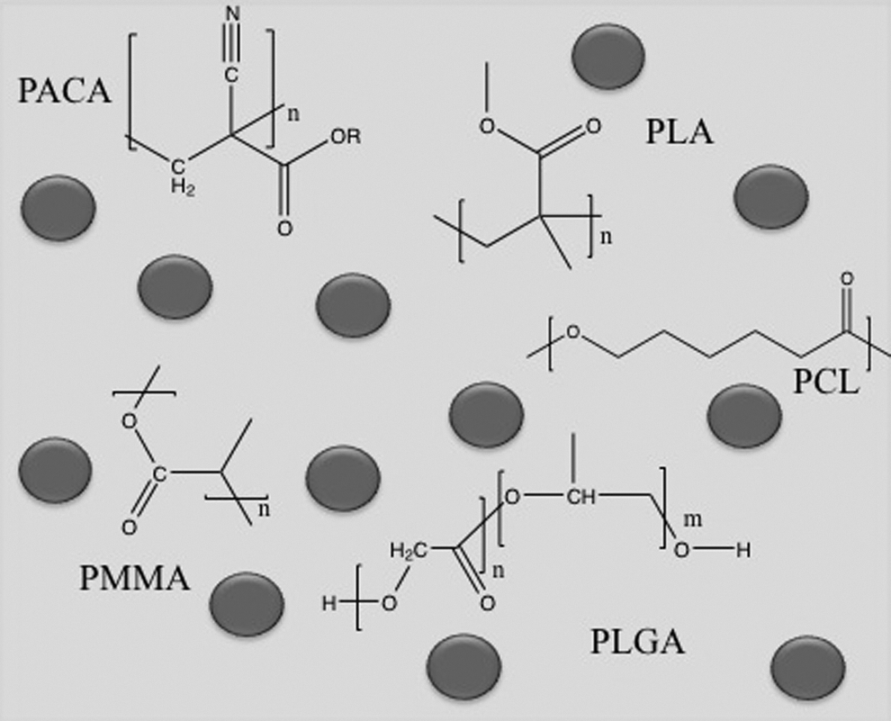
Diagram showing the chemical structure of typical polymers of interest in nanoparticle delivery to the eye: poly(lactic-co-glycolic acid) (PLGA); poly(lactic acid) (PLA); poly(methyl methacrylate) (PMMA); poly(caprolactone) (PCL); poly(alkylcyanoacrylate) (PACA).
The nature of the vitreous humor plays a key role in charge-related particle distribution effects due to the anionic medium created by its structure. 45 Due to the overall anionic charge of the vitreous, it is anticipated that anionic particles will distribute through the vitreous without any major restriction, whereas cationic particles may bind to the anionic structures of the vitreous humor, hindering particle movement. It is therefore important to understand the optimal nanoparticle size and surface charge to ensure nanoparticle movement through the vitreous to the retina for the treatment of the posterior eye disease.
Effect of Nanoparticle Size
Studies directly investigating the impact of nanoparticle size on distribution following intravitreal injection are limited. Although various distribution studies of individual particles have been carried out, it is difficult to directly compare distribution data from different studies. In the majority of studies available, the nanoparticles under investigation were compositionally different and as a result will have different surface characteristics exposed to the vitreous, potentially impacting on movement and penetration pathways. That being said, the impact of size on nanoparticle movement in the vitreous post-intravitreal injection has been directly studied using polystyrene particles tagged with sodium fluorescein. Particles were generated to create a range of particles with particle sizes of 2 μm, 200 nm, and 50 nm. 46 In this instance, the impact of particle size on the residence time of the particles in the vitreous humor was shown to be important. Elimination half-life from the vitreous was shown to increase with decreased particle size, with t1/2=5.4 h for 2 μm, t1/2=8.6 h for 200 nm, and t1/2=10.1 h for 50 nm. Movement of particles from the vitreous into the surrounding ocular tissues also differed depending on size with larger microparticles shown to travel to the trabecular meshwork, whereas smaller nanoparticles moved to the trabecular meshwork and to the retina. In addition to this work, a recent study by Raju et al. (2012) demonstrated differences in the elimination of iron oxide particles after intravitreal injection. 47 Larger (4 μm) iron oxide particles coated with polystyrene were shown by magnetic resonance imaging (MRI) to remain in the rat eye 5 weeks following administration, whereas smaller (50 nm) iron oxide particles coated with dextran were not found to reside in the eye after 5 weeks. This data seems somewhat conflicting with the work by Sakurai and colleagues (2001), 46 where the residence time increased with decreased size. A potential reason for this could be the functionalization of the iron oxide particles and also the small sample size investigated. It is difficult to directly compare the movement of these iron oxide particles as a function of size, due to the difference in the applied polymer coating and its role in the elimination rate of the particles. 47 Since nanoparticles formulated for drug delivery are regarded to be within the size range of 10 nm up to 1000 nm, 48 the impact of size alone on nanoparticle movement to the posterior eye is unlikely to be the main controlling factor, with a range of nanoparticles from 50 nm 46 up to 643 nm 47 of different composition shown to penetrate through the retina following intravitreal injection. It is likely that movement through the vitreous involves a combination of both the right size and the optimal surface characteristics. Further studies involving nanoparticles of the same composition in a range of sizes would be of benefit in advancing our understanding the direct impact of nanoparticle size on nanoparticle movement through the vitreous into the retina.
Effect of Nanoparticle Charge
Nanoparticle surface charge is an important determinant of drug movement through the vitreous humor. Negatively charged anionic nanoparticles, in general, appear to move through the vitreous with little restriction. Polymer-based polyactide nanoparticles, with an average size of 140 nm and an average ζ-potential of −60 mV, have been shown to move through the vitreous humor unrestricted, to reach the posteriorly positioned retina and also the anteriorly positioned lens, iris, and ciliary body. One hour following administration, nanopartcle (NP) were found to settle on the ILM of the vitreal–retinal barrier before penetrating through to the deeper layers of the retina 6 h postinjection. 22 Similar effects on distribution were seen using another polymeric-based system of poly(lactic-co-glycolic acid) (PLGA), to generate larger anionic NP for gene delivery of red fluorescent protein. PLGA-NP with an average particle size of 643 nm and an average ζ-potential of −7.4 mV were shown to travel through the vitreous humor to penetrate through to the retina. The result was successful gene expression of red fluorescent protein in the retinal pigmented epithelium after 4 days following intravitreal injection. 49 Fluorescent particles generated using a core of fluorescent polystyrene nanoparticle (200 nm), coated with the polymethoxy polyethylene glycol cyanoacrylate-co-hexadecyl cyanoacrylate copolymer, were also found to distribute from the vitreous and accumulate on the ILM and also the ciliary body between 8 and 24 h post-intravitreal injection. The particles were then phagocytosed by the astrocytes of the ILM 1 day following injection and then were found in the RPE 3 days following injection. 50 Although the ζ-potential of these particles was not determined, based on previous NP work carried out using the copolymer ratio, the particles were likely to be anionic in physicochemical nature. 51 Similar effects were seen with another copolymer-based nanoparticle system of PLA/PLA-poly(ethylene oxide) [PEO]. Nanoparticles were produced with a bimodal size range of 114–126 nm and 302–367 nm and carried an average ζ-potential of −38 mV. The nanoparticles were able to move freely in the vitreous and were found to penetrate to the retina and, additionally, were found in the trabecular meshwork and ciliary body. 52 Nanoparticles designed to deliver ganciclovir, composed of albumin (290 nm, −27.8 mV) and biotin (304 nm, −25.1 mV), were also shown to diffuse well through the vitreous. Both particles were found to distribute through the vitreous and accumulate as a thin layer on the retina. In addition, the biotin-based particles appeared to have a broader distribution and were also seen anteriorly in ciliary body and at blood–aqueous barrier. 53
Compared with anionic nanoparticles, data from distribution studies following intravitreal injection of positively charged particles are more limited. Unlike anionic nanoparticles, for cationic particles, the vitreous humor has been shown to hinder nanoparticle movement from the vitreous into the retina. Positively charged polymeric nanoparticle systems of polyethyleneimine (PEI) and 1,2 dioley-3-trimethylammonium-proprane, designed for the delivery of DNA, experienced retinal cell uptake of 35%–42% and 20%, respectively, in vitro. However, in the presence of vitreous humor, this uptake was reduced to <2% for both carriers, suggesting a potential association of the NP with the vitreous humor, thus preventing movement into the retinal cells. 54
The direct relationship of charge on particle movement was investigated by Kim and colleagues using human serum albumin (HSA) nanoparticles. 55 Two types of particles containing Alexa fluorescent dye were produced from HSA to generate anionic NP and also cationic NP, created through the coupling of ethylenediamine to carboxyl groups on HSA. Anionic NP averaged at 114 nm and had an overall ζ-potential of −33.3 mV, whereas cationic NP were slightly larger with an average diameter of 175.5 nm and mean ζ-potential of 11.7 mV. Using epifluorescence microscopy on vitreous cross sections, at 5 h postinjection, the signal intensity from the cationic NP in the vitreous remained high, as cationic NP were unable to freely penetrate through the vitreous into the other ocular tissues. On the other hand, the signal intensity generated from anionic NP was much lower as the majority of anionic particles were able to penetrate through the vitreous and were located in the retina at 5 h postinjection. The direct relationship between charge and distribution was then taken further using PEI, glycol chitosan (GC), hyaluronic acid (HA), and HSA to generate NP of similar molecular size (between 213 and 345 nm) containing either a single polymer or polymer complexes of PEI/GC, HSA/GC, and HSA/HA. 56 Using confocal microscopy to image distribution in the rodent eye, cationic PEI NP could not penetrate the vitreous barrier and were found to aggregate. GC NP were also determined to be cationic (average ζ-potential 16.4 mV) and although they were able to move through the vitreous and accumulate on the ILM, the particles were unable to penetrate the ILM to move into the retina. Movement of these particles through the vitreous was thought to be due to the ability of the glycol groups in NP structure to mask amine groups. This would have resulted in an anti-fouling effect, masking the cationic charge and thus preventing direct interactions with the components of the vitreous humor. A similar effect was seen for PEI/GC NP (mean ζ-potential 20.7 mV), where NP accumulation was seen in the vitreous alongside some NP penetration into ILM; however, particles were unable to penetrate any deeper. Anionic particles were able to penetrate through the retina more effectively than cationic particles. HA (average ζ-potential −26.2 mV) penetrated through the vitreous into the retina; however, the majority of the NP administered were cleared quickly over 72 h following injection. In addition, HSA (average ζ-potential of −20.9 mV) penetrated through to the nuclear layer and outer plexiform layer of the Müller cells of the retina and HSA/HA (average ζ-potential of −23.3 mV) reached the outer retinal structures, including the photoreceptor layer and retinal pigmented epithelium. Although it has been generally accepted that positive functionality of NP promotes cellular uptake, 57 the barrier the vitreous humor creates in the distribution of positivity charged NP represents a major drawback to their use in the treatment of posterior eye disease.
Conclusions
The vitreous creates a significant barrier to nanoparticles delivered intraocularly to target the retina. The structure of this tissue will impact the rate and extent of penetration of nanoparticles through the vitreous to reach the ILM. In addition, the influence of flow systems operating within the eye, age-related structural changes, and modifications associated with inflammation will also potentially influence the free movement of nanoparticulate delivery systems. To successfully penetrate the vitreous, the physical properties, namely, the size and charge, of the nanoparticles are key attributes of the formulation and should be taken into account when designing an effective ocular drug delivery system.
Footnotes
Disclosure Statement
No competing financial interests exist.