
Abstract
Abstract
Purpose:
To characterize nanoparticles produced by self-assembly of oppositely charged polymers, cationic chitosan (CS), and anionic dextran sulfate (DS), for drug delivery to the ocular surface. The goal is to overcome the short residence time of topical drugs through their sustained release from mucoadhesive nanoparticles.
Methods:
Chitosan–dextran sulfate nanoparticles (CDNs) were produced by mixing CS and DS; polyethylene glycol-400 was used as a surface stabilizing agent. Fourier transform infrared spectroscopy (FTIR) spectra of CS, DS, and CDNs were determined in the wavenumber range of 4,000–700 cm−1 to assess the ionic interactions in the formation of CDNs. The physicochemical properties, entrapment efficacy, and dissolution profile of CDNs were investigated using Rhodamine B (RhB) and Nile Red (NR) as drug analogs. The mucoadhesiveness of the CDNs was assessed by imaging the retention of the fluorescein isothiocyanate-labeled CDNs on the cornea ex vivo, which was subjected to shear stress by a steady stream of saline solution.
Results:
CDNs were obtained by the polyelectrolyte complexation technique. The FTIR spectra of CDNs showed spectral shifts in the amine and sulfate regions, confirming an involvement of electrostatic interactions between cationic CS and anionic DS. The CDNs were spherical in shape and segregated. They possessed a particle size of ∼400 nm with a polydispersity index of 0.3 and exhibited a zeta potential of ∼40 mV. A high entrapment efficacy of up to 80% was observed with both RhB and NR. In the dissolution experiments, NR was released from CDNs within 60 min, but RhB was not released. This indicates that the release of drugs could depend on their molecular interactions with the particle. Exposure of CDNs to lysozyme, which is found in tears, had no effect on the mean particle size or the surface charge. Instillation of NR, RhB, and FITC in the presence of saline irrigation resulted in their rapid disappearance (<5 min) from the corneal surface. In contrast, fluorescent CDNs showed retention on the cornea even after 60 min.
Conclusions:
Cationic and biocompatible mucoadhesive CDNs have been developed for sustained drug delivery to the ocular surface. The CDNs were stable to lysozyme and showed prolonged adherence to the corneal surface.
Introduction
For purposes of drug delivery, nanoparticles (100–1,000 nm in diameter) can be produced with drug molecules either entrapped in the particle or bound to their surface. Preparation of such nanoparticles starting from water-insoluble polymers usually involves heat, organic solvents, and/or high shear force; all of which can be detrimental to the stability of the drug. Moreover, the numerous steps required to completely remove the solvent to avoid irritation to the ocular surface may add to the complexity of the preparation of the nanoparticles. In contrast, nanoparticles can also be prepared by the polyelectrolyte complexation technique 16 using water-soluble polymers. This approach does not require the use of organic solvents, and involves relatively mild conditions during preparation. In this study, we have examined chitosan–dextran sulfate nanoparticles (CDNs) produced in an aqueous medium by self-assembly of 2 polyelectrolytes: cationic chitosan (CS) and anionic dextran sulfate (DS). Polyethylene glycol-400 (PEG-400) was used to prevent self-aggregation during storage. Many studies reported the development of CDNs for pharmaceutical applications, including oral and parenteral, drug/gene delivery systems.17–21 In addition, polycationic CS is known to possess mucoadhesive and permeation enhancing properties that are suitable for topical ocular delivery. Nevertheless, the use of CDNs for topical ocular administration is not reported. Therefore, in this study, the potential for CDNs for use in topical ocular drug delivery has been investigated. The results show that CDNs possess mucoadhesiveness and can release fluorescent dyes in a manner corresponding to their aqueous solubility.
Methods
Materials
Chitosan shrimp (CS, MW 30 kDa with 95% deacetylation) was obtained from Aquapremier, Inc. (Bangkok, Thailand). DS (MW 500 kDa) and lysozyme were purchased from Sigma-Aldrich Chemie GmbH (Steinheim, Germany). Nile Red (NR) was purchased from Invitrogen (Carlsbad, CA). Rhodamine B (RhB) was purchased from BioBasic, Inc. (Markham, Canada). Fluorescein isothiocyanate (FITC) was purchased from Sigma-Aldrich Chemie GmbH. All other chemicals and solvents were of analytical grade. Deionized (DI) water was used in the preparation of solutions and dispersion of CDNs.
Preparation of CDNs
The CDNs were prepared by polyelectrolyte complexation as reported earlier by Tiyaboonchai and Limpeanchob 21 with minor modifications. NR and cationic RhB were incorporated into the CDNs as drug analogs. The CDNs were also covalently labeled with FITC to investigate their mucoadhesive properties.
First, 1.25 mL of the CS solution (1%, w/v; dissolved in 1.75% acetic acid) was mixed with DI water (total volume: 20 mL). Then, 0.75 mL of the DS aqueous solution (1%, w/v) was added to the CS solution with homogenization at ∼8,000 rpm for 15 min. Subsequently, 0.07 mL of PEG-400 was added with mixing over a period of 5 min. The contents were then mixed for another 30 min. To prepare RhB-loaded CDNs, RhB (0.1, 0.25, 0.5, and 1 mg/mL) was mixed with the DS solution before complexation with the CS solution. Similarly, for NR-loaded CDNs, NR (0.0025, 0.005, 0.015, and 0.025 mg/mL) was mixed with CS before complexation with DS. All the steps were performed at room temperature (RT) and in the dark. All CDNs were produced in triplicates.
The CDNs were labeled with FITC based on the reaction of its isothiocyanate group with the primary amino group of chitosan. 22 For this purpose, 10 mg of FITC (in 5 mL methanol) was added to 10 mL of 1% w/v CS solution with mild stirring. Following the addition of 10 mL of methanol, the reaction was allowed to occur for 6 h at RT and in the dark. The FITC-labeled CS was precipitated by adding 25 mL of 0.2 M NaOH, centrifuged (5,000 rpm, 20 min), and washed 3 times with methanol. The FITC-labeled CS was then washed 3 times with DI water and stored after freeze drying. The resulting FITC-labeled CS in the powder form was dissolved in a 1.75% acetic acid solution to produce 1% v/v solution. For preparing FITC-labeled CDNs, 1.25 mL of the FITC-labeled CS solution was mixed with DS, PEG-400, and DI water as mentioned above.
Characterization of CDNs
Morphology
The morphology of the nanoparticles was characterized by scanning electron microscopy (SEM) (1455VP; LEO Electron Microscopy Ltd., Cambridge, United Kingdom). CDNs were dropped on studs with a carbon tape, sputter-coated with gold, and then observed at magnification of 10,000×.
Particle size and zeta potential
The mean particle size and size distribution was measured by dynamic light scattering (DLS) using ZetaPAL/90plus (Brookhaven Instrument, Holtsville, NY). This instrument was equipped with a 35 mW HeNe laser diode operating at 632.8 nm (JDS Uniphase, San Jose, CA) and a BI-200SM Goniometer with an EMI-9863 photomultiplier tube connected to a BI-9000AT digital correlator. An aliquot of CDNs was diluted in DI water. The particle size of each sample was measured at 25°C at a detection angle of 90° for 10 repeated measurements. 23 Raw data were analyzed to obtain an average mean size by cumulative analysis. 23
The zeta potential was determined using phase analysis light scattering with ZetaPAL/90plus (Brookhaven Instrument). Measurements were carried out at 25°C at 14.8° to the incident light. Samples were prepared by redispersing the CDNs in DI water. The zeta potential was calculated using the electrophoretic mobility based on the Smoluchowski approximation. 24
Entrapment efficiency
The entrapment efficiency (EE) of the CDNs for NR and RhB was determined by 2 different approaches. For NR, NR-CDNs were centrifuged at 14,000 rpm for 30 min. After discarding the supernatant, NR was extracted from CDNs using ethanol and sonication for 20 s at RT. Then, the sample was centrifuged at 14,000 rpm for 30 min. The supernatant was analyzed using a UV-Vis spectrophotometer at 575 nm. EE was calculated as [(Amount of NR extracted)×100]/(Initial amount NR). For RhB, RhB-CDNs were centrifuged at 14,000 rpm for 30 min. The supernatant was collected for detecting the free-RhB content using the spectrophotometer at 556 nm. The EE was calculated as [(Initial amount of RhB−Free RhB in the supernatant)×100]/(Initial amount of RhB). Ethanol and DI water were used as blanks for NR and RhB, respectively. The EE is reported as the mean of 3 independent trials.
Dissolution studies in vitro
The shake-flask method was employed to evaluate the release profile of NR and RhB from the corresponding CDNs. Since NR has poor aqueous solubility, a normal saline solution (NSS, pH 6.5), containing 1% (v/v) Tween 80, was used as the dissolution medium to provide sink condition. On the contrary, RhB, which has a relatively higher aqueous solubility, required only NSS alone as the dissolution medium. A known amount of the fluorescent-loaded CDNs (2.4 mg for RhB-CDNs and 16.8 μg for NR-CDNs) were mixed into 10 mL of the dissolution medium and shaken at 100 rpm with an orbital shaker at 34°C in the dark. An aliquot (1 mL) of sample was taken at predetermined time intervals of 5, 15, 30, and 60 min in triplicates. The samples were then centrifuged at 14,000 rpm for 30 min and the supernatant was assayed for the amount of dye released using the UV-Vis spectrophotometer.
Fourier transform infrared spectroscopy
The Fourier transform infrared spectroscopy (FTIR) spectra were obtained using a Spectrum GX series (Perkin Elmer, Waltham, MA) equipped with a KBr beam splitter and MIRTGS detector. Spectra were obtained at a 4 cm−1 resolution, under a dry air purge, and accumulation of 16 scans. The IR spectra of CS, DS, and CDNs were obtained in the wavenumber range of 4,000–700 cm−1 region at RT. The KBr disc was used as a sample holder. The spectrum of the KBr disc was subtracted from each sample spectrum.
Stability of CDNs in the presence of lysozyme
The stability of CDNs was analyzed following their incubation at 37°C in a solution of lysozyme in purified water (1 mg/mL) with continuous stirring. Samples were taken out at 5, 30, 60, 120, 240 min to assess the mean particle size and zeta potential. CDNs in DI water were used as control.
Mucoadhesion study ex vivo
The retention of CDNs on the corneal surface, which is a measure of mucoadhesiveness, was determined ex vivo using an experimental setup previously described by Choy et al., 12 but with some modifications. Porcine eyes were obtained from the local slaughterhouse. The eyes were held in ice-cold phosphate-buffered saline with a 1% (v/v) antibiotic–antimycotic solution until used (<8 h after death). First, corneal buttons (6 mm), cut out with a trephine from porcine eyes, were held on a glass slide support as shown in Fig. 1. Fluorescent CDNs (10 μL) were instilled on the surface of the trephined cornea. Next, the tissue was exposed to a continuous stream of NSS at a rate of 10 mL/min for 5 and 60 min to induce shear stress mimicking blink action. 25 Finally, the corneal cryostat sections were prepared and imaged to visualize the fluorescent CDNs adhered to the tissue with a fluorescence microscope at 10× magnification (AxioImager Z1; Zeiss, Oberkochen, Germany).
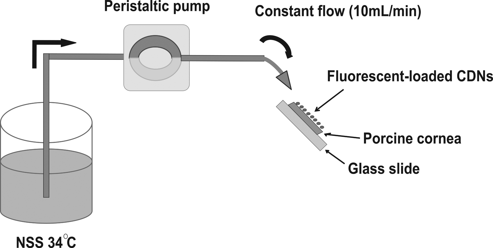
Schematic drawing of the apparatus for ex vivo mucoadhesion tests (based on modifications of Choy et al. 12 ). We employed corneas trephined (6 mm diameter) from freshly isolated porcine eyes.
Statistical analysis
The data are expressed as mean±standard deviation and analyzed using the Student's t-test for comparison of the means. Differences are considered significant at P<0.05.
Results
Physicochemical properties of CDNs
As shown by the SEM micrographs (Fig. 2), the particle sizes of CDNs were found to be within the range of 170–350 nm and spherical. The DLS indicated a slightly bigger particle size with an average diameter of 400 nm (Table 1), a polydispersity index of 0.3, and exhibited a strong positive charge with a zeta potential of ∼40 mV. The entrapment of the 2 NR and RhB produced only a small effect on the mean size and surface charge of CDNs. Specifically, increasing NR loading led to an increase in the mean particle size to 500–700 nm, but showed no effect on the zeta potential. On the other hand, RhB loading produced a negligible effect on both the mean particle size and zeta potential.
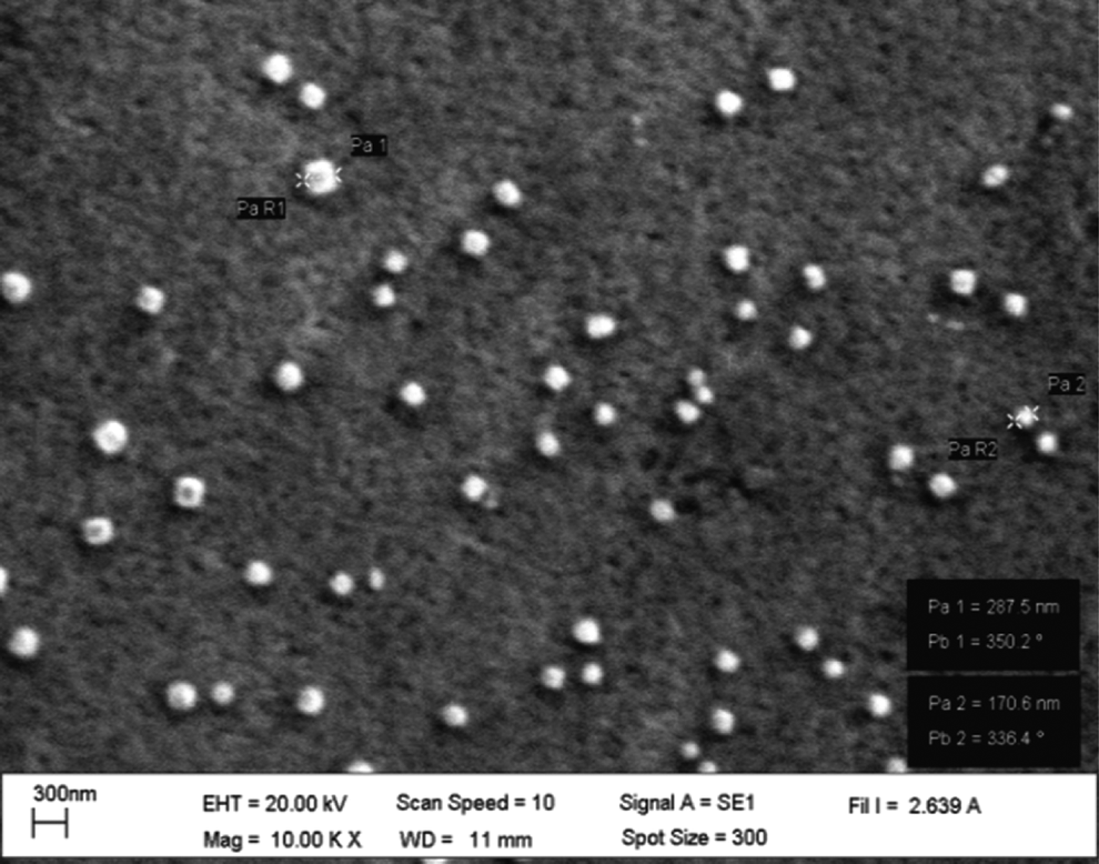
Micrographs showing scanning electron microscopy of chitosan–dextran sulfate nanoparticles (CDNs), which were prepared at chitosan (CS):dextran sulfate (DS) of 1:1.67 (by weight) and with 0.35% v/v polyethylene glycol-400 (PEG-400).
Particle size exceeded 1,000 nm.
SD, standard deviation; ZP, zeta potential; PI, polydispersity index; EE, entrapment efficiency; CDNs, chitosan–dextran sulfate nanoparticles; NR, Nile Red; RhB, Rhodamine B.
Entrapment efficacy and dissolution study of the fluorescent dyes
NR and RhB were successfully entrapped in CDNs with slightly different efficiency. The maximum EE for NR and RhB were 83.75% and 82.68% at 0.0042 and 0.4134 mg/mL of NR and RhB, respectively (Table 1). In the in vitro release experiments, NR release from CDNs was found to occur rapidly. More than 60% and 90% of NR was released within 5 and 30 min, respectively (Fig. 3). In contrast, there was no measurable release of RhB (Fig. 3).
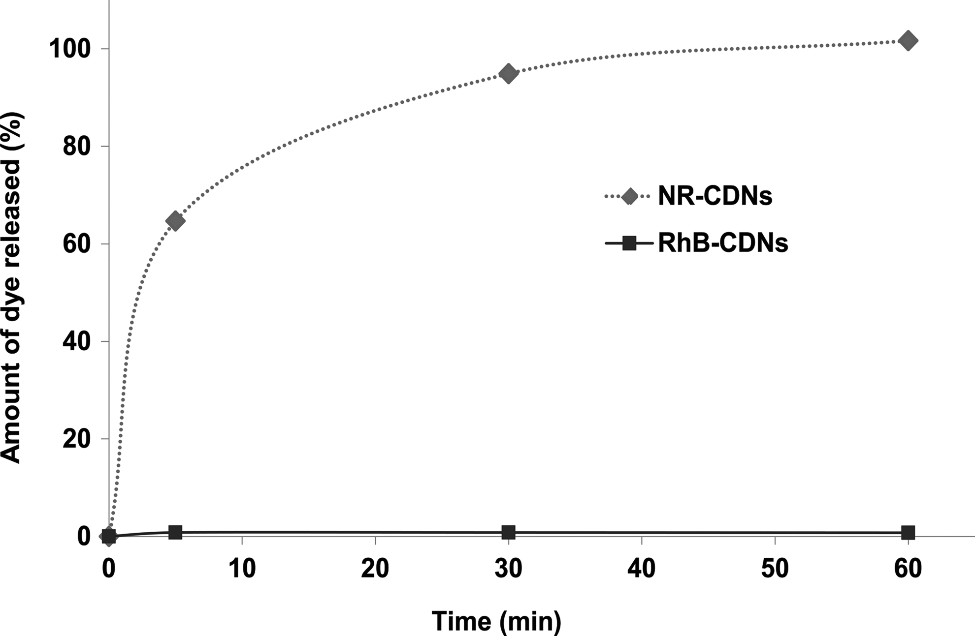
In vitro dissolution profiles for Nile Red (NR) and Rhodamine B (RhB) from CDNs. The dotted and continuous lines refer to NR-CDNs and RhB-CDNs, respectively. Normal saline solution (NSS) and NSS containing 1% (v/v) Tween 80 were used as dissolution media for RhB-CDNs and NR-CDNs, respectively. Error bars indicate standard deviation (SD) for n=3.
Interaction between CS and DS
The FTIR spectra obtained for CS, DS, and CDNs are shown in Fig. 4. The DS spectrum 26 shows an asymmetrical S=O stretching vibration band at wave number 1,260 cm−1. However, CS shows a very low IR absorption at 1,260 cm,−1 but indicated characteristic N–H bending at 1,631 and 1,598 cm−1, respectively.26,27 Therefore, changes in absorbance at 1,260, 1,631, and 1,598 cm−1 could be used to detect potential interactions of CS and DS in the CDNs. The spectrum of the CDNs, also shown in Fig. 4, exhibited altered absorbance at the S=O and N–H groups. The asymmetrical S=O stretching vibration band at 1,260 cm−1 is split into 2 well defined bands at 1,256 and 1,240 cm−1, and the N–H bending absorption band at 1,598 cm−1 is shifted to 1,533 cm−1. These changes indicate that the CDNs are formed by ionic interactions between the positively charged amine groups of CS with the negatively charged sulfate group of DS.
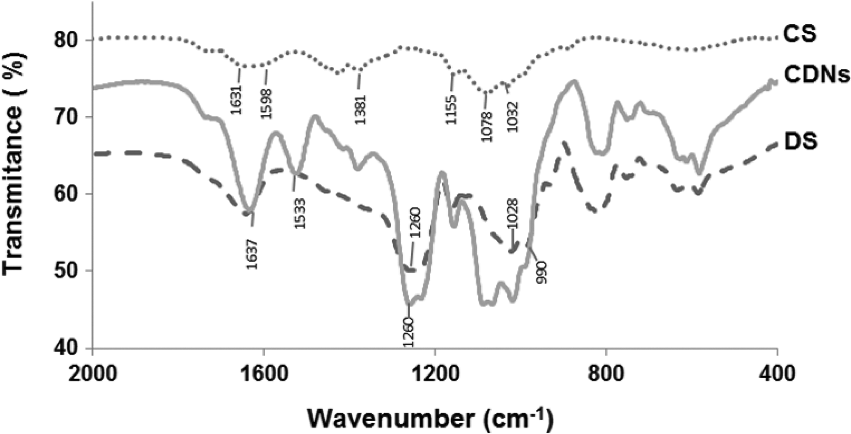
Fourier transform infrared spectroscopy spectra of CS, DS, and CDNs produced at CS:DS of 1:1.67 (by weight) and with 0.35% v/v PEG-400. KBr disc was used for holding the samples.
Stability of CDNs in the presence of lysozyme
To assess the physicochemical stability of CDNs on the ocular surface, the impact of lysozyme on the mean particle size and zeta potential was examined. As shown in Fig. 5A, the mean particle size was slightly reduced upon incubation of CDNs with the enzyme, but this difference was not statistically significant. The slight reduction in particle size can be attributed to partial hydrolysis of some surface bound CS molecules by lysozyme. In addition, lysozyme showed no effect on the surface charge of nanoparticles (Fig. 5B).
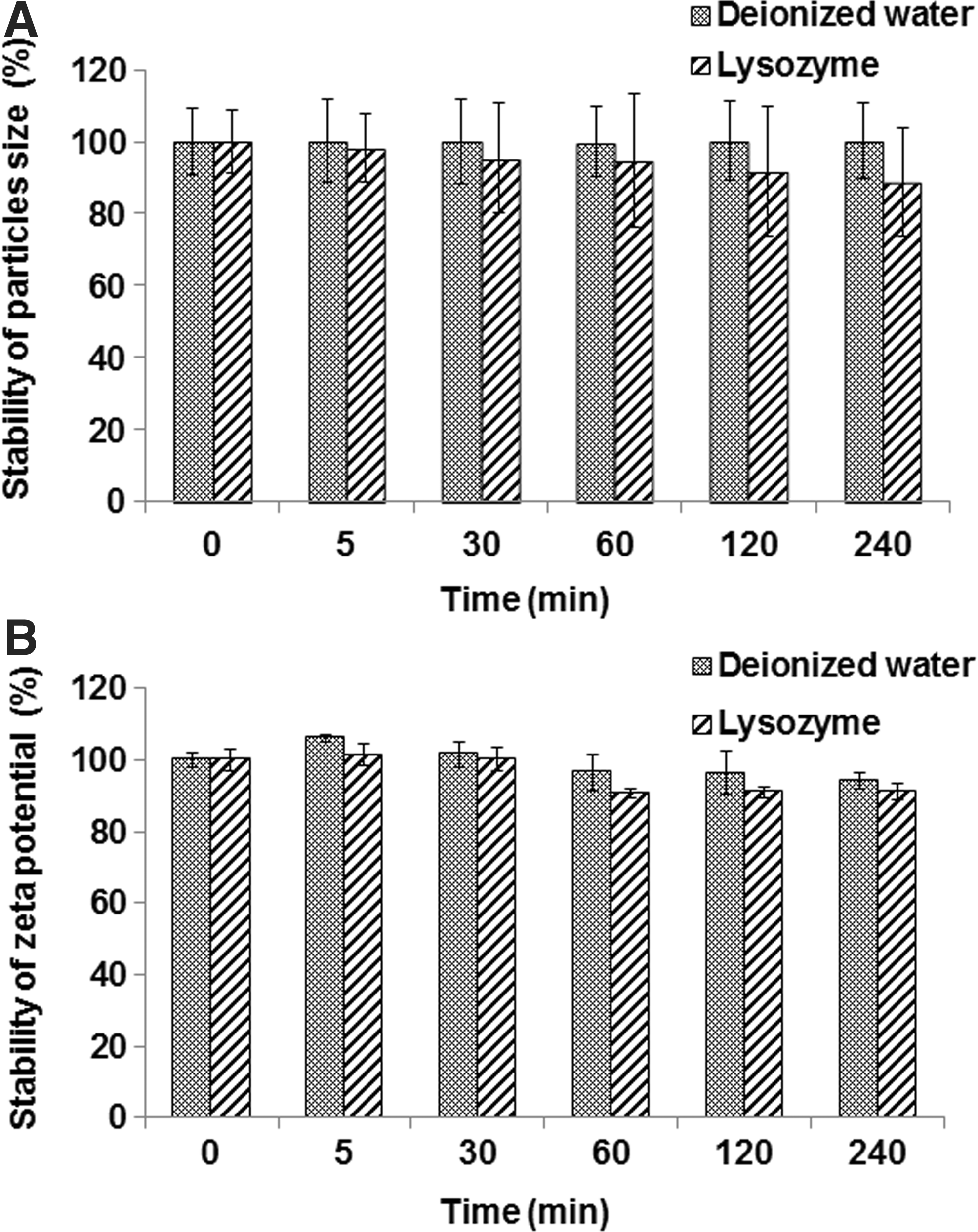
Stability of CDNs in the presence of lysozyme for 4 h at 37°C.
Mucoadhesiveness
The adhesion of the CDN corneal surface was examined ex vivo to assess the mucoadhesive properties of CDNs. As shown in Fig. 6A, C, and E, instillation of NR, RhB, and FITC in NSS on the cornea did not result in significant levels of fluorescence after 5 min of fluid flow. However, instillation of NR- and RhB-CDNs produced significant fluorescence could be noticed at 5 and 60 min of fluid flow, indicating mucoadhesiveness of the CDNs (Fig. 6B, D). As expected, FITC-CDNs also showed significant retention even after 60 min under fluid flow (Fig. 6F).

Ex vivo mucoadhesion tests: Porcine corneas were trephined (6 mm diameter) and held on glass slides (Fig. 1). Fluorescent CDNs were instilled on the corneal surface and fluorescence images were recorded after 0, 5, and 60 min. During the entire time, the corneal surface was irrigated with a stream of NSS at a rate of 10 mL/min at 34°C.
Discussion
A major requirement of ophthalmic formulations for topical administration is that their components are nontoxic and nonirritant to the sensitive cells/tissues of the ocular surface and its adnexa. In the case of nanoparticle-based formulations, the particle size, size distribution, and surface charge are also crucial parameters that are likely to influence the safety and efficacy of the formulation. In this study, a mucoadhesive CDN prepared using biocompatible polymers, CS and DS, was investigated for potential application to enhance drug delivery to the ocular surface.
In this study, we first showed that CDNs are formed rapidly by phase separation as a result of ionic interactions between the oppositely charged CS and DS. FTIR spectra of CDNs demonstrated the spectral shifts in the S=O and N–H absorption bands of CS and DS, respectively, confirming the existence of the electrostatic interactions between the sulfate groups of DS and the amine groups of CS. Since CS and DS mixing produced CDNs, which showed significant aggregation, we subsquently included PEG-400 as a stabilizer during the complexation. This manuever prevented aggregation of CDNs, possibly through steric hindrance.
A structure of CDNs is proposed in Fig. 7A. Consistent with the main requirement for the sensitive ocular surface, the components of CDNs, CS, and DS, are not only biocompatible, but are also nontoxic, biodegradable, and water soluble.12,21,25,28,29 In addition, PEG-400, a nonionic surfactant, is commonly used in the formulation of lubricating eye drops.30–32 Thus, the CDNs offer many advantages, specifically, only simple steps were employed in the production without the need for high temperatures or organic solvents.

Schematic illustration of proposed structure model of
The developed CDNs were aimed at enhancing the resident time of drugs on the ocular surface. Consequently, their particle size and surface charge can be expected to play an important role in their ability to interact with the ocular mucosa. 29 The mean particle size was found to be dependent upon the nature of the model drug. When loading with NR, a hydrophobic molecule, the mean particle size of NR-CDNs was slightly larger compared with blank CDNs. On the contrary, when loading RhB, a positively charged hydrophilic molecule, the particle size of CDNs was unaffected. Nevertheless, at optimal conditions, the mean particle sizes of ∼400–500 nm were succesfully prepared with a positively charged zeta potential of ∼40 mV. Thus, the CDNs produced in this study should be able to adhere onto the negatively charged ocular surface. It is also interesting to note that Katas and Wen have reported incorporation of SR-101 (sulforhodamine) into CS-DS nanoparticles. 33 They have indicated that zeta potential as high as 60 mV is nontoxic. Similarly, Sharma et al. prepared immunoglobulin A-loaded CS–DS nanoparticles that were positively charged (53 mV) and possessed an average particle size of ∼300 nm. They have claimed that their CS–DS nanoparticles were efficacious and nontoxic as an immunological adjuvant for vaccine delivery. 34 Overall, CS–DS nanoparticles are thought to be nontoxic and it remains to be seen if the positive charge could cause irritation to the ocular surface.
Interestingly, we found that the nature of the model drug also affects the dissolution profile depending on its interactions with the CDNs. Since NR is poorly water soluble, we propose that it is held in the CDNs by weak hydrophobic forces (Fig. 7B). This phenomenon possibly explains the initial burst release preceding a relatively slower release for 60 min (Fig. 3). On the other hand, RhB could not be extracted from RhB-CDNs using water and/or solvents, such as ethanol, methanol, and dimethyl sulfoxide. This could be due to the interaction of the positively charged nitrogen of RhB with the negatively charged sulfate groups of DS (modeled in Fig. 7B). Consistent with this result, the EE of RhB in the CDNs was much higher when RhB was initially mixed with anionic DS as compared to when the dye was first mixed with the cationic CS (data not shown). In contrast, mixing of NR with the DS solution led to a lower EE as compared to when the dye was first mixed with the CS solution (data not shown). These order-of-mixing effects imply the hydrophilic/hydrophobic nature of the drug in determining EE.
The stability of nanoparticles in tear fluids containing important amounts of proteins and enzymes is a crucial issue. The CDNs are constructed from CS, which can be hydrolyzed by the lysozyme present in tear fluids. Therefore, the particle size and zeta potential of CDNs were determined upon their incubation in the presence and absence of lysozyme. The incubation of CDNs with lysozyme did not modify either their surface charge or their size (Fig. 5). In the studies presented here, isoelectric point of lysozyme is 11.35, and hence, the enzyme is positively charged at the pH employed in the study. Therefore, an electrostatic interaction of lysozyme and CDNs did not significantly affect the CDNs zeta potential. A similar observation has been previously reported for CS-coated poly(lactic acid–glycolic acid) nanoparticles. 29 Therefore, the major conclusion from this stability study is that the integrity of CDNs is not significantly compromised by the presence of lysozyme in tears.
An important finding in this study is that CDNs are retained for a prolonged period on the freshly isolated corneal surface, indicating their mucoadhesiveness. The shear stress at the high flow rate of the saline at 10 mL/min on the corneal button mimics the higher end of the shear stress on the ocular surface, which is induced during period blink action. 35 We note that the fluorescence at 60 min in Fig. 6B and D indicate mucoadhesion rather than absorption of NR/RhB into the corneal epithelium. This is further confirmed by the staining following instillation of FITC-labeled CDNs at 60 min (Fig. 6F). Overall, the CDNs formulated in this study possess strong mucoadhesion, and hence, fulfill the main requirement for prolonged delivery of drugs to the ocular surface. This strong mucoadhesion can be attributed to the net charge on the CDNs, which was found to be 40 mV (Table 1) despite DS being negatively charged. The mucosal surface is negatively charged,25,36,37 and hence, CDNs can be predicted to have significantly high mucoadhesivity. In addition, PEG-400 associated on the CDNs surface may also be responsible for the enhanced mucoadhesiveness, since the surfactant is known to promote mucoadhesion possibly via hydrogen bonding with mucin.
The increase in residence time of topical drugs to the eye has relevance for several therapeutic applications, including dry eye, glaucoma, intra/extraocular inflammation, and infectious diseases. In all of these situations, CDNs can be used to deliver drug-relevant drugs. However, the extent of bioavailability will depend not only on the residence time, but also on the partition coefficient of the drug in question. In principle, an increase in residence time is useful for ocular surface treatment as in the case of infection and dry eye. Since trans-corneal penetration is key for enhanced anterior segment drug delivery (e.g., glaucoma drugs), the residence time on the corneal surface would increase net drug influx into the cornea. The increase in residence time on the ocular surface can also increase posterior segment drug delivery through penetration across the conjunctiva and sclera.
In summary, our major finding is that CS and DS undergo self-assembly rapidly via ionic interactions under mild conditions resulting in uniformly sized nanoparticles with a positive surface charge and significant EE. The prepared CDNs proved to be stable to lysozyme and showed mucoadhesiveness to the ocular surface. Thus, the CDNs are useful in the treatment of ocular surface diseases, including dry eye and ocular surface infections. In addition, this nanoparticle system can be adopted for other drugs to improve residence time, and thereby, intraocular bioavailability for diseases such as glaucoma.
Footnotes
Acknowledgments
The authors would like to thank Kulasub Distes for her assistance in conducting the some of the experiments. Financial support from the Agricultural Research Development Agency, Thailand, the Center of Excellence for Innovation in Chemistry (PERCH-CIC), Commission on Higher Education, Ministry of Education, Thailand and the Thailand Research Fund (TRF) for Wanachat Chaiyasan under the Royal Golden Jubilee Ph.D. Program (Grant No. PHD54K0094) is gratefully acknowledged.
Author Disclosure Statement
No competing financial interests exist.