
Abstract
Abstract
As an effective compound found mainly in the honeybee product propolis, caffeic acid phenethyl ester (CAPE) has been commonly utilized as a medicine and remedial agent, in a number of countries. Specifically, it might inhibit nuclear factor kappa B at micromolar concentrations and demonstrate antioxidant, antineoplastic, antiproliferative, cytostatic, antiviral, antibacterial, antifungal, and anti-inflammatory features. This review article summarizes the recent progress regarding the favorable effects of CAPE on a number of eye disease models, including cataract and posterior capsule opacification, corneal diseases, retina and optic nerve-related diseases, ischemia/reperfusion injury of retina, inflammation and infection-related diseases. CAPE has been found to exhibit promising efficacy, with minimal adverse effects, in animal and cell culture studies of several eye diseases.
Introduction
C
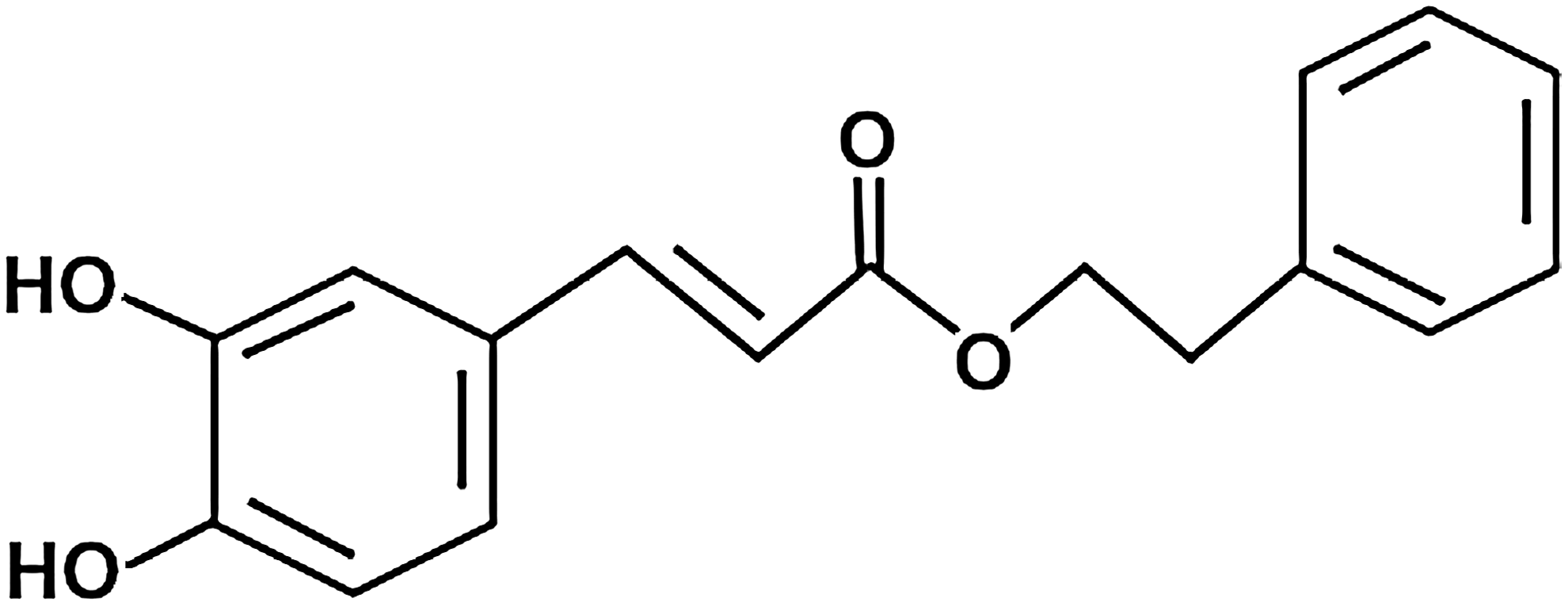
The chemical structure of caffeic acid phenethyl ester (CAPE).
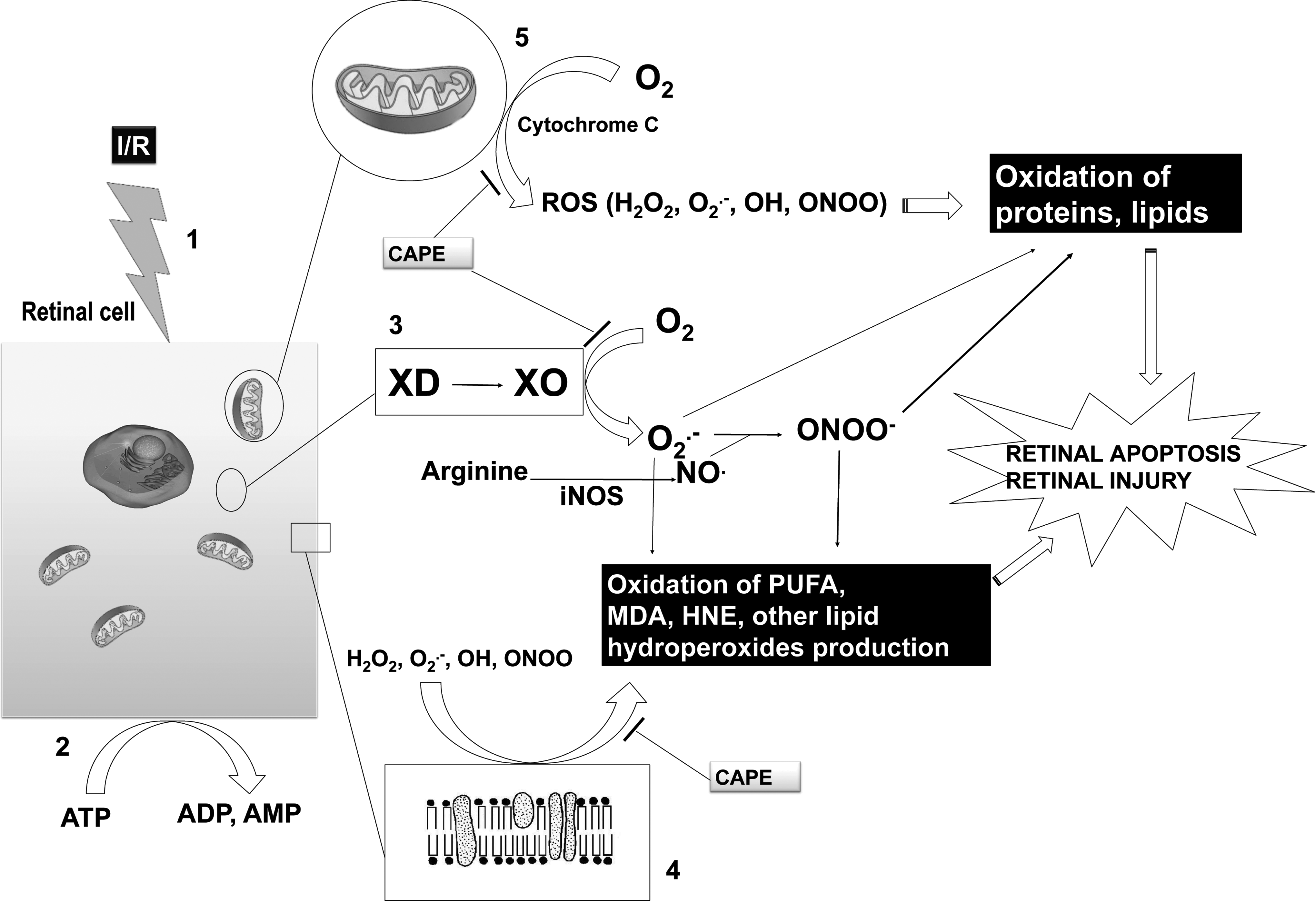
The schematic representation of free oxygen radical generation sites after ischemia/reperfusion. 66 (1) Ischemia/reperfusion triggers several destructive pathways in retinal cells. (2) First of all, because there is a shortage of molecular oxygen, cells consume all ATP reserves for energy need. The hydrolytic removal of phosphate from AMP produces adenosine. Adenosine is further deaminated to inosine by adenosine deaminase. Hypoxanthine formed from inosine is metabolized to xanthine. (3) Xanthine is metabolized to uric acid by xanthine oxidase (XO). Before this conversion, XO is formed from xanthine dehydrogenase (XD) in this ischemic condition. Normally XD does not generate free radicals, but the proteolytic product XO generates lots of superoxide radicals (O2·−) and it is accepted as a major potential source of reactive oxygen species (ROS), especially O2·−. After adding nitric oxide (NO), O2·− is converted to one of the most potent oxygen radicals peroxynitrite (ONOO). It attacks proteins, lipids, and nucleic acids. (4) The most vulnerable structure subjected to free radical attack is cell membrane. ROS attack polyunsaturated fatty acids (PUFA) on membranes and produce oxidation end products such as malondialdehyde (MDA), 4-hydroxy-2-nonenal (HNE), and other hydroperoxides. (5) After reperfusion, molecular oxygen (O2) is converted to ROS leading to protein and lipid destruction. H2O2, hydrogen peroxide; iNOS, inducible nitric oxide synthase; OH, hydroxyl radical.

The proposed mechanism for CAPE to diminish or block inflammation by different pathways. Note that among a variety of transcription regulators, NF-κB has been shown to play a critical role in regulating the expression of large numbers of genes such as cytokines, chemokines, and other mediators involved in inflammatory responses. 67 CAPE is a potent and specific inhibitor of NF-κB. GM-CSF, granulocyte macrophage-colony stimulating factor; IL-2,6,8, interleukin-2,6,8; IFγ, interferon γ; IL-1α,β, interleukin-1α,β; LPS, lipopolysaccharides; PMNL, polymorphonuclear leukocytes; TNFα, tumor necrosis factor α.
A number of review articles focused on the protective role of CAPE against general in vivo and in vitro neoplasm models, 3 melanomas, lung and prostate cancers, 4 chemotherapy-induced and radiotherapy-induced toxicities, 5 systemic mycoses, 6 and heart diseases 7 have been published in the literature. However, to the best of our knowledge, this is the first review article focusing on the protective effects of CAPE toward certain major eye diseases. This article systematically reviews and summarizes the protective role of CAPE on major eye diseases, including cataract and posterior capsule opacification (PCO), corneal diseases, retina and optic nerve-related diseases, ischemia/reperfusion (I/R) injury of retina, inflammation and infection-based diseases.
The Usage of CAPE in Cataract and PCO
Hepsen et al. previously studied whether CAPE prevents PCO by suppressing the transformation of the epithelial cells of lens. 8 Pigmented island rabbits that underwent phacoemulsification were divided into 2 groups. Afterward 10 μg/mL of CAPE was added to the anterior chamber irrigation solution of the first group and 1% of CAPE solution was injected to the subconjunctival area for 3 weeks postoperatively. Weekly assessment was performed for the development of PCO. Three months following surgery, histological analyses were performed. The rats that underwent CAPE application showed clear capsules or presented minor PCO. Rats treated with the irrigation solution without CAPE showed severe PCO and complete opacification. CAPE was determined to be effective in suppressing PCO in rabbits, which suggests its favorable role in clinical use because of being free from documented harmful effects against normal cells.
12-O-tetradecanoylphorbol-13-acetate (TPA) induces the production of hydrogen peroxide (H2O2) in mouse skin and causes oxidation of DNA bases in vivo as well. 9 In addition, agents showing in vivo antitumor-promoting properties suppress inflammation processes. These are infiltration of polymorphonuclear leukocytes (PMNL), ROS production, oxidation of DNA bases, induction of ornithine decarboxylase, and edema.10,11 TPA was reported to induce oxidative stress and also to cause opacity in bovine eye lens. 12 Certain molecules used in tumor promotion studies, such as epigallocatechin gallate and sacrophytol A, were effective inhibitors of TPA-mediated lens opacification and H2O2 production as well. 12 Frenkel et al. performed in vivo and in vitro experiments to study whether CAPE prevents TPA-mediated ROS generation and lens opacity, properties that may be useful in the prevention of cataract. 13 The in vivo part of the study revealed that the TPA-induced increase in epidermal ornithine decarboxylase, ear edema, TPA-induced H2O2 production, and formation of 5-hydroxymethyl-2′-deoxyuridine (HMdUrd) and 8-hydroxy-2′-deoxyguanosine (8-OHdGua) in mouse skin were all inhibited by CAPE. The in vitro experiment showed that CAPE inhibited TPA-mediated H2O2 production by human PMNL, and inhibited TPA-mediated induction of H2O2 in bovine lens, which means that CAPE protected bovine lens from TPA-induced opacity. TPA-treated lens generates large amounts of H2O2, which is known to contribute to cataract formation. This involves a change in the homeostasis of calcium ions, which when elevated damage the lenses. TPA-induced H2O2 formation, as well as opacity of lenses, was observed to respond to the chemopreventive agent, CAPE.
The Usage of CAPE in Corneal Diseases
The ability of topically applied CAPE in comparison to known steroidal and nonsteroidal topical agents in terms of reducing corneal neovascularization induced by silver nitrate cauterization was studied in male rats. 14 The authors scored burn stimulus intensity from 0 to +3 in respect of the height of blister from the corneal surface, and recorded the extent of neovascularization as from 0 to +6 according to the distance from the limbus to the endpoint of corneal neovascularization toward the central corneal burn site. CAPE was detected to be more effective than indomethacin and almost as effective as dexamethasone in terms of reducing corneal neovascularization, which indicates the inhibitory effect of topical CAPE on corneal neovascularization.
Defensins are cationic antimicrobial peptides responsible for a broad spectrum of antimicrobial activities since they are effective against many gram-positive and negative bacteria, certain fungi, and enveloped viruses. 15 A total of 6 human beta defensins (hBD-1 through 6) has been defined. Human corneal epithelial cells (HCECs) were shown to express hBD-1 constitutively whereas the expression of hBD-2 was variable 16 and might be upregulated by bacterial products. In this respect, hBD-1 is known to be constitutively expressed, yet the expression of hBD-2 is upregulated during reepithelialization of wounded corneal areas in organ culture. 17 Therefore, McDermott et al. speculated that the upregulation of hBD-2 following injury might contribute to the protection of the ocular surface when it is particularly vulnerable to infection and that hBD-2, with its nonmicrobicidal activities, may improve the wound healing process per se. They investigated the effects of exposure to proinflammatory cytokines on expression hBD-2 by corneal epithelial cells. 18 To examine the role of intracellular signaling pathways, CAPE (90 μM) was added to the cells 30 min before the step of adding 10 ng/mL interleukin (IL)-1β. Three hours after the addition of IL-1β, the cells were collected for real-time PCR (RT-PCR). CAPE, as a nuclear factor kappa B (NFκB) inhibitor, blocked the effect of IL-1β on expression of hBD-2 mRNA. Inhibitors of NFκB activation by CAPE completely blocked the effects of IL-1β, which indicates the importance of the activation of this transcription factor for the upregulation of hBD-2 in HCECs.
A study dealing with the anti-allergic efficacy of 1% CAPE was also performed. 19 As a first step, experimental allergic conjunctivitis was provoked in New Zealand rabbits using a mast cell activator. The edema and hyperemia in the eyes with conjunctivitis and in those of controls were then scored. The scores of edema and hyperemia were significantly different between these groups. Furthermore, the histopathological scores of the provocation and postprovocation terms were evaluated and CAPE was observed to block the allergic process. CAPE at 1% concentration was found to be as clinically efficient as 0.1% dexamethasone and olopatadine.
The Usage of CAPE in Retina and Optic Nerve-Related Diseases
The stimulation of human retinal pigment epithelium (hRPE) causes the release of a number of chemokines, including IL-8 and monocyte chemotactic protein (MCP)-1. IL-8 is a commonly known chemoattractant, neutrophil and eosinophil activator, and a mediator of angiogenesis. The increase of IL-8 in the vitreous of eyes with proliferative diabetic retinopathy was previously described.20,21 Since MCP-1 activates lymphocytes and monocytes causing monocyte/macrophage infiltration in the tissue, 22 it could be considered as an inflammatory stimulator involved in the tissue repair process. On the other hand, enhanced protein glycation might be encountered in experimental diabetes mellitus. Glycated albumin has effects on cellular functions and thus may play a role in the pathophysiology of diabetes, namely the microvascular complications associated to diabetic retinopathy.23,24 Cohen et al. and Schalkwijk et al. also investigated the signal mediators taking place in glycated human serum albumin (GHSA) stimulation of the secretion of IL-8 and MCP-1 in hRPE cells. Incubation of hRPE cells with GHSA revealed rapid activation of Raf-1, p38, extracellular signal-regulated protein kinases, and the transcription factor NFκB. After coincubation of hRPE cells with NFκB inhibitor, CAPE largely eliminated the production of IL-8 and MCP-1, thus showing the role of Raf/mitogen-activated protein kinase (MAPK) pathway in GHSA signaling.
Literature indicates that excessive exposure to white light, ROS production and inflammatory stress might result in the development of age-related macular degeneration and progression of retinitis pigmentosa. Following these damaging processes, retinal degeneration along with death of light-sensitive photoreceptor cell and retinal pigment epithelium cell may take place irreversibly.25–27 CAPE was also reported to be protective for retinal cells against oxidative and inflammatory stress-induced damage and might be used as an augmentative therapy to prevent or delay the onset of retinal degeneration in humans. 28 To examine the protective role of CAPE, a H2O2-mediated cell death model of mouse photoreceptor-derived 661W cells and acute or chronic bright light-induced retinal degeneration models of albino Sprague Dawley rats were used by Chen et al. 28 The expression of certain genes was measured in the retina using real-time quantitative reverse transcriptase PCR (real-time qRT-PCR). Cone photoreceptor function by electroretinography and retina outer nuclear layer thickness from rats treated for 8 weeks were also studied. Furthermore, the relative mole percentage of fatty acid composition from rat retina fed by CAPE or control diet under dim or bright cyclic light rearing was determined. Among rats maintained under cyclic dim light, CAPE was observed to activate the antioxidant gene expression pathway in 661W cells and in rat retinas, and enhanced the scotopic electroretinogram (ERG) A/B wave amplitude and altered fatty acid profile in the retinas. To summarize, related studies revealed that CAPE protected retina-derived 661W cells against H2O2-induced cell death in vitro, which was accompanied by changes in the expression of certain antioxidant genes and proteins. On the other hand, the expression of a series of antioxidant genes and proteins in albino rat retinas could be modulated by the supplementation with CAPE in the diet (Fig. 4).
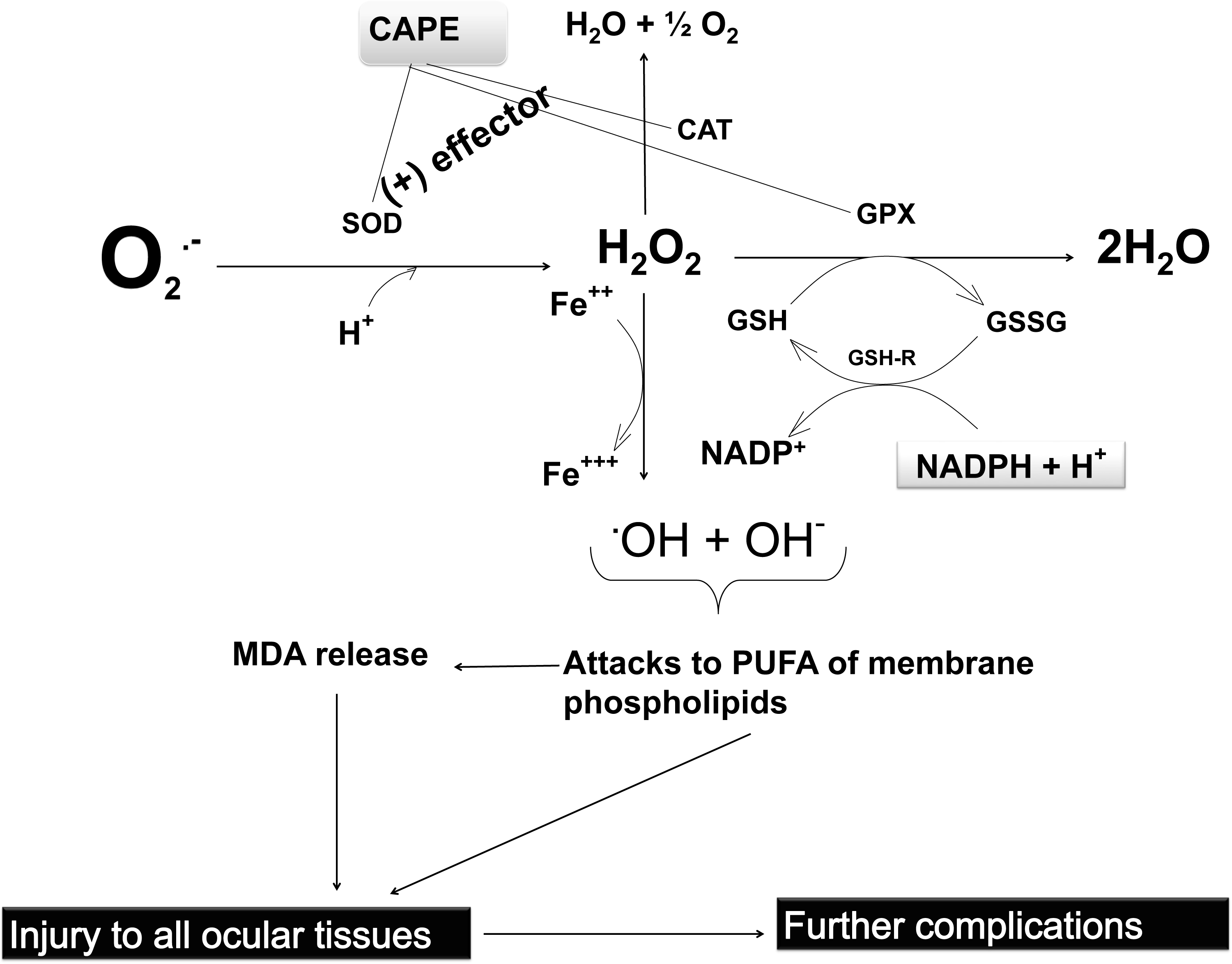
Proposed mechanism of how oxidative stress is blocked by antioxidant enzymes in several parts of ocular cells and how CAPE shows its protective effects against oxidative stress. CAT, catalase; Fe++, ferrous iron; GPx, glutathione peroxidase; GSH, reduced glutathione; GSH-Red, glutathione reductase; GSSG, oxidized glutathione; H+, hydrogen ion, proton; H2O, water; NADP+, oxidized nicotinamide adenine dinucleotide phosphate; NADPH+H+, reduced nicotinamide adenine dinucleotide phosphate; O2, molecular oxygen; O2·−, superoxide anion radical; OH−, hydroxyl ion; .OH, hydroxyl radical; PUFA, polyunsaturated fatty acid; SOD, superoxide dismutase.
Several angiogenic factors, the most known among, which is vascular endothelial growth factor (VEGF), are involved in the onset and development of choroidal and retinal neovascularization. VEGF is an endothelial cell-specific mitogen promoting vascular permeability. 29 In diabetic subjects, increased VEGF immunoreactivity was observed in retinal vascular endothelium, blood vessel walls, choriocapillaris endothelium, choroidal neovascular endothelium, and migrating human RPE cells. 30 Expression of VEGF might be stimulated by a number of stimuli such as hypoxia, mechanical stress, vasopressor hormones, cytokines, transforming growth factor-β (TGF-β), and platelet-derived growth factor. Recent studies revealed that all TGF forms might induce VEGF expression in several cell types, including hRPE cells. 31 The effects of TGF-β2 alone and in combination with tumor necrosis factor-α or VEGF on hRPE VEGF mRNA expression and protein secretion, as well as the signaling pathways involved in VEGF expression in hRPE cells, were investigated by Bian et al. 32 ELISA and qRT-PCR for VEGF and western blot analysis of human RPE cells were utilized as methods. Treatment of hRPE cells with TGF-β2 for 24 and 48 h, when compared with the 8 h period, showed significant increases in VEGF secretion by 5-fold and 9-fold, respectively. Induced VEGF expression was completely abrogated by CAPE, an NFκB inhibitor, and by N-acetylcysteine, a ROS inhibitor. They concluded that mitogen-activated protein kinase/ERK kinase (MEK), p38, c-Jun N-terminal kinase (JNK), phosphoinositide 3-kinase (PI3K), and NFκB, as well as multiple essential signaling mediators, including PKC, PTK, and ROS are involved in hRPE VEGF upregulation by TGF-β2.
Tuberculosis is treated with a combination of antibiotics, including isoniazid (INH), rifampin, pyrazinamide, streptomycin, and ethambutol (ETM). Among them, INH and ETM are commonly used. However, the use of INH and ETM may cause optic neuropathies.33,34 Notably, INH-induced cytotoxicity has been attributed to oxidative stress and ROS. Sahin et al. hypothesized that toxic effects of INH and ETM in retina and optic nerve are partially caused by oxidative stress they induce. 35 They aimed to investigate the protective effects of CAPE on retina and optic nerve tissue subjected to oxidative stress induced by INH and/or ETM studying a rat model. Furthermore, an in silico solution was utilized to understand the mechanism underlining the protective effect of CAPE based on the CAPE-superoxide dismutase (SOD) interaction. Both INH and ETM were orally administered at a dose of 50 mg/kg/day for 30 days. CAPE was applied daily intraperitoneal (i.p.) 10 μmol/kg for 30 days, as well. The antioxidant enzyme SOD, malondialdehyde (MDA), total antioxidant status (TAS), and total oxidative status (TOS) were measured in retina and optic nerve homogenate. Protein Data Bank entry of SOD isoforms were downloaded and analyzed for SOD-CAPE protein and ligand structure for docking. The authors also performed a histopathological examination. According to in silico calculations, what they found was that ETM, INH, and CAPE can bind active sites of SOD isoforms SOD1, SOD2, and SOD3. It was suggested that INH and ETM may present their oxidant potential by binding the active site of SOD isoforms and they can inhibit the conversion of superoxide radicals (O2·−) to H2O2 through blocking all forms of SOD enzymes. Since CAPE may bind the active sites of SOD enzymes stronger than INH and ETM, they proposed that CAPE binding to the SOD enzymes might not inhibit the functions of SOD isoforms and, at the same time, block the side effects of INH and ETM. On the other hand, CAPE cotreatment was found to reduce ganglion cell loss caused by INH and ETM. Increased TOS and MDA, and decreased TAS and SOD activity were observed after INH and ETM application, whereas they were all normalized with coadministration of CAPE. Since the preventive role of CAPE toward retinal ganglion cell (RGC) loss was determined to be due to its strong antioxidant activity, they concluded that it might be considered as a promising agent to protect the optic nerve, retina, and possibly other organs from INH and ETM-induced oxidative damage in tuberculosis patients. 35
Methanol poisoning is a life-threatening event that results in severe metabolic disturbances, neurological dysfunction, blindness, and even death. Methanol is absorbed by the digestive system easily 36 and is responsible for severe morbidity and mortality. Methanol intoxication specifically leads to blurred vision, diplopia, and blindness in the visual system based on the severity of exposure. 37 Since CAPE was detected to be an effective agent in diazinon intoxication38,39 and prevents ocular toxicity secondary to isoniazid and ethambutol treatment, 35 Sahin et al. hypothesized that CAPE may be deployed as an antidote in methanol poisoning as well. 40 Therefore, they intended to find out the protective role of CAPE on retina and optic nerve in methanol-induced toxicity in a rat model. The rats were given methotrexate, at a dose of 0.3 mg/kg/day i.p. for 7 days, to generate a rat model of methanol intoxication. 41 At the 8th day, i.p. methanol (3 g/kg) was administered. Four hours after methanol administration, 10 μmol/kg i.p. CAPE was given. The TAS, TOS, histological examinations, and preparation of the protein and the ligand structure for docking were performed to test the hypothesis. CAPE administration prevented the increase of TOS levels and preserved TAS in the retina and optic nerve. Moreover, as effective as ethanol, CAPE treatment prevented cell loss of RGC layer in acute methanol toxicity. CAPE was shown to have an affinity to the active site of alcohol dehydrogenase, which may support a role as an antidote in acute methanol intoxication.
The literature reported evidences that mobile phones emitting electromagnetic radiation (EMR) of 900 MHz stimulate ROS formation in most organs.42–44 Ozguner et al. aimed to compare the potential effects of CAPE against oxidative stress in rat retina exposed to 900 MHz EMR emitting mobile phones on long-term basis. 45 Rats were exposed to EMR 60 days as 30 min/day and a group of rat was given 10 μM/kg/day of CAPE during 60 days of study. The retinas of rats were dissected and homogenized for biochemical analyses, at the end of the study. EMR was found to increase the retinal MDA and nitric oxide (NO) levels, whereas it caused a decrease in SOD, catalase (CAT), and glutathione peroxidase (GPx) activities; CAPE administration decreased retinal MDA and NO levels, and it increased SOD, CAT, and GPx activities (Fig. 4) keeping them in the normal ranges. The changes in the activities of antioxidant enzymes, as well as MDA and NO in EMR-applied group, may be considered as an index of increased ROS production because of the pathological process driven by mobile phone in rat retina. This was prevented by CAPE as an efficient free radical scavenger.
A number of inflammatory cytokines and growth factors are involved in proliferative vitreoretinopathy (PVR). This is known to be the most common cause of failure during the treatment of retinal detachment and other vitreoretinal procedures. Retina pigment epithelial cell migration should be blocked for an efficient treatment of PVR. If it is achieved, then epithelial cell proliferation can be prevented. In this respect, the preventive role of CAPE in the pathophysiology of experimental PVR was investigated. 46 The rabbits were given 0.15 mL of platelet-rich plasma to the left eye. One of the groups was given 15 μmol/kg of CAPE, i.p., for 3 days and the other was treated with 4 mg/mL of cortisone, intravitreally. The eyes were examined by ophthalmoscopy to assess the proliferation during a 15-day follow-up period. Significant decreases in glutathione (GSH) levels, increase in MDA content, and increase in NO level in ocular tissue were observed during the inflammatory process. As a consequence of suppression of inducible nitric oxide synthase (iNOS) activity by CAPE, NO level was detected to be decreased in the CAPE-treated group. In summary, CAPE was found to be effective, at least partly, in the inhibition of PVR experimentally. Clinical results of CAPE were correlated with NO, MDA, and GSH levels.
Diabetic retinopathy is among the major complications of diabetes that leads to blindness. It is a microvascular disease, in which the blood–retinal barrier breakdown is a hallmark. 47 The underlying pathological mechanism for diabetic retinopathy, and particularly for other diabetic complications, has been attributed to increased production of ROS and increased oxidative stress in tissues. 48 Previously conducted studies revealed important evidences for the ROS-mediated injuries in streptozotocin (STZ)-induced diabetic animals; for example, the levels of lipid peroxidation and the activities of antioxidant enzymes were found to be increased in STZ-induced diabetic rats.49,50 In this regard, a protective role of CAPE against oxidative stress was investigated in the retinas of STZ-induced diabetic rats. 51 CAPE at a dose of 10 μmol/kg/day was administered 3 days after 1 dose of 35 mg/kg body weight STZ treatment and continued until the sacrificing of rats. The levels of MDA and NO, and the activities of SOD and GPx were examined in retinal tissues. MDA levels in the retina were increased in untreated diabetic rats and CAPE application caused a significant decrease in MDA levels. The SOD and GPx activities were found to be decreased in diabetic untreated group, whereas these were normal or a little increased in CAPE-administered rat group. This was attributed to the beneficial effects of CAPE in the prevention or decreasing oxidative stress in the diabetic retina.
The Usage of CAPE in I/R Injury of Retina
The I/R related injury of retina has been reported to be a common problem that results in loss of vision in a number of ocular diseases such as retinal vascular occlusion, acute glaucoma, diabetic nephropathy, and prematurity related retinopathy (Fig. 2).52–54 On the other hand, ROS was shown to be involved in the process of retinal apoptosis caused by I/R related injury. 55 A significant number of in vivo studies have revealed that antioxidant molecules have protective properties on eye tissues and compartments following I/R-induced damage. 56 The protective effects of CAPE against I/R-induced retinal cell damage were also examined in a rat retinal I/R model and its possible antioxidant mechanism was studied. 57 Authors determined whether the alterations in retinal tissues were associated with MDA, SOD, CAT, GPx, as well as apoptosis of retinal cells, histopathological changes, and the electroretinogram of the retina. Retinal ischemia was induced by increasing the intraocular pressure to 110 mmHg for 60 min, which was confirmed by fundus examination. After this procedure, the cannulating needle was removed and the intraocular pressure normalized. CAPE was administered to an assigned group before reperfusion and once a day for 1 or 7 days after I/R. The study indicated the protective effects of CAPE on the retinal damage secondary to I/R in studied rat model. They found that CAPE significantly decreased retinal cell injury secondary to I/R as demonstrated by a reduction in both the histological damage (fluorescence detection of retinal cell apoptosis showed that control rats were negative in terminal deoxynucleotidyl transferase dUTP nick end labeling (TUNEL) staining, I/R group has yellow fluorescent retinal cell nuclei, and only a few yellow fluorescent nuclei were found on TUNEL staining of rat retina in CAPE group) and the functional changes (control group showed typical ERG records when I/R group showed a decrease in a- and b-wave amplitudes and finally CAPE inhibited the reduction of the a- and b-wave amplitudes of the ERG). Histopathological examination confirmed that CAPE clearly protected retina against retinal ischemic damage; the retina appeared much more normal with thicker inner retinal layer when compared with I/R group.
The Usage of CAPE in Case of Inflammation and Infection-Based Diseases
Endotoxin-induced uveitis (EIU) was utilized as an animal model of acute ocular inflammation (Fig. 3). It can be easily induced by systemic injection of lipopolysaccharide (LPS) in rats and mice. 58 Upon application, LPS causes acute inflammation mainly in the anterior chamber of the eye through cellular infiltration and protein extravasation. At the same time, vitreous humor and retina might also be infiltrated by the inflammatory cells. 59 The anti-inflammatory and antioxidant effects of CAPE in EIU were investigated in the model of EIU by administering 200 μg of LPS in rats. 60 CAPE was injected i.p. just after LPS administration. Cardiac blood and 15–20 μL of aqueous humor were obtained from rats by anterior chamber puncture and they were analyzed in terms of total antioxidant and myeloperoxidase (MPO), as well as protein concentration and infiltrating cells in aqueous humor immediately. Furthermore, enucleated eyes were histopathologically examined. LPS administration caused severe uveitis with massive PMNL infiltration in all sections, but CAPE coadministration significantly reduced the number of infiltrating PMNLs and severity of uveitis. Although total antioxidant levels remained in the normal range, serum MDA and MPO levels were detected to be elevated in rats subjected to only LPS. CAPE normalized these alterations effectively. CAPE administration caused a significant decrease in protein concentration and MPO activity in the aqueous humor of rats, in contrast to the significant increases in both parameters in the LPS-administered group. In summary, the biochemical and histological study revealed that systemic administration of CAPE effectively suppressed the development of EIU.
In spite of the recent approach to treat postoperative and penetrating traumatic bacterial endophthalmitis, of which almost 85% linked to gram-positive aerobic bacteria with strong antibiotics and microsurgical techniques, the prognosis still remains poor.61,62 Besides the direct effects of bacteria, the host inflammatory reaction might also worsen the retinal damage and cause vision loss. 63 The protocols of intravitreal corticosteroid use in endophthalmitis treatment remain controversial. The efficacy of the combination therapy of anterior subtenon injection of CAPE with intravitreal vancomycin and dexamethasone was studied. 64 New Zealand albino rabbits were treated with 0.1 mL of 4.7×10⁴ colony forming unit of Methicillin-resistant Staphylococcus aureus suspension by intravitreal injection after 1% atropine sulfate and 2.5% phenylephrine hydrochloride ophthalmic solution to dilate the pupil. Groups were treated with 1 mg vancomycin, 1 mg vancomycin+400 μg of dexamethasone, or 1 mg vancomycin+10 mg of CAPE. First, the eyes of all rabbits were examined with an indirect ophthalmoscope and slit-lamp biomicroscopy; vitreal aspirates were then obtained using a needle attached to a tuberculin syringe for microbiological analyses and eyes were enucleated. Analyses showed that the pathological scores of CAPE-administered group were lower than all other groups. All treatment groups were significantly less inflamed compared with the disease control group and CAPE was found to be comparable to steroids. In all examinations following treatment, the CAPE group showed the lowest, but statistically insignificant, clinical score among the treatment groups.
Conclusion
CAPE seemingly has superior effects on in vivo and in vitro eye disease models and should be considered as a promising agent for major eye diseases. The majority of its biological effects are attributed to its anti-inflammatory and antioxidant activities. CAPE was administered to rabbits through intracameral and subconjunctival routes, which indicated that CAPE had no known harmful effects on normal nonproliferative cells.8,65 Therefore, CAPE can be administered at doses relatively higher than those of other alternative medicine-based agents without any adverse effects. It can, therefore, be concluded from the literature that CAPE prevents certain eye disease processes and can be considered as a potential candidate compound for some eye diseases after being tested in preclinical and clinical trials.
Footnotes
Author Disclosure Statement
The authors have no conflicts of interest to declare.