
Abstract
Abstract
Purpose:
To examine whether butyroyloxymethyl-diethyl phosphate (AN-7), a histone deacetylase inhibitor, inhibits chemically induced corneal neovascularization (NV) in mice.
Methods:
Corneal NV was induced in the right eye of male C57BL mice by application of a mixture of 75% silver nitrate and 25% potassium nitrate to the corneal center. Immediately thereafter, the mice were randomized into 2 groups, receiving an intraperitoneal injection of AN-7 or saline, which served as control. Corneal NV was evaluated at constant time intervals from the corneal injury by corneal photographs and the area of corneal NV was measured. Centricity and density of the corneal vascularization were graded. Corneal flat mounts blood vessels staining and histological studies were performed on day 10. Unpaired t-test was used for group comparisons.
Results:
The corneal neovascular area was statistically significantly reduced by AN-7 treatment on days 10 and 14 postinjury and compared with the untreated group. The centricity and density of the corneal NV between treated and untreated groups showed no significant difference at any time point.
Conclusions:
Systemic treatment with AN-7 had a significant inhibitory effect on chemical burn-induced corneal NV in mice. These results suggest that AN-7 should be further evaluated for its therapeutic potential for the treatment of corneal NV.
Introduction
C
Histone acetylation status plays a pivotal role in the epigenetic modulation of gene expression in health and in disease. The status of acetylation is maintained by a dynamic balance between the activities of histone acetyl transferases and histone deacetylases (HDACs).1–3 We have described a family of HDAC inhibitory (HDACI) prodrugs that are metabolized to butyric acid and formaldehyde and possess antineoplastic properties with low toxicity. 15
Butyroyloxymethyl-diethyl phosphate (AN-7), a second generation HDACI in this family of prodrugs, has been shown to possess even lower toxicity and to mediate cell apoptosis and inhibition of tumor growth and metastasis. In addition, it effectively induced reduction in vascularization in a variety of tissues in vitro, ex vivo, and in vivo.16–20 Treatment with AN-7 in combination with doxorubicin showed cell-type selective changes of key proteins involved in the control of viability, inflammation, and angiogenesis. 21
These findings raise the possibility that AN-7 may serve as an inhibitor of pathological corneal angiogenesis, a process known to involve cell proliferation as well as inflammation. The effect of AN-7 on ocular tissue has not yet been examined. The purpose of this study was to determine whether AN-7 has an inhibitory effect on corneal NV in a mouse model.
Methods
Animals and procedure
Eight-week-old male C57BL/6J mice (Harlan Laboratories Ltd., Jerusalem, Israel) were handled according to the recommendations of the Association for Research in Vision and Ophthalmology statement for the Use of Animals in Ophthalmic and Visual Research and the Institutional Animal Care and Use Committee of Rabin Medical Center.
General anesthesia was induced with an intraperitoneal (IP) injection of ketamine (40 mg/kg; Vetoquinol, Lure, France) and xylazine (8 mg/kg; Eurovet Animal Health BV, The Netherlands), supplemented with topical anesthesia with oxybuprocaine hydrochloride (0.4%; Fischer Pharmaceutical Labs Ltd., Israel). Corneal NV induction was performed by a chemical burn according to the description of Mohoney and Waterbury 22 Under an operating microscope, an applicator with an end diameter of 2.5 mm coated with a combination of silver nitrate (75%) and potassium nitrate (25%) (Graham Field Health Products, Atlanta, GA) was applied on the central cornea of the right eye of each mouse for 10 s, followed by a thorough irrigation of the eye with saline. All burns were made by a single investigator to minimize interinvestigator variability. The area of the acute chemical burn measured ∼2 mm in diameter. Mice with a corneal burn larger than 2 × 2 mm, 1 day after the procedure, were excluded from further analysis.
AN-7 administration
After corneal chemical burn induction, mice were randomized to 2 groups treated with IP injection of AN-7 (n = 10) or control group receiving IP injection of saline (untreated; n = 6). The first treatment was administered immediately after the chemical burn.
AN-7 was prepared as previously described. 16 Its structure and metabolic products are shown in Table 1. Each 1 μL of the prepared stock solution contained 1 mg of AN-7 before further dilution. AN-7 (20 mg/kg) formulated in saline just before injection was administered twice weekly (total 4 injections per mouse).
Clinical evaluation of corneal NV
To measure corneal vascularization, digital images were taken with a 5.0 megapixel digital camera (Canon PC1099, Beijing, China) on days 1, 3, 7, 10, and 14 after chemical burn induction. At each time point, 2 photographs, each representing 180° of the cornea, were analyzed to ensure that the entire limbal area was included.
The amount of vascularization was measured as a percentage of the total corneal area using an image processing and analysis software program (ImageJ 1.31v; National Institute of Health, Bethesda, MD). Corneal vascularization and total corneal area were delineated and measured. Percentage of vascularized area out of the total corneal area was calculated.
In addition, 2 blinded examiners received all the photographs taken during the study, as already described, and were asked to analyze the pictures for 2 parameters: the first parameter was centricity, measured by the examiner as the millimeters of extension of the blood vessels from the limbus into the central cornea. The scoring for centricity was a 3-point scale as follows: maximum 2 mm from limbus, 1 point; 2–4 mm from the limbus, 2 points; and within the central 3 mm of cornea, 3 points. The second parameter was density of the vascularized area specifically, graded on a 4-point scale as follows: no more than 1 blood vessel, 1 point; more than 1 blood vessel but less than a half of the area, 2 points; more than half of the area but not completely, 3 points; vascularized area completely covered with blood vessels, 4 points.
Fluorescein isothiocyanate–dextran perfusion and corneal flat mount
To validate our clinical assessment, we induced corneal NV to 14 additional mice, 7 of them were treated with IP injection of AN-7 (20 mg/kg as already described) and 7 with IP injection of saline, which served as a control (untreated). Injections were administered on days 0, 4, and 8 postcorneal chemical burn.
On day 10, mice were anesthetized. Fluorescein isothiocyanate–dextran conjugate (FITC–dextran, MW 500k; Sigma Aldrich, Rehovot, Israel) was dissolved in saline to a concentration of 25 mg/mL, and 0.1 mL was injected to the left ventricle of the mouse heart. Five minutes after FITC–dextran injection, eyes were enucleated, fixed for 2 h in 4% formalin, washed with phosphate-buffered saline (PBS), and corneas were isolated.
For positive assessment of FITC–dextran perfusion, retinas were isolated as well. Left eye corneas (no chemical burn) were used as a negative control to demonstrate no corneal NV.
Approximately 4 radial incisions were used to flatten each cornea and retina. Slides were covered using Prolong Gold antifade reagent (Invitrogen, Carlsbad, CA). Images of flat corneas (with their respective retinas positive for FITC–dextran staining) were captured digitally using standard microscope and camera settings (Olympus Optical Co., Tokyo, Japan). Quantification of FITC–dextran area was performed using NIH ImageJ software. Corneal vasculature area was delineated, measured, and calculated.
Histochemical and FITC–dextran staining
Corneal NV was induced by chemical burn in 9 mice, 5 were treated with IP injection of AN-7 and 4 with IP injection of saline. Injections were applied on days 0, 4, and 8 postcorneal chemical burn. On day 10, mice were anesthetized, FITC–dextran perfusion was performed as already described, and eyes were enucleated. The eyes were fixed with 4% paraformaldehyde for 2 h and cryopreserved in optimal cutting temperature embedding medium.
Serial sagittal cryosections of 10 μm thickness were prepared. For FITC–dextran staining analysis, cryosections were washed with PBS, stained with DAPI, and covered with Prolong Gold antifade reagent. Images were captured digitally using a standard fluorescence microscope and camera settings (Olympus Optical Co.). Areas with retinal vessels were used as positive control to validate the FITC–dextran perfusion in the eyes.
Slides with comparable regions to FITC–dextran staining were counterstained with hematoxylin and eosin (H&E; American MasterTech Scientific®, Lodi, CA) and examined under a light microscope (Olympus Optical Co.).
Statistical analysis
Findings based on the study parameters are presented as mean ± standard deviation. Unpaired t-test was used to compare values among the study groups. Differences were considered significant at P < 0.05.
Results
New vessels were observed in both AN-7-treated and untreated groups starting on day 3 after corneal injury. The vessels originated in the corneal limbus and progressed toward, but did not always reach, the center of the cornea by days 7 to 10 postinjury.
Figure 1A shows the mean corneal NV area calculated as a percentage of the whole corneal surface using slit lamp photographs and digital images (ImageJ) for each group, at different time points. The difference in corneal NV between the AN-7-treated and untreated groups was statistically significant on days 10 (P = 0.04) and 14 (P = 0.03). Figure 1B shows representative corneal slit lamp images (taken on day 10 postchemical burn) with NV formation in AN-7-treated and untreated groups.
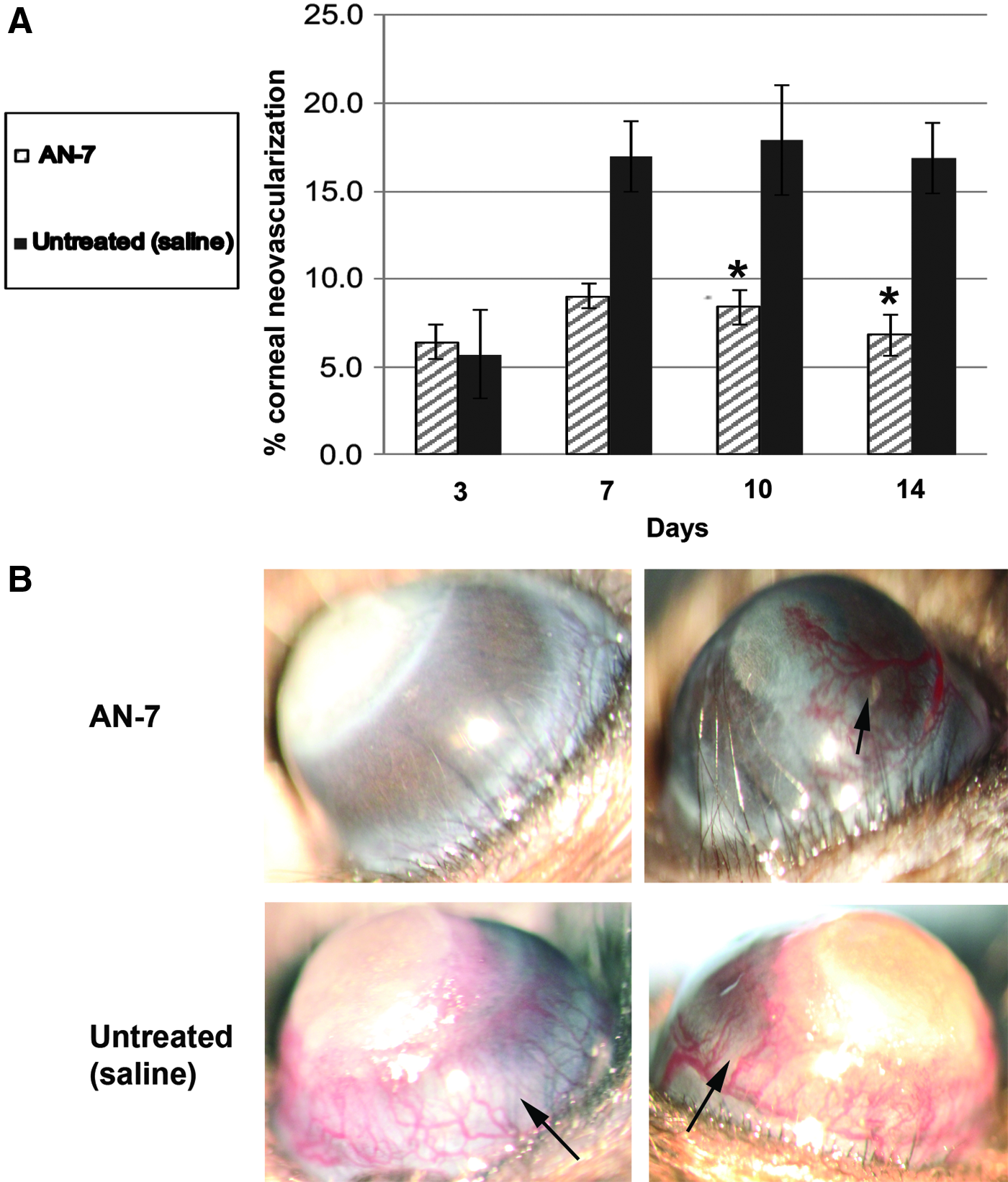
No difference in centricity or density of the new blood vessels graded by masked observers was found among the groups at any time point, yet, although it was not statistically significant, a trend for benefit of AN-7 over the control group could be detected (Fig. 2).
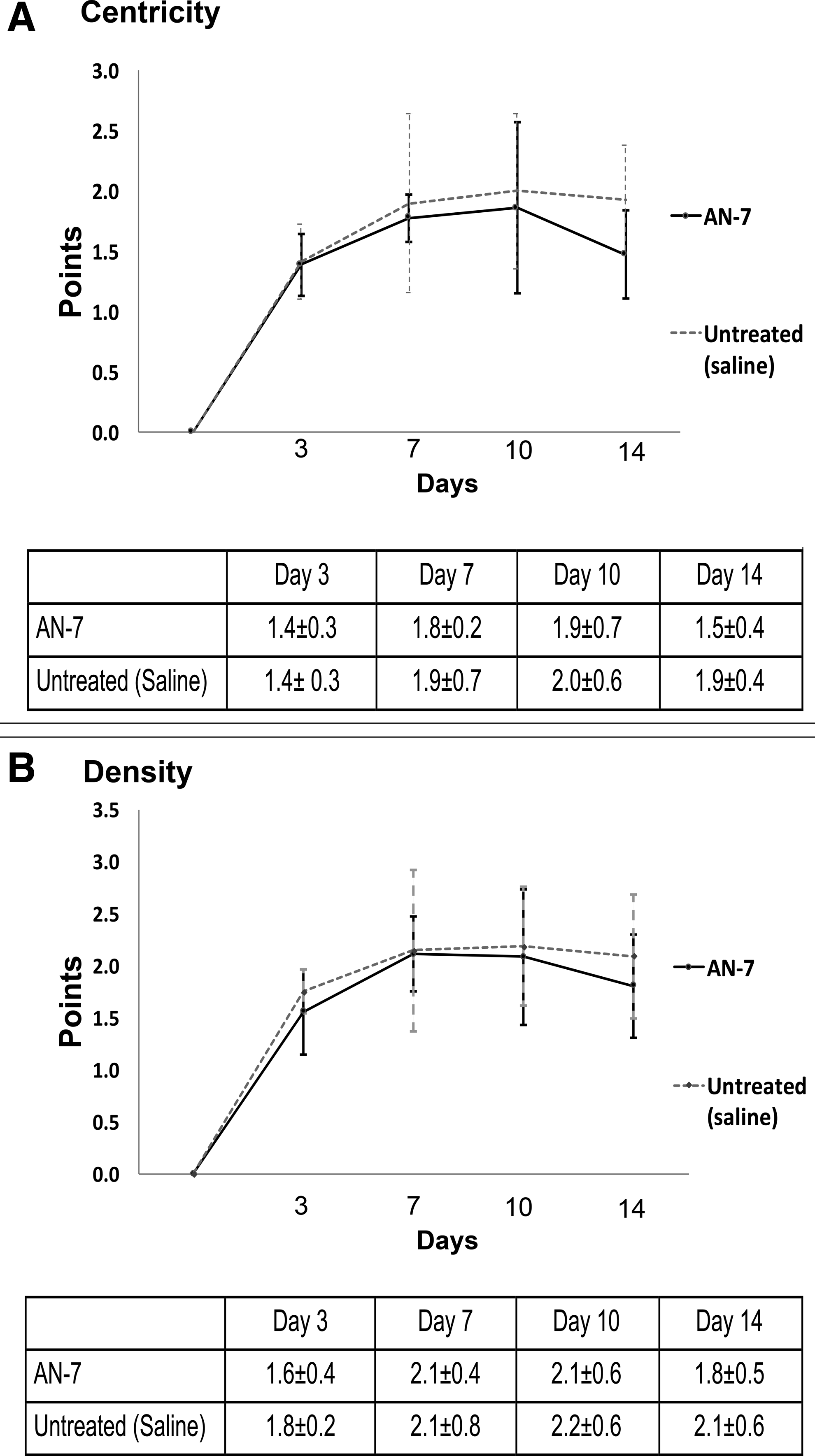
Graphs comparing blinded grading of the corneal photographs, for centricity of the corneal vascularization
Based on the mentioned results of corneal vascularization, we further explored the ability of AN-7 to reduce corneal NV, using FITC–dextran perfusion.
Two of the AN-7-treated mice retinas showed no fluorescent staining; hence their respective corneas were not included in the experiment, with a total of 5 AN-7-treated corneas included in the statistics.
Figure 3A shows representative images of corneal flat mounts stained with FITC–dextran of AN-7- and saline-treated mice, 10 days postchemical burn. Quantification of blood-vessel areas on day 10 after chemical burn (Fig. 3B) shows that AN-7 treatment dramatically reduced NV area (0.04 ± 0.07 μm2) in comparison with saline-treated control (0.52 ± 0.23 μm2) (P = 0.0005). All corneas without induction of chemical burn showed no staining.
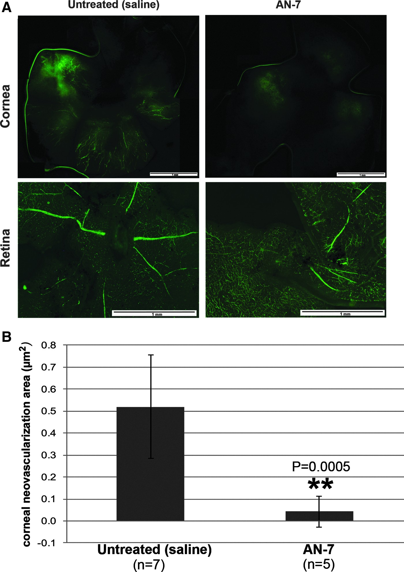
Intraperitoneal injection of AN-7 decreases corneal NV in flat-mounted corneas.
H&E and FITC–dextran staining in consecutive sections of samples from AN-7-injected and untreated group, 10 days after chemical burn (Fig. 4), showed NV and presence of blood vessels (arrows) in the untreated group but not in the AN-7-injected group.

Histological and FITC–dextran staining in consecutive sections of samples from AN-7-treated and untreated group, 10 days after chemical burn. NV and presence of blood vessels (arrows) were observed in the untreated control, but not in the AN-7-injected group. Asterisk indicates the corneal chemical burn.
Discussion
The purpose of the study was to determine whether AN-7 given as IP injections exerts a protective effect against corneal NV induced by a chemical burn. To the best of our knowledge, this is the first report on the use of AN-7 for this purpose.
A mouse model was used, as described in previous reports on corneal NV.11,12 The results of the control group in those studies were comparable to ours. We detected new corneal vessels as early as 3 days after injury. Corneal NV peaked on days 7–10, as was also reported in the earlier studies using the same model.11,12
Many previous reports have focused on the ability of anti-VEGF agents, usually bevacizumab, administered by different routes, to inhibit corneal NV. Manzano et al. 9 found that topical bevacizumab inhibited corneal NV after chemical burn injury in a rat model. Periocular and intraocular injections yielded similar results.10–12 Successful treatment with anti-VEGF agents was also reported in several studies in humans. 13 However, the effect of anti-VEGF is known to be temporary, mandating repeated applications or injections to maintain remission.
Recently it was reported that trichostatin A (TSA), a known potent HDACI, attenuated choroidal NV. 23 In vivo application of TSA predominantly resulted in an impairment of meiosis because of increased apoptosis, demonstrating genotoxicity. 24 In addition, treatment with TSA is limited by its low oral bioavailability and relatively short half-life. 25
AN-7, an orally bioavailable, water soluble, antimetastatic agent with very low toxicity, is a member of the HDACI family, having the ability to downregulate basic fibroblast growth factor and VEGF in tumors. 15 Besides the ability of AN-7 to exert antitumor, antiangiogenic (in tumors), and anti-inflammatory activities,16–19 it was shown to suppress NV. After AN-7 administration, a decrease in the amount and diameter of newly created blood vessels and an increase in their homogeneity were observed. 20
In this study, we showed that AN-7 administered as an IP injection induced a significant inhibition of corneal NV. The effect on days 10 and 14 after corneal injury was statistically significant relative to the untreated group. The ability of AN-7 to inhibit corneal NV development was assessed clinically and was further confirmed by blood-vessel quantification on flat-mount corneas and histology.
However, while examining the centricity and density of the corneal NV, no significant differences were observed between groups. A possible cause may be that although AN-7 inhibits growth of blood vessels, those already formed carry the same characteristic as in untreated mice. Therefore, the blood vessels in the areas of corneal NV are not different in density and centricity. It is also possible that the ImageJ program is a more sensitive method for calculating the total area of corneal NV. However, further analysis by blinded observers of the centricity and density of the new blood vessels is warranted to support the benefit of AN-7 treatment.
We conclude that systemic administration of AN-7 has the potential to attenuate corneal NV. Further preclinical testing with long-term follow-up to determine the optimal dosage of AN-7 and its additive effect together with other antiangiogenic agents is planned.
Footnotes
Acknowledgments
The authors thank the Young Investigator Funds, Rabin Medical Center, Petah Tikva, Israel, for supporting this study. We also acknowledge the contribution of Dr. Udi Lebel, Mrs. Dalia Sela, and Mr. Amy Sharon in treating the animals and providing the animal housing facilities.
Author Disclosure Statement
No competing financial interests exist.