
Abstract
Abstract
Purpose:
Aldose reductase (ALR), the first and rate-limiting enzyme involved in polyol pathway plays a central role in diabetes and its related complications, including diabetic retinopathy (DR). Inhibition of ALR may also be an ideal target for reducing the deleterious effects of DR. Therefore, the purpose of the present study was to investigate the protective effect of epalrestat (EPL), ALR inhibitor on glucose-induced toxicity in ARPE-19 cells.
Methods:
ARPE-19 cells were challenged with normal glucose (NG, 5 mM) and high glucose (HG1, 25 mM and HG2, 50 mM) in the presence or absence of EPL. ALR and VEGF165 expression in retinal pigment epithelial (RPE) cells under experimental conditions were quantified by real-time polymerase chain reaction using SYBR Green chemistry. Vascular endothelial growth factor (VEGF) secretion in the cell supernatant was measured by Sandwich ELISA. Cytotoxicity of EPL was assessed by MTT assay. ALR inhibitory activity, apoptosis, and sorbitol accumulation were also investigated.
Results:
EPL at studied concentration did not show any toxicity to RPE cells and showed as maximum as 65% ALR inhibition under high glucose condition (HG1). The presence of EPL significantly reduced ALR expression and VEGF levels as induced by high glucose in ARPE-19 cells.
Conclusion:
Inhibition of ALR appeared to be beneficial in reducing diabetes-related complications in RPE cells under high glucose condition.
Introduction
D
Aldose reductase (ALR), a member of aldo-keto reductase superfamily is the key enzyme in the polyol pathway, which catalyzes the conversion of excessive glucose to sorbitol. 7 Acceleration of intracellular polyol metabolism as a consequence of hyperglycemia leads to the accumulation of sorbitol, an osmolyte responsible for the development of vascular lesions associated with hyperglycemia. 8
ALR is widely distributed throughout the body, including the target organs that are affected by diabetes, including lens and retina.9,10 Chronic hyperglycemia causes retinal edema and increased apoptosis in retinal endothelial cells, pericytes, and retinal pigment epithelial (RPE) cells.11–15 Previous studies have demonstrated that blockade of ALR by genetic or pharmacological intervention prevents the onset and progression of many diabetic-related retinal complications such as pericytes loss, capillary degeneration, oxidative stress, and retinal inflammation.16–20 These complications could be overcome by the administration of aldose reductase inhibitors (ARIs).
Several ARIs had been tried for treating diabetic-related complications over the past 2 decades. However, only epalrestat (EPL) is given marketing approval as a therapeutic adjunct for the clinical management of diabetic neuropathy.
EPL is a carboxylic acid derivative, noncompetitive and reversible ARI, which inhibits ALR, a rate-limiting enzyme of polyol pathway. Several clinical trials reported that EPL was efficacious in patients with diabetic neuropathy and nephropathy. Previous studies demonstrated that EPL reduces sorbitol accumulation in the sciatic nerve, erythrocytes, and ocular tissues in animals, and in erythrocytes in humans. 21 Moreover, EPL reduced the expression of high glucose-induced neutrophil–endothelial cell adhesion and surface expression of intracellular adhesion molecule-1, P-selectin, and E-selectin on HUVEC cells. 22 EPL inhibited smooth muscle cell (SMC) migration in human coronary artery 23 and reduces 3-Deoxyglucosone and AGEs in erythrocytes. 24 However, its role in protecting the retinal cells under stress is not well studied. Therefore, we aimed to demonstrate the protective role of EPL in alleviating glucose-induced toxicity in RPE cells.
Since RPE cells secrete several growth factors, including vascular endothelial growth factor (VEGF) for the maintenance of the structural integrity of retina, 25 we have also investigated the effect of EPL on preventing VEGF expression and secretion by RPE cells under high glucose condition. Our data indicate that EPL treatment to cultured RPE cells significantly reduced ALR expression, sorbitol and VEGF levels as induced by high glucose in ARPE-19 cells, and thus offered protection against glucose toxicity in RPE cells.
Methods
Materials
Dulbecco's modified Eagle's medium/Nutrient Mixture F12 (DMEM/F12 medium, Invitrogen; Gibco, Grand Island, NY). Fetal bovine serum, Penicillin–Streptomycin (10,000 U/mL and 10 mg/mL) and
Cell culture
Adult human RPE (ARPE-19) cells were purchased from the American Type Culture Collection (ATCC, Mantissa, VA). ARPE-19 cells were cultured in DMEM/F12 medium supplemented with 10% FBS and penicillin–streptomycin (100 U/mL and 100 μg/mL) at 37°C in a 5% CO2 atmosphere. The medium was changed every other day. Cells at passage 19–30 were used for this study unless otherwise mentioned. For stimulation with glucose experiments, the confluent cells were serum starved for 3 h, and then exposed to different concentrations of glucose (normal glucose [NG, 5 mM] or high glucose 1 [HG1, 25 mM] and high glucose 2 [HG2, 50 mM]) in the presence or absence of 0.1 μM EPL for 72 h.
Cytotoxicity of EPL
The cytotoxicity of EPL was assessed in ARPE-19 cells using the In Vitro Toxicology Assay Kit; MTT based (Sigma-Aldrich) as per the manufacturer's instructions. ARPE-19 cells seeded onto 96-well plates at a density of 104 cells/well were allowed to grow for 1 week. After serum starvation for 3 h, cells were exposed to different concentrations of EPL (0.001–1,000 μM). Cells without EPL were named as EPL untreated group. After treatment with 20 μL of 3-(4,5-dimethylthiazol-2-yl)-2,5-diphenyl tetrazolium bromide (MTT) for 3 h, 200 μL solubilization solution was added to each well. Levels of formazan reaction product were determined by measuring the absorbance at 570 nm using the SpectraMax M3 Multiplate Reader (Molecular Device, CA).
Apoptosis (terminal deoxynucleotidyl transferase dUTP nick end labeling) assay
High glucose-induced DNA damage was assessed with a Colorimetric Apoptosis Detection Kit as per the manufacturer's instructions (Titer TACS; R&D System, Inc., Minneapolis, MN). Briefly, cells (2 × 10⁴ cells/well) cultured in a 96-well plate were serum starved for 3 h and exposed with different concentrations of glucose in the presence or absence of EPL for 72 h. Cells were then fixed with 3.7% buffered formaldehyde for 5 min followed by permeabilization with 100% methanol for 20 min. Cells were labeled as per the manufacturer's instructions. The reaction was stopped with 2 N HCl after 30 min of substrate addition and the absorbance was measured at 450 nm using the multiplate reader. For comparison, a positive control (nuclease-treated control) was used to confirm the permeabilization and labeling reaction.
Sorbitol content assay
The accumulated sorbitol in ARPE-19 cells under high glucose condition was estimated using the
ALR and VEGF mRNA expression by qRT-PCR
Total RNA was isolated from cell pellet obtained after glucose treatment procedure using TRIzol reagent (Sigma-Aldrich) as per the manufacturer's protocol. The RNA was quantified using a NanoDrop Spectrophotometer (NanoDrop Technologies, Inc., Wilmington, DE). For qPCR analyses, 2,000 ng of individual RNA samples were reverse transcribed into cDNA using the High-Capacity cDNA Reverse Transcription Kit (Applied Biosystems, Carlsbad, CA). ALR and VEGF mRNA expressions were quantified using SYBR Green chemistry (ThermoScientific, Waltham, MA) in 5 μL volume reaction using a 7900 HT Real-Time PCR system (Applied Biosystems) in triplicate. The product specificity was ascertained by melting curve.
Primers (Bioserve, Hyderabad, India) sequences used were:
ALR (Fwd: TTTTCCCATTGGATGAGTCGG, Rev: ACGTGTCCAGAATGTTGGTGT)—60 bp;
VEGF165 (Fwd: GCTACTGCCATCCAATCGAG, Rev: TCTTTCTTTGGTCTGCATTCAC)—255 bp;
GAPDH (Fwd: GTCTCCTCTGACTTCAACAGCG, Rev: ACCACCCTGTTGCTGTAGCCAA)—131 bp;
The quantitative real-time Polymerized Chain Reaction (qRT-PCR) cycle parameters consisted of an initial denaturation step of 95°C for 10 min followed by 40 cycles of 95°C for 15 s and 60°C for 1 min. Relative gene expression was calculated as described previously 26 using GAPDH as the housekeeping gene.
ALR activity assay
ARPE-19 cells after treatment with different concentrations of glucose in the presence or absence of EPL were lysed and the protein concentration was estimated by standard protocol. ALR activity was measured spectrophotometrically by the method as described previously. 27 The assay mixture (300 μL) contained 100 mM of sodium phosphate buffer (pH.7), 0.2 mM LiSO4, 5 mM DL-glyceraldehyde, and enzyme preparation. The assay was initiated with the addition of 0.15 mM NADPH and decrease in optical density was measured at 340 nm at 37°C for 20 min in the presence or absence of 5 mM DL-glyceraldehyde as a substrate. For ALR inhibition experiments, EPL was added to the culture medium at a concentration of 0.1 μM. Enzyme activity was normalized to supernatant protein content. The percentage ALR inhibition was calculated using the following formula:
%AR Inhibition = (Δ absorbance of Negative control–Δ absorbance of treated)/Δ absorbance of Negative control) × 100
Immunofluorescence
Coverslip-cultured ARPE-19 cells (90% confluent) were fixed with ice-cold methanol at −20°C for 20 min and washed twice with PBS for 10 min each. After treatment with avidin–biotin blocking solution for 10 min, the cells were incubated with goat anti-ALR antibody (sc-17732; Santa Cruz Biotechnology), at 1: 50 dilution. After overnight incubation at 25°C, the cells were washed twice with PBS and incubated with biotinylated rabbit anti-goat IgG secondary antibody (sc-2774; Santa Cruz Biotechnology) at 1:100 dilutions, for 1 h in dark at 25°C. Visualization was carried out with streptavidin-FITC. The cells were treated with RNase at 37°C for 30 min and nuclei stained with Propidium Iodide (PI) and mounted using Vectashield fluorescence mounting medium (H-1000; Vector Laboratories, Inc., Burlingame, CA).
Image acquisition was carried out using LEICA SP 8 Confocal Microscope (LEICA Microsystems, Heidelberg, Germany). The emission (bandwidth) for FITC ranged from 496 to 535 nm and from 560 to 600 nm for PI. Using Leica confocal software (version 4.3), the mean fluorescence intensity for FITC and PI were calculated after reconstructing the z-stack images to a 2D maximum projection along a fixed axis (3 fields per slide). The ALR fluorescence intensity was then quantified by normalization to PI fluorescence intensity.
VEGF secretion by ELISA
ARPE-19 cells seeded at a density of 105 cells/well on a 12-well plate were allowed to reach near confluence and grown in conditions as described above. The cell supernatant was collected for the estimation of VEGF by Sandwich ELISA (R&D System, Inc.) according to the manufacturer's protocol. This kit recognizes VEGF165 isoform. Total protein concentrations were measured by Lowry's method.
Statistical analysis
A Mann–Whitney U Test (SPSS, version 20.0, Chicago, IL) was used to compare difference in cytotoxicity, apoptosis, ALR and VEGF165 mRNA expression and VEGF secretion studies. Student's t-test was used to compare specific ALR activity and relative fluorescence intensity between treated and control. Difference was considered significant when P < 0.05.
Results
Cytotoxicity of EPL
The presence of EPL did not show any toxicity in ARPE-19 cells under experimental condition upto 10 μM at 72 h time point (Fig. 1). EPL at 100 and 1,000 μM concentration showed 77% and 92% toxicity, respectively and its effect was found to be statistically significant (P < 0.005; 0.001 μM vs. 100 and 1,000 μM). Therefore, in the present study, we have selected 0.1 μM EPL concentration for further experiments.
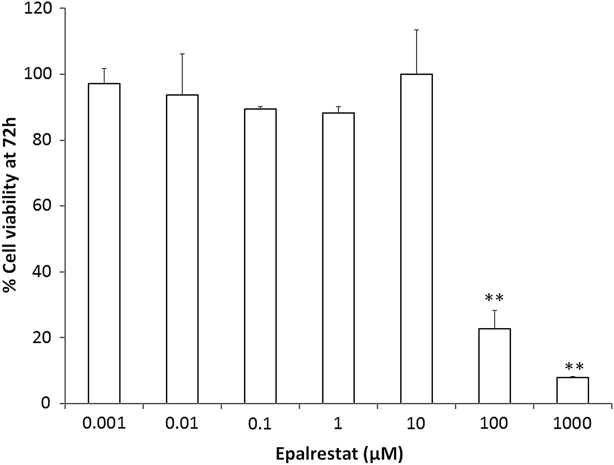
Effect of EPL on cell viability. ARPE-19 cells were cultured under NG in the presence or absence of EPL (0.001–1,000 μM). Viability was assessed by MTT assay for 72 h. The presence of EPL did not show any toxicity up to 10 μM. EPL at 100 and 1,000 μM concentration showed 77% and 92% toxicity, respectively. Data shown are mean ± SEM (n = 3). **P < 0.005 (0.001 μM vs. 100 and 1,000 μM). EPL, epalrestat; NG, normal glucose.
EPL protects ARPE-19 cells from apoptosis and decreases sorbitol accumulation under high glucose conditions
EPL at the studied concentration (0.1 μM) showed protection against HG-induced apoptosis at 72 h (Fig. 2A). The presence of EPL significantly reduced apoptosis in ARPE-19 cells under HG2 condition as compared with untreated (P = 0.026).
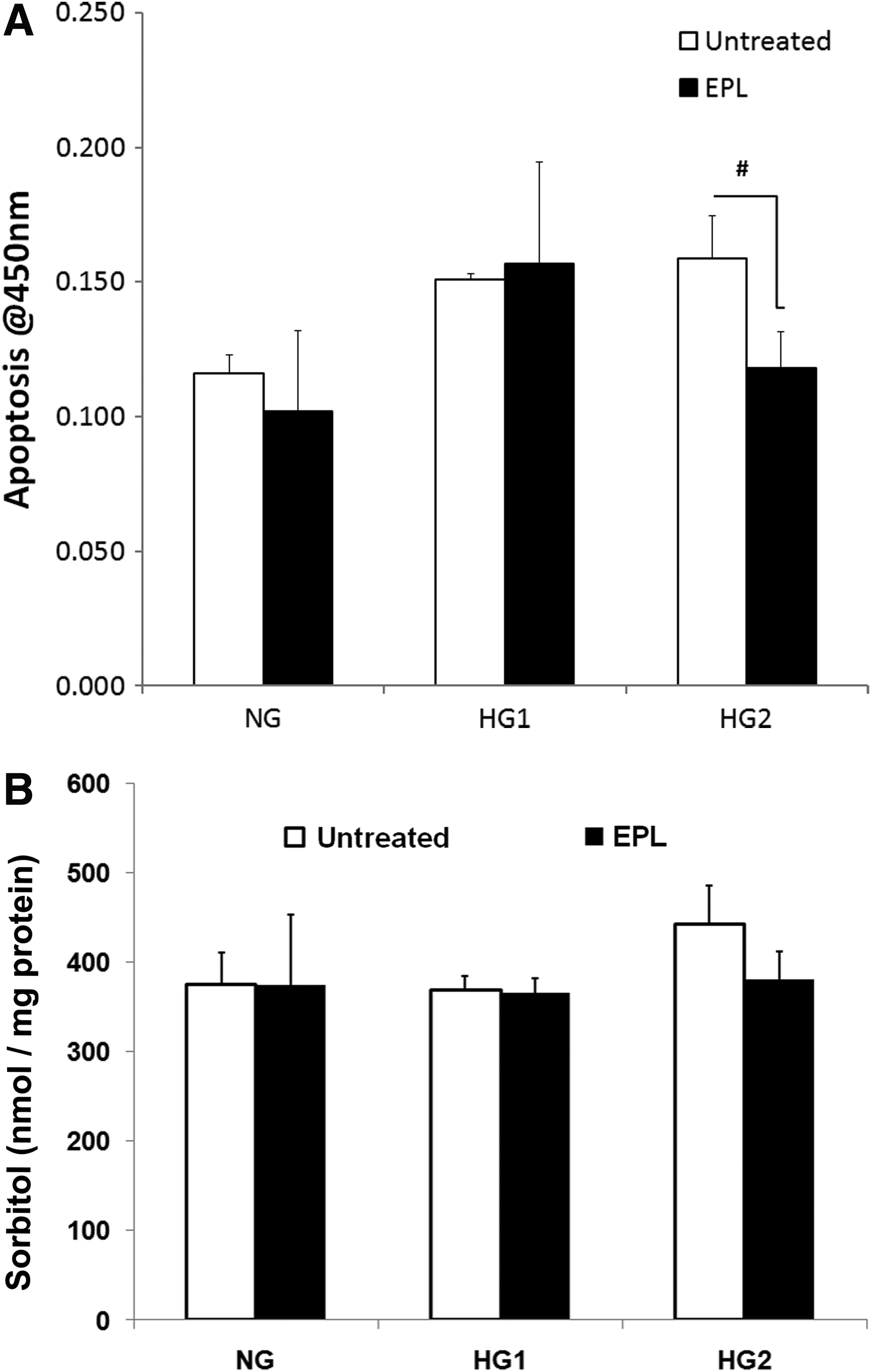
Effect of EPL on apoptosis and sorbitol accumulation in ARPE-19 cells under high glucose
Increased accumulation of sorbitol was observed at 72 h with high glucose (HG2-443 ± 42.65 nmol/mg protein) compared with NG (375.16 ± 35.04 nmol/mg protein); however, the difference was found to be statistically not significant (P = 0.24). Presence of EPL showed only marginal reduction in sorbitol level and under experimental condition (Fig. 2B).
EPL reduces HG-induced expression of ALR and ALR activity
ARPE-19 cells under high glucose conditions showed significant increase in ALR mRNA expression at 72 h. The presence of EPL significantly reduced ALR expression in both HG conditions (P = 0.049; HG1-untreated vs. HG1-EPL) and (P = 0.024; HG2-untreated vs. HG2-EPL) (Fig. 3A).
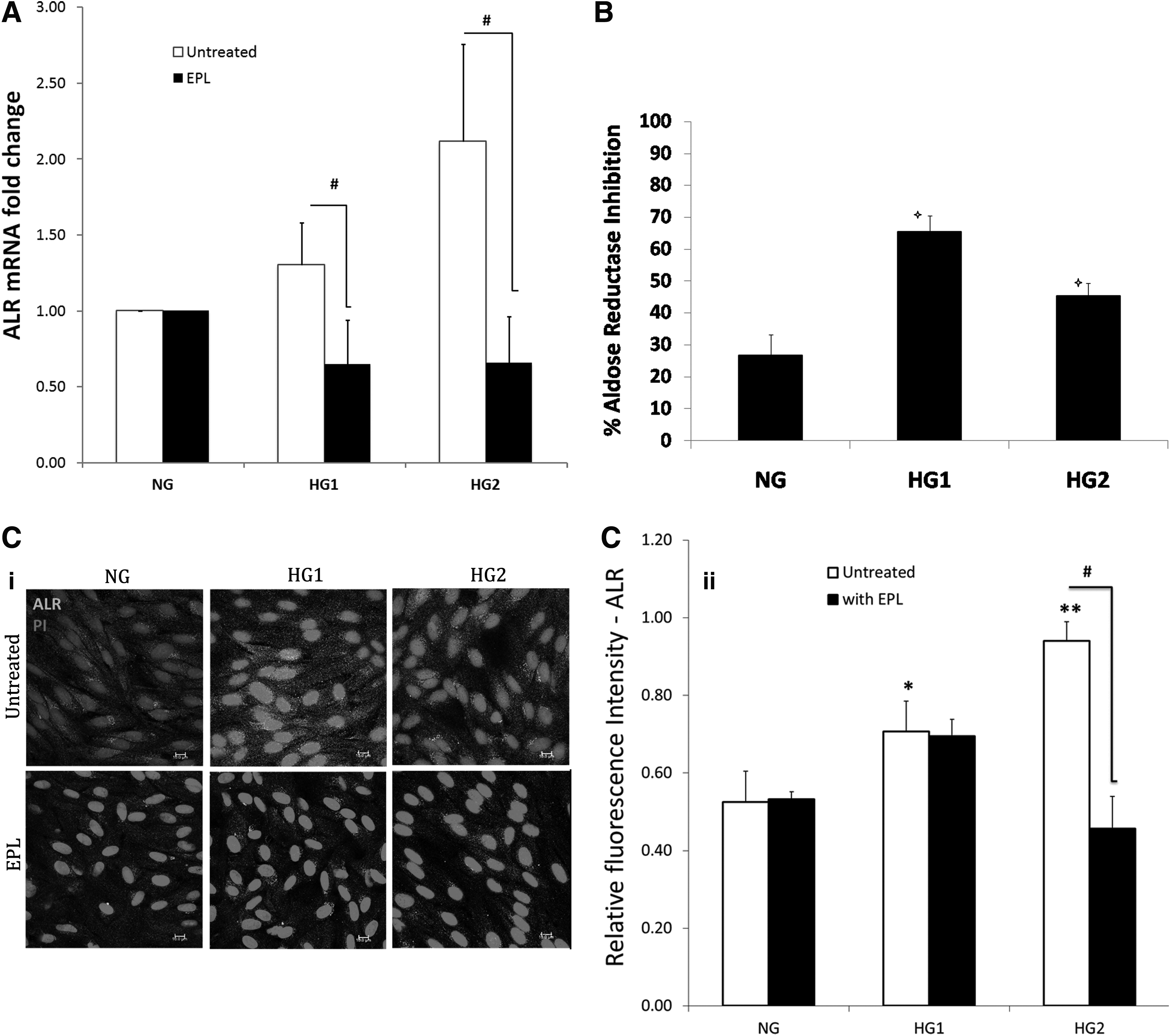
ALR mRNA expression was quantified by qPCR using SYBR Green and the expression was normalized to GAPDH expression (housekeeping gene)
In case of ALR enzyme activity, the presence of EPL showed 26.7%, 65%, and 45% inhibition with NG, HG1, and HG2, respectively as compared with EPL untreated at 72 h (Fig. 3B).
Immunofluorescence analysis of ALR showed that the intensity of ALR increased with increase in glucose concentration. The presence of EPL significantly reduced ALR in HG2 condition (P = 0.0010, Fig. 3C).
ALR inhibitor prevents HG-induced VEGF expression and VEGF secretion
In this study, we have also investigated the effect of high glucose on VEGF165 mRNA expression. Significant increase in VEGF165 expression was observed with high glucose at 72 h as compared with NG (Fig. 4A). The presence of EPL significantly reduced VEGF expression at both HG conditions (P = 0.0097; HG1-untreated vs. HG1-EPL) and (P = 0.0005; HG2-untreated vs. HG2-EPL).
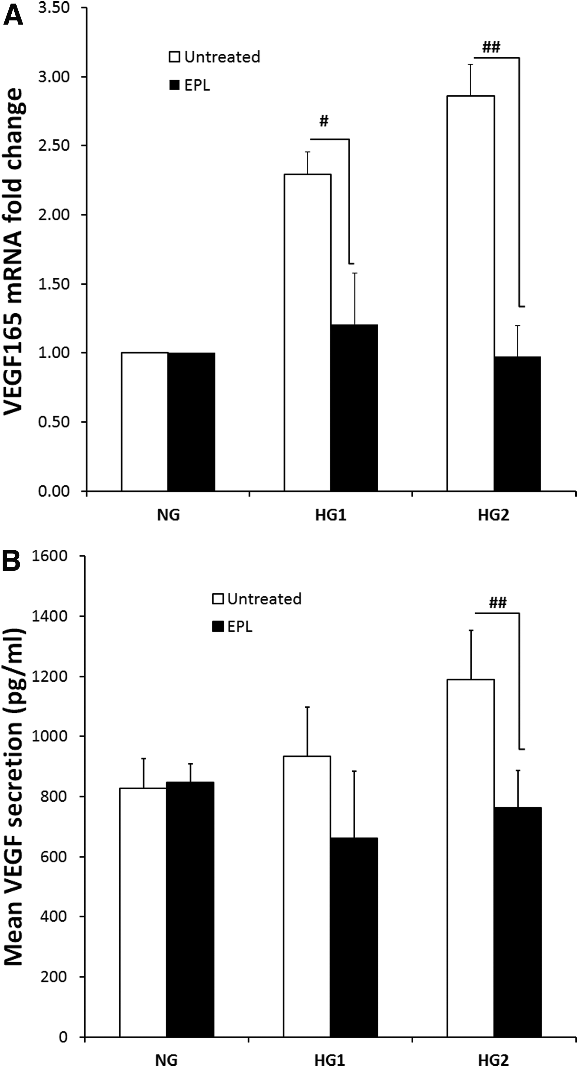
VEGF165 mRNA expression was quantified by qPCR using SYBR green and the expression was normalized to GAPDH expression (housekeeping gene)
In case of VEGF secretion, the presence of EPL significantly reduced VEGF secretion at 72 h under HG2 condition (P < 0.05) as compared with EPL untreated group (P = 0.0058) (HG2-untreated vs. HG2-EPL) (Fig. 4B).
Discussion
Our present study was to demonstrate the protective role of EPL in alleviating glucose-induced toxicity in RPE cells. RPE is a monolayer of pigmented cells situated between the neural retina and choroid. It plays a vital role in transport of essential nutrients, reisomerization of all transretinal for visual function, protection from phototoxicity, and phagocytosis of photoreceptor outer segments. Apart from these functions, RPE secretes many factors like pigment epithelium-derived growth factor, VEGF, and ciliary neurotrophic factor, which are essential for the structural integrity of neural retina. 28 Several evidences suggest that, the functional and structural changes of RPE occur in experimental and clinical diabetes. Especially the RPE layer of the human eye has been shown immunohistochemically to contain large amounts of ALR that are increased in DR. 29 Understanding the metabolism of glucose metabolism in the RPE cells might give insight into in-depth knowledge on pathogenesis of DR.
ARPE-19 is a spontaneously arising RPE cell line derived from the normal eyes of a 19-year-old male. 30 ARPE-19 cells have been widely used, in studies on oxidative stress, retinal pathogenesis, signaling pathways related to high glucose, as well as in drug and toxicity-related studies.31–35
In our present study, we have used ARPE-19 cell line under high glucose as an in vitro model system to study the effect of EPL on HG-induced ALR mRNA expression, activity, and sorbitol accumulation. In addition, we have also studied reduction of HG-induced VEGF mRNA expression and secretion with EPL. We have observed significant increase in mRNA expression of ALR, which indicated the acceleration of glucose metabolism through polyol pathway.
In many of the previously reported in vitro studies, micromolar (μM) concentrations (10–50 μM) of EPL has been tried to investigate its efficacy against high glucose-induced complications, but only very few studies utilized EPL in nanomolar concentrations. 36 EPL at 0.07 μmol/L were reported to inhibit ALR activity in human lens epithelial cells 37 and also in human coronary artery SMC. 23 Therefore in the present study, we have investigated efficacy of EPL at nanomolar concentration in alleviating HG-induced toxicity in retinal pigment epithelium with 0.1 μM concentration of EPL. The findings of the present study indicated that, EPL at the studied concentration showed significant reduction of glucose-induced high expression of ALR and enzyme activity at a maximum of 52% inhibition in retinal pigment epithelium under experimental conditions. However, the present study aimed to address pharmacological inhibition with less toxicity.
Sorbitol, an osmolyte accumulation as a result of activation of polyol pathway is considered to play a key role in mediating diabetic-related microvascular complications. In the present study, we found time-dependent accumulation of sorbitol in human retinal pigment epithelium under high glucose conditions (25 and 50 mM). The sorbitol concentration of 101.2 nmol/mg protein and 96.3 nmol/mg protein concentrations was observed in HG1 and HG2 conditions at 24 h, respectively (data not shown), whereas increase in the concentration of sorbitol was observed at 72 h (Fig. 2B). This observation indicates that the polyol pathway is accelerated in retinal pigment epithelium under high glucose condition. This is in agreement with the findings from other studies that, 30 mM of glucose resulted 110 mmol/mg protein of sorbitol and the presence of Beta-glucogallin reduced its accumulation by 44%. 19 However, in the present study, the presence of EPL at nanomolar concentration did not show any significant reduction in sorbitol levels. This could be due to the fact that the EPL at the studied concentration might not be sufficient enough to reduce sorbitol accumulation for the extended condition. However, the presence of EPL significantly reduced the mRNA expression of ALR under experimental conditions.
VEGF has been recognized as a critical mediator of DR in experimental and clinical studies for several decades.27,38–40 VEGF is primarily expressed in the retina, including Muller cells, endothelial cells, astrocytes, retinal pigment epithelium, and ganglion cells. 10 The cellular or vitreous level of VEGF was reported to be highly correlated with retinal neovascularization and edema. 41 Our studies correlate with the previous reports, where EPL decreases polyol pathway activity and lead to restoration of NADPH, prevents hypoxia-like responses under high glucose, leading to VEGF downregulation and thereby reduce intracellular oxidative stress. 42
It was demonstrated in a diabetic rat model by the other group that increased ALR activity as a result of hyperglycemia could induce retinal VEGF overexpression and administration of fidarestat, a highly specific ARI, ameliorated the same. 43 It has been reported that the high glucose-induced upregulation of VEGF expression and secretion is due to the involvement of carbohydrate response element-binding protein, which activates HIF-1α under high glucose normoxia condition in human retinal pigment epithelium. 44 ALR inhibitor not only decreases the high glucose-induced VEGF expression and secretion, but also prevent VEGF-induced growth and tube formation in human retinal endothelial cells. 45 In the present study also, we have observed a significant increase in HG-induced expression and secretion of VEGF in RPE cells and the presence of EPL significantly reduced the same. However, further studies are necessary to understand the role of EPL in regulation of VEGF under glucose stress in retinal pigment epithelium.
To conclude, glucose-induced toxicity in RPE cells was effectively inhibited with EPL at nanomolar concentration at 72 h. Also high mRNA expression of angiogenic marker VEGF165 and its secretion level was significantly reduced by EPL in vitro. This further confirms that ALR inhibition is not only an attractive strategy for the treatment of diabetic complications, but also ameliorates retinal neovascularization in proliferative DR.
Footnotes
Acknowledgments
The authors sincerely thank their biostatistician Mr. Vijayakumar for his assistance in performing statistics, Aravind Eye Hospital, Madurai. The authors sincerely acknowledge Prof. Colin Willoughby, University of Liverpool, United Kingdom for his assistance in language editing of the article.
Author Disclosure Statement
No competing financial interests exist.