
Abstract
Abstract
Purpose:
To explore the inhibitory effect of insulin-like growth factor binding protein-related protein 1 (IGFBP-rP1) on retinal angiogenesis and its underlying molecular mechanisms in the mouse model of oxygen-induced retinopathy (OIR).
Methods:
C57BL/6J mice were classified into three groups as control group, OIR nonintervention group, and OIR intervention group. Postnatal day 12 (P12) mice in OIR intervention group were received recombinant mouse IGFBP-rP1 (50, 100, and 200 ng/mL) intravitreal injection. Five days later, the proliferative neovascular responses were estimated by quantifying the new vessel areas in flattening retinal tissues stained by high molecular fluorescein isothiocyanate-dextran and counting the numbers of neovascular cell nuclei breaking through the internal limiting membrane in cross sections. Expressions of phospho-extracellular signal-regulated kinase 1/2 (p-ERK1/2), ERK1/2, and vascular endothelial growth factor (VEGF) proteins in retinal tissues were assessed by western blot analysis.
Results:
Irregular neovascularization, nonperfusion region, and fluorescence leakage were observed in OIR models. The expression of retinal p-ERK1/2 and VEGF proteins were significantly upregulated in OIR nonintervention group compared with control group. The area ratio of retinal new vessels and the number of neovascular cell nuclei in OIR intervention group both decreased significantly, following the downregulation of retinal p-ERK1/2 protein expression and VEGF protein expression in a dose-dependent manner. Moreover, there was no significant difference in retinal ERK1/2 protein expression.
Conclusions:
IGFBP-rP1 inhibits retinal angiogenesis by blocking ERK signaling pathway and downregulating VEGF expression in the mouse model of OIR. It highlights the potential importance of IGFBP-rP1 serving as a target of gene therapy for retinal neovascularization in the future.
Introduction
A
Retinal neovascularization is regarded as a common pathophysiological basis of irreversible vision loss in several retinal neovascularization dependent diseases, such as retinopathy of prematurity (ROP) as well as proliferative diabetic retinopathy.2–4 During the retinal pathological process of uncontrolled angiogenesis, nonperfusion areas and fragile vessels quickly form, which lead to retinal blood vessels leakage. At last, irreversible vision loss and ocular pain caused by neovascular glaucoma comprise the devastating end-stage characteristics of the retinal neovascularization-dependent diseases.
Recent researches have shown vascular endothelial growth factor (VEGF) is one of the critical factors associated with retinal neovascularization dependent diseases. 5 Therefore, anti-VEGF therapy can inhibit retinal neovascularization significantly. However, there are still some special clinical conditions that anti-VEGF therapy is unable to perform its therapeutic effect such as treatment nonresponse or resistance. Furthermore, some studies showed that anti-VEGF therapy could cause retinal damages,6,7 so that new antiangiogenic strategies are needed to be discovered to supplement the current therapeutic schedules.
Our preliminary study discovered insulin-like growth factor binding protein-related protein 1 (IGFBP-rP1), a 36 kDa secreted glycoprotein of retinal endothelial cells (RECs), was able to inhibit proliferation, migration, and capillary-like tube formation of RECs induced by VEGF. These results revealed IGFBP-rP1 might serve as an endogenous angiogenesis inhibitor, which can inhibit VEGF-induced retinal angiogenesis in vitro in a dose-dependent manner. 8 However, the biological roles of IGFBP-rP1 are still unclear in vivo.
The mouse model of oxygen-induced retinopathy (OIR) has demonstrated two phases involved in the pathophysiological course of ROP. 9 Phase 1 is the vaso-obliterative phase, also named as the hypovascular phase. Exposing the postnatal day 7 (P7) mice to high oxygen levels (75%) (hyperoxia) to postnatal day 12 (P12) results in abnormal oxygen dynamics, which inhibits the expression of VEGF and lead to delayed retinal vascular growth as well as the loss of vascular density in retina.10,11 Phase 2 is the proliferative phase, also known as the hypervascular phase. Transferring mice to room air turns the retinal hyperoxic condition into a relative hypoxic condition, which results in the upregulation of proangiogenic factors such as VEGF, leading to severe pathological neovascularization and causing the maximum neovascularization at postnatal day 17 (P17). 11 The mouse model of OIR is highly reliable and quantifiable for ROP as well as other retinal neovascularization-dependent diseases. This model is easily established, highly repeatable, and widely used for analyzing vascular leakages, vessel loss, and pathologic neovascular proliferations.
In this study, we hypothesized that IGFBP-rP1 might have an inhibitory role during the procedure of retinal neovascularization. Due to the advantage of OIR model, we used this model in C57BL/6J mice to explore the procedure of retinal neovascularization and the intervention caused by IGFBP-rP1.
Methods
Animals
P7 C57BL/6J mice and their dams were fed in the cleaning degree experimental animal room (constant temperature: 26°C; humidity: 40%–50%; lighting: 8 hours/day). All animal procedures used in this study were performed in accordance with the Association for Research in Vision and Ophthalmology (ARVO) Statement for the Use of Animals in Ophthalmic and Vision Research.
OIR model
One hundred twenty C57BL/6J mice were classified into three groups randomly as control group (n = 24), OIR nonintervention group (n = 24), and OIR intervention group (n = 72). The mouse model of OIR was established referring to the improved Smith method. 12 Mouse pups were exposed to 75% O2 from P7 to P12 and transferred to room air subsequently. Next, mice of OIR intervention group (n = 72) were divided equally into three subgroups and received intravitreal injection of 1.0 μL recombinant mouse IGFBP-rP1 (R&D, Minneapolis, MN) of different concentrations (50, 100, and 200 ng/mL). After the injection, the mice were fed to P17 in room air according to the previous studies.13,14
Retinal vessels perfusion
Intracardiac perfusions of 1 mL high molecular fluorescein isothiocyanate-dextran (HM FITC-dextran) (Sigma, St. Louis, MO) dissolved in 4% paraformaldehyde were performed in P17 mice of each group. Eyeballs of the sacrificed mice were removed and incubated in 4% paraformaldehyde at 4°C. Then retinal tissues were recovered and flat-mounted on glycerol/gelatin-coated glass slides. Whole retinas were observed and imaged at × 4 by a fluorescence microscope (Olympus, Tokyo, Japan) as described previously.12,15 The vessel morphology and distribution such as regions of nonperfusion areas, vascular tortuosities, and irregular expansions were observed as morphological changes. Meanwhile, the areas of retinal fluorescein leakage section were measured and analyzed by Image-Pro Plus 6.0 Analyzer software (Media Cybernetics, Bethesda, MD). Ratios of the fluorescein leakage areas to the whole retinal areas were considered as the assessment criteria.
Histology analysis
Five days after the intravitreal injection, six P17 mice of each group were sacrificed and the eyeballs were subjected to 55%, 65%, 75%, 85%, 95%, and 100% (v/v) ethanol for 30 min successively for dehydration. Subsequently, the eyeballs were infiltrated in dimethylbenzene for 5 min before embedded in paraffin. Then the tissues were sectioned into 4 μm serial section sagittally. Ten uncontinuous sections chosen from each eyeball were stained by hematoxylin and eosin. Two skilled pathologists separately observed and counted the numbers of neovascular cell nuclei breaking through the internal limiting membrane (ILM) in cross sections excluding these ones existed in the vitreous cavity, as previously described.16,17
Western blot analysis
The retinal tissue homogenates were lysed by 50 mM Tris-HCl (pH 8.0), 150 mM NaCl, 0.1% Nonidet P-40, 0.5% sodium deoxycholate, and phenylmethylsulfonyl fluoride (Beyotime Biotechnology, Shanghai, China). The whole tissue lysates were centrifugated at 12,000 rpm 4°C for 15 min. Protein levels were detected by bicinchoninic acid assay (Beyotime Biotechnology) and degenerated for western blotting to detect the expression of Phospho-extracellular signal-regulated kinase 1/2 (p-ERK1/2), ERK1/2, and VEGF proteins. Protein samples (30 μg) were run on 10% sodium dodecyl sulfate-polyacrylamide gels and transferred onto polyvinylidene fluoride membranes (Millipore, Billerica, MA). After blocked with 5% blocking liquid diluted in Tris-buffered saline with 0.1% Tween-20 for 1 h at 37°C, the membranes were incubated with specific primary antibodies overnight at 4°C. Next day, the membranes were washed three times and subsequently incubated with horseradish peroxidase (HRP)-conjugated secondary antibody for 1 h at room temperature. Signals were detected by enhanced chemiluminescence.
According to the area of each stripe and gray value determined by Gel-Pro Analyzer software (Media Cybernetics), the integrated optical density (IOD) of each sample was detected and compared with the internal reference to get the sample's relative protein expression level. The primary antibodies were p-ERK1/2 rabbit anti-mouse polyclonal antibody (CST, Danvers, MA) at a dilution of 1:1,000, ERK1/2 rabbit anti-mouse polyclonal antibody (CST) at a dilution of 1:1,000, VEGF rabbit anti-mouse polyclonal antibody at a dilution of 1:1,000 (Abcam, Cambridge, MA), and glyceraldehyde-3-phosphate dehydrogenase (GAPDH) rabbit anti-mouse monoclonal antibody (ProteiTech, Chicago, IL) at a dilution of 1:1,000. The second antibody was HRP-conjugated goat anti-rabbit antibody (R&D) at a dilution of 1:5,000.
Statistical analysis
The experimental data are expressed as means ± standard deviation. Statistical analysis was performed with the statistical software program SPSS 16.0 for windows (SPSS, Inc., Chicago, IL). P values <0.05 were considered statistically significant in all cases. The results between the control group and OIR nonintervention group were analyzed by using independent-samples t test. The results between OIR nonintervention group and OIR intervention group were analyzed by using one-way analysis of variance. Fisher's least significant difference (LSD)-t test was applied to compare the difference between each OIR intervention group during the experiments.
Results
Alternation of the new vessels in flattening retinas stained by HM FITC-dextran
The retinal tissues in the control group were normally vascularized, showing up as great vessels erupting from the optic disks and branching equally to the peripheral retinas radially (Fig. 1A). In contrast, the normal radial vessel networks were replaced by expanding and circuitous vessels in OIR nonintervention group. They were characterized by numerous microaneurysms, nonperfusion regions, and neovascularization at the junctions of perfusion and nonperfusion areas, which resulted in significant fluorescence leakage (Fig. 1B). To determine whether IGFBP-rP1 might have a role in inhibiting retinal angiogenesis in vivo, we performed intravitreal injection of IGFBP-rP1 in OIR intervention group and observed the alteration of retinal neovascularization. It turned out that the range of nonperfusion and fluorescence leakage gradually reduced, which were relevant to the increasing concentration of the rmIGFBP-rP1 injected into vitreous chamber (Fig. 1C–E).

Inhibiting effect of IGFBP-rP1 on retinal angiogenesis of OIR mice ( × 4, fluorescence microscope). The retinal tissues of control group developed maturely, without vessel occlusion or neovascularization
Ratio of retinal new vessels relative areas in different OIR intervention group (50, 100, and 200 ng/mL) compared with OIR nonintervention group was (76.53 ± 1.35)%, (34.27 ± 2.18)%, and (19.72 ± 2.79)%, respectively. This significantly descending tendency (P < 0.05) revealed that IGFBP-rP1 might have the ability to inhibit retinal neovascularization.
Number of mice retina neovascular cell nuclei
The numbers of neovascular cell nuclei breaking through the ILM were counted to confirm whether IGFBP-rP1 might act as an antiangiogenic factor in vivo. No neovascular cell nucleus was observed in control group (Fig. 2A). However, a cluster of neovascular cell nuclei breaking through ILM were found in OIR nonintervention group (Fig. 2B). With the concentration of rmIGFBP-rP1 injected into vitreous cavity growing, the number of neovascular cell nuclei decreased (Fig. 2C–E). The number of neovascular cell nuclei was (28.72 ± 4.68) per tissue slice in OIR nonintervention group, whereas the numbers of the OIR intervention group were (11.62 ± 2.54), (6.07 ± 2.47), and (3.25 ± 1.50) per slice, respectively (P < 0.05). Similar to the result of retinal new vessels relative areas assay, it confirmed that IGFBP-rP1 might act as an angiogenesis inhibitor in the prevention of retinal neovascularization. Furthermore, we discovered the neovascular cell nuclei breaking through ILM were mainly located in peripheral retina rather than central retina. Since there is no accepted standard to define the boundary of the peripheral and central retina, we were unable to analyze the statistical difference.
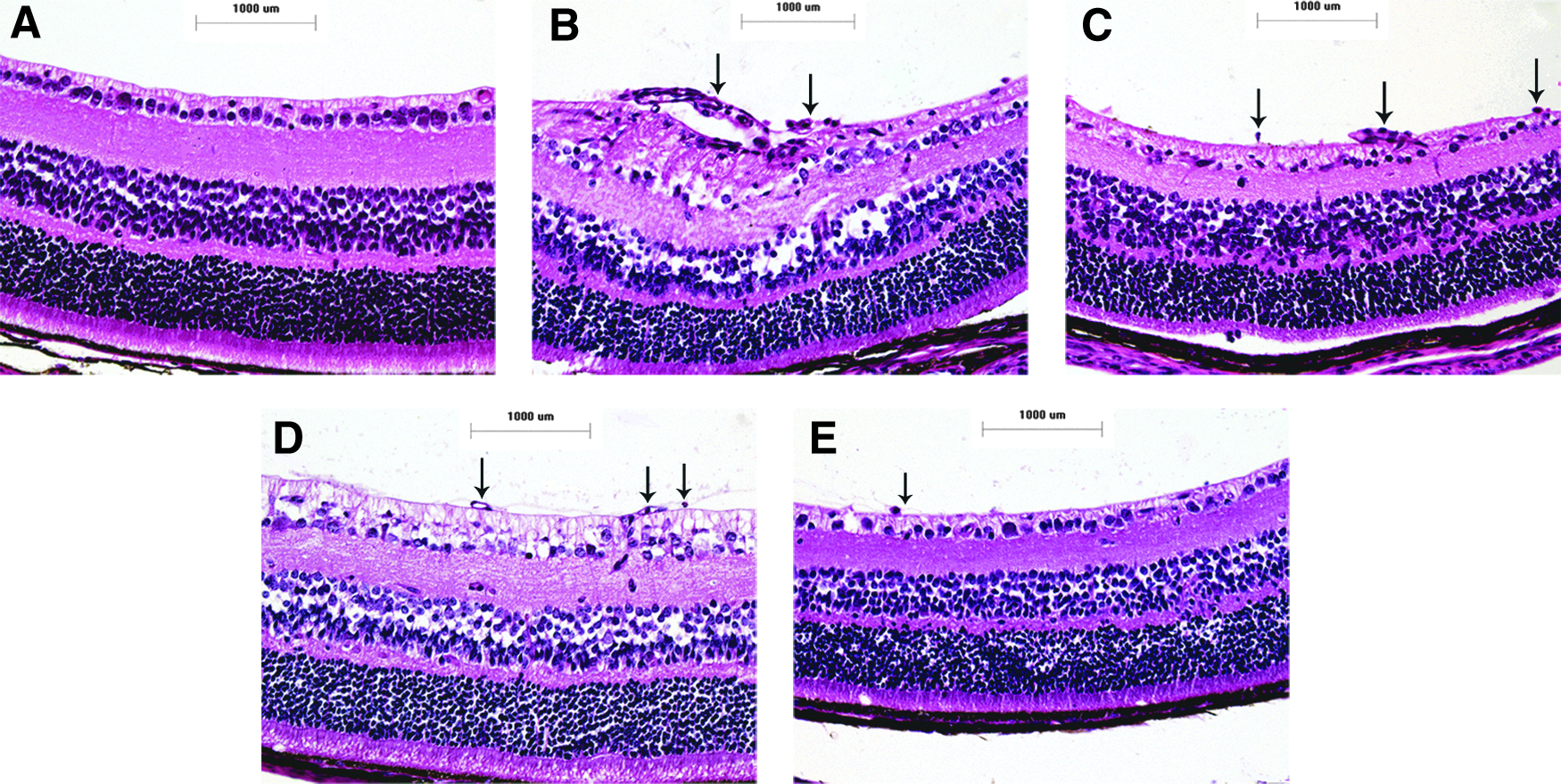
Inhibiting effect of IGFBP-rP1 on retinal angiogenesis of OIR mice (histology analysis). The retinal tissues of control group show no retinal neovascular endothelial cell nuclei
Expression of p-ERK1/2, ERK1/2, and VEGF in mice retina tissue
In light of the ERK1/2 signaling pathway which stimulated VEGF expression might be involved in the procedure of IGFBP-rP1 inhibiting retinal angiogenesis, we performed western blotting analysis to detect the distinct expressions of p-ERK1/2, ERK1/2, and VEGF protein and evaluated their differences by IODs comparing. The expressions of p-ERK1/2 and VEGF protein in OIR nonintervention group significantly increased than control group (P < 0.001). Furthermore, the expressions of p-ERK1/2 and VEGF protein in OIR intervention group reduced dose-dependently compared with OIR nonintervention group (P < 0.001) (Figs. 3 and 4). However, there was no significant difference in the expression of ERK1/2 protein between OIR nonintervention group and control group or OIR intervention group (P > 0.05). It proved that IGFBP-rP1 inhibited retinal angiogenesis, in part, by blocking ERK signaling pathway and downregulating VEGF expression in the mouse model of OIR.
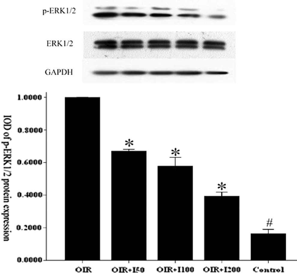
ERK1/2 signaling pathway suppressed by IGFBP-rP1. Equal proteins from the mice retinal tissue were loaded and probed with ERK1/2, p-ERK1/2, and GAPDH antibody. Representative bolts showed that hypoxia stress upregulated p-ERK1/2 protein expression in OIR nonintervention group, however, this upregulation was inhibited by IGFBP-rP1 dose-dependently. Values are means ± standard deviations of three independent experiments with similar results and showed as the IOD of p-ERK1/2 relative to the IOD of GAPDH. #P < 0.05 versus the control group and *P < 0.05 versus the OIR nonintervention group. (Control: control group, OIR: OIR nonintervention group, OIR+I50: 50 ng/mL IGFBP-rP1 intervention group, OIR+I100: 100 ng/mL IGFBP-rP1 intervention group, and OIR+I200: 200 ng/mL IGFBP-rP1 intervention group). IGFBP-rP1, insulin-like growth factor binding protein-related protein 1; GAPDH, glyceraldehyde-3-phosphate dehydrogenase; IOD, integrated optical density; OIR, oxygen-induced retinopathy; p-ERK1/2, phospho-extracellular signal-regulated kinase 1/2.
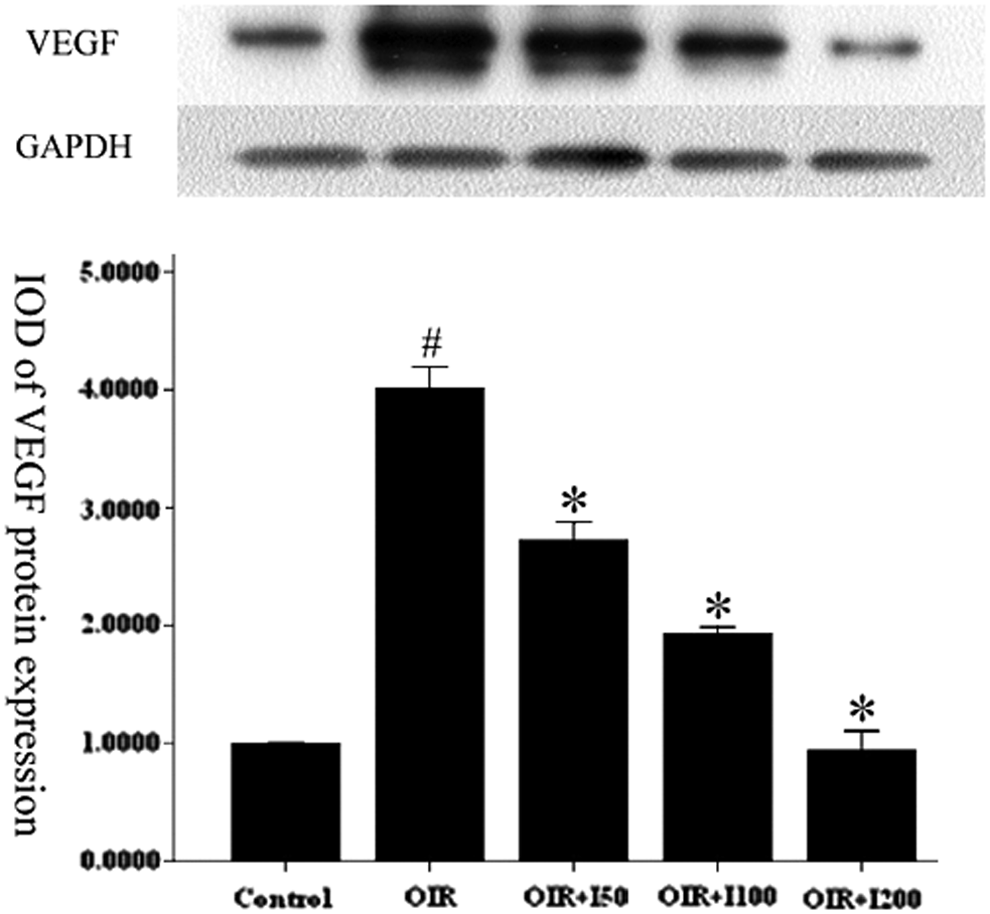
IGFBP-rP1 downregulates VEGF protein expression. Similar to the former methods, the VEGF protein and GAPDH protein were detected by western blotting. The result showed that VEGF higher expressed in OIR intervention group relative to control group. Nevertheless, this overexpression was suppressed by IGFBP-rP1 dose-dependently. The figure below showed the IOD of VEGF relative to the GAPDH. #P < 0.05 versus the control group and *P < 0.05 versus the OIR nonintervention group. (Control: control group, OIR: OIR nonintervention group, OIR+I50: 50 ng/mL IGFBP-rP1 intervention group, OIR+I100: 100 ng/mL IGFBP-rP1 intervention group, and OIR+I200: 200 ng/mL IGFBP-rP1 intervention group). IGFBP-rP1, insulin-like growth factor binding protein-related protein 1; IOD, integrated optical density; OIR, oxygen-induced retinopathy; VEGF, vascular endothelial growth factor.
Discussion
IGFBP family can be divided into two groups according to the affinity with IGF: high-affinity IGFBPs (IGFBP-1 to IGFBP-6) and low-affinity IGFBP-rPs (IGFBP-rP1 to IGFBP-rP10). Moreover, IGFBP-rP1 is distinct from other low-affinity IGFBP-rPs because it can highly bind to insulin. Therefore, IGFBP-rP1 exerts its biological functions such as controlling cell proliferation, differentiation, apoptosis, capillary-like tube formation, stimulating prostacyclin synthesis, and participating in the development of female reproductive system 18 through IGF-independent pattern. Current researches discovered downregulation of IGFBP-rP1 might participate in the tumorigenesis, acting as a cancer suppressive factor in several cancers, including breast cancer, 18 colorectal cancer, 19 as well as prostate cancer. 20
Our research group previously discovered that IGFBP-rP1 was capable of inhibiting REC phenotype activation induced by VEGF in vitro. 8 However, the role of IGFBP-rP1 on retinal neovascularization and its underlying molecular mechanisms was still unclear. To explore the effect of IGFBP-rP1 on retinal neovascularization and its potential mechanisms, we established the mouse model of OIR to observe retinal neovascularization. Meanwhile, we utilized intravitreal injection of recombinant mouse IGFBP-rP1 in a dose-dependent manner to observe the manifestations related to retinal neovascularization, such as fluorescein leakage and neovascular cell nuclei breaking through ILM, to confirm our hypothesis that IGFBP-rP1 might have an inhibitory effect on retinal neovascularization.
Our research indicated that intravitreal injection of recombinant mouse IGFBP-rP1 obviously reduced the range of fluorescein leakage caused by retinal neovascularization and the number of neovascular endothelial cell breaking through the ILM in a dose-dependent manner partly by blocking ERK signaling pathway and downregulating VEGF expression. The discovery prompted IGFBP-rP1, one of REC secretory proteins, could inhibit retinal neovascularization in the mouse model of OIR.
The pathophysiologic mechanisms of retinal neovascularization-dependent diseases are still confused. Numerous cytokines and signaling pathways may be involved in the neovascularization. So far, researchers have discovered that aberrant neovascularization triggered by ischemia 21 and hypoxia 22 may be the main reason for the retinal neovascularization-dependent diseases. In view of retinal neovascularization playing a critical role in the pathogenic process of diabetic retinopathy (DR) and ROP, we considered that IGFBP-rP1 might have a potential therapeutic effect in these neovascularization-dependent diseases.
VEGF plays a pivotal role in retinal neovascularization as a central mediator and a powerful permeability factor in complex angiogenesis cascade reaction. 23 VEGF family has multiple isoforms, such as VEGF121, VEGF145, VEGF165, VEGF185, and VEGF206, owing to alternative splicing of messenger RNA. Some of these isoforms are secretory proteins secreted into vitreous cavity, which are overexpressed in vitreous humors of DR and ROP patients.24–26 The others located in retinal tissue play roles in promoting angiogenesis locally. Our study confirmed that VEGF overexpressed in mice retinal tissues in OIR nonintervention group, in accordance with the previous findings.26–28 Moreover, intravitreal injections of recombinant mouse IGFBP-rP1 could obviously inhibit VEGF expressions in OIR mice retinal tissues and subsequently restrain retinal neovascularization in OIR intervention group, which indicated that IGFBP-rP1 acted as an angiogenesis inhibitor partly through suppressing VEGF generating.
The Ras-Raf-MEK-ERK signaling pathway plays a key role in regulating cell division, proliferation, and apoptosis during pathologic and physiologic process. 29 This pathway was highly associated with tumorigenesis and overactivated in a mount of neoplastic diseases. Persistent activation of this pathway was discovered in the majority of cancers,30–32 leading to uncontrolled cell proliferation and apoptosis inhibition or escapement. Recently, this pathway was discovered to perform an important function in regulating VEGF expression in diabetic rat retinal tissues. 33 Our study discovered that intravitreal injection of recombinant mouse IGFBP-rP1 obviously inhibited p-ERK1/2 expression in OIR mice retinal tissues. Moreover, our research group previously had confirmed that IGFBP-rP1 regulated REC phenotype activation induced by VEGF by means of restraining B-Raf expression. 8
Based on these findings, we hypothesized that B-Raf-MEK-ERK signaling pathway was associated with IGFBP-rP1 inhibiting VEGF expression in OIR mice retinal tissues. B-Raf-MEK-ERK signaling pathway performs a critical biological function in cancer tissues. 34 When the pathway is activated, it is able to stimulate oncogene protein expression. As a result, the overexpression of VEGF protein promotes cancer tissue neovascularization, leading to tumor cell apoptosis inhibition, migration, and organization invasion 35 Likewise, IGFBP-rP1 may act as an antiangiogenic factor through the intervention of B-Raf-MEK-ERK signaling pathway to downregulate VEGF expression and inhibit angiogenesis. In addition, because of its high affinity to insulin, IGFBP-rP1 can restrain combination between insulin and its receptor to inhibit its biological effect, which can stimulate VEGF expression in retinal tissue.36–38 Thus, IGFBP-rP1 might indirectly downregulate VEGF expression in OIR mice retinal tissues by way of inhibiting biological effect of insulin. These two pathways both reflect that IGFBP-rP1 plays its biologic role through the IGF-independent pathway, which is distinct from other IGFBP family members.
In conclusion, our discoveries confirmed that IGFBP-rP1 served as an angiogenesis inhibitor in vivo, which we had uncovered in the previous reports. Furthermore, we revealed that IGFBP-rP1 could inhibit retinal angiogenesis in vivo in a dose-dependent manner by blocking ERK1/2 signaling pathway and downregulating VEGF expression. Therefore, IGFBP-rP1 might become a potential therapeutic target in retinal neovascular disorders and present promising application in the future. However, our research was only focusing on the anatomic abnormalities of retinal neovascularization. The retinal biological changes of OIR mice need to be further explored by functional assessments such as electroretinogram. Despite the significant antiangiogenic effect of IGFBP-rP1 in mice retinas, there are still doubts whether IGFBP-rP1 can also perform well in human retinal tissue as well as its effect on the other ocular tissues.
Footnotes
Acknowledgments
This research was supported by grants from the National Natural Science Foundation for Young Scholars of China (No. 81200701) and Shanghai Municipal Health Bureau Scientific Research Project for Young Scholars (No. 20114Y054).
Author Disclosure Statement
No competing financial interests exist.
References
Supplementary Material
Please find the following supplemental material available below.
For Open Access articles published under a Creative Commons License, all supplemental material carries the same license as the article it is associated with.
For non-Open Access articles published, all supplemental material carries a non-exclusive license, and permission requests for re-use of supplemental material or any part of supplemental material shall be sent directly to the copyright owner as specified in the copyright notice associated with the article.